Essentials of Human Anatomy Physiology Elaine N Marieb

Essentials of Human Anatomy & Physiology Elaine N. Marieb Chapter 7 The Nervous System Copyright © 2003 Pearson Education, Inc. publishing as Benjamin Cummings

Functions of the Nervous System · Sensory input – gathering information · To monitor changes occurring inside and outside the body · Changes = stimuli · Integration · To process and interpret sensory input and decide if action is needed Copyright © 2003 Pearson Education, Inc. publishing as Benjamin Cummings Slide 7. 1 a

Functions of the Nervous System · Motor output · A response to integrated stimuli · The response activates muscles or glands Copyright © 2003 Pearson Education, Inc. publishing as Benjamin Cummings Slide 7. 1 b
 · Brain ·")
Structural Classification of the Nervous System · Central nervous system (CNS) · Brain · Spinal cord · Peripheral nervous system (PNS) · Nerve outside the brain and spinal cord Copyright © 2003 Pearson Education, Inc. publishing as Benjamin Cummings Slide 7. 2
 division · Nerve fibers")
Functional Classification of the Peripheral Nervous System · Sensory (afferent) division · Nerve fibers that carry information to the central nervous system Figure 7. 1 Copyright © 2003 Pearson Education, Inc. publishing as Benjamin Cummings Slide 7. 3 a
 division · Nerve fibers")
Functional Classification of the Peripheral Nervous System · Motor (efferent) division · Nerve fibers that carry impulses away from the central nervous system Figure 7. 1 Copyright © 2003 Pearson Education, Inc. publishing as Benjamin Cummings Slide 7. 3 b
 division · Two subdivisions")
Functional Classification of the Peripheral Nervous System · Motor (efferent) division · Two subdivisions · Somatic nervous system = voluntary · Autonomic nervous system = involuntary Figure 7. 1 Copyright © 2003 Pearson Education, Inc. publishing as Benjamin Cummings Slide 7. 3 c

Organization of the Nervous System Figure 7. 2 Copyright © 2003 Pearson Education, Inc. publishing as Benjamin Cummings Slide 7. 4
 · Astrocytes · Abundant, star-shaped cells · Brace neurons")
Nervous Tissue: Support Cells (Neuroglia) · Astrocytes · Abundant, star-shaped cells · Brace neurons · Form barrier between capillaries and neurons · Control the chemical environment of the brain Figure 7. 3 a Copyright © 2003 Pearson Education, Inc. publishing as Benjamin Cummings Slide 7. 5

Nervous Tissue: Support Cells · Microglia · Spider-like phagocytes · Dispose of debris · Ependymal cells · Line cavities of the brain and spinal cord · Circulate cerebrospinal fluid Figure 7. 3 b, c Copyright © 2003 Pearson Education, Inc. publishing as Benjamin Cummings Slide 7. 6

Nervous Tissue: Support Cells · Oligodendrocytes · Produce myelin sheath around nerve fibers in the central nervous system Figure 7. 3 d Copyright © 2003 Pearson Education, Inc. publishing as Benjamin Cummings Slide 7. 7 a

Nervous Tissue: Support Cells · Satellite cells · Protect neuron cell bodies · Schwann cells · Form myelin sheath in the peripheral nervous system Figure 7. 3 e Copyright © 2003 Pearson Education, Inc. publishing as Benjamin Cummings Slide 7. 7 b

Nervous Tissue: Neurons · Neurons = nerve cells · Cells specialized to transmit messages · Major regions of neurons · Cell body – nucleus and metabolic center of the cell · Processes – fibers that extend from the cell body Copyright © 2003 Pearson Education, Inc. publishing as Benjamin Cummings Slide 7. 8

Neuron Anatomy · Cell body · Nissl substance – specialized rough endoplasmic reticulum · Neurofibrils – intermediate cytoskeleton that maintains cell shape Figure 7. 4 a Copyright © 2003 Pearson Education, Inc. publishing as Benjamin Cummings Slide 7. 9 a


Nerve Fiber Coverings · Schwann cells – produce myelin sheaths in jelly-roll like fashion · Nodes of Ranvier – gaps in myelin sheath along the axon Figure 7. 5 Copyright © 2003 Pearson Education, Inc. publishing as Benjamin Cummings Slide 7. 12

Neuron Cell Body Location · Most are found in the central nervous system · Gray matter – cell bodies and unmylenated fibers · Nuclei – clusters of cell bodies within the white matter of the central nervous system · Ganglia – collections of cell bodies outside the central nervous system Copyright © 2003 Pearson Education, Inc. publishing as Benjamin Cummings Slide 7. 13
 neurons · Carry impulses from the sensory")
Functional Classification of Neurons · Sensory (afferent) neurons · Carry impulses from the sensory receptors · Cutaneous sense organs · Proprioceptors – detect stretch or tension · Motor (efferent) neurons · Carry impulses from the central nervous system Copyright © 2003 Pearson Education, Inc. publishing as Benjamin Cummings Slide 7. 14 a
 · Found in neural pathways in")
Functional Classification of Neurons · Interneurons (association neurons) · Found in neural pathways in the central nervous system · Connect sensory and motor neurons Copyright © 2003 Pearson Education, Inc. publishing as Benjamin Cummings Slide 7. 14 b

Neuron Classification Figure 7. 6 Copyright © 2003 Pearson Education, Inc. publishing as Benjamin Cummings Slide 7. 15

Structural Classification of Neurons · Multipolar neurons – many extensions from the cell body Figure 7. 8 a Copyright © 2003 Pearson Education, Inc. publishing as Benjamin Cummings Slide 7. 16 a

Structural Classification of Neurons · Bipolar neurons – one axon and one dendrite Figure 7. 8 b Copyright © 2003 Pearson Education, Inc. publishing as Benjamin Cummings Slide 7. 16 b

Structural Classification of Neurons · Unipolar neurons – have a short single process leaving the cell body Figure 7. 8 c Copyright © 2003 Pearson Education, Inc. publishing as Benjamin Cummings Slide 7. 16 c

Fire your synapses Build a Neuron!

Build a Motor Neuron! 1. Using the materials at hand build a motor neuron 2. Be sure to include: - dendrite cell body axon myelin sheath schwann cell nodes of Ranvier axon terminal synapse neurotransmitter 3. Include a description of the role each of the above structures plays in nerve cell function. 4. Surround your nerve cell with: astrocytes, microglial cells, and Oligodendrocytes. 5. Explain the supporting role these cells play in nerve tissue

Functional Properties of Neurons · Irritability – ability to respond to stimuli · Conductivity – ability to transmit an impulse · The plasma membrane at rest is polarized · Fewer positive ions are inside the cell than outside the cell Copyright © 2003 Pearson Education, Inc. publishing as Benjamin Cummings Slide 7. 17

Starting a Nerve Impulse · Depolarization – a stimulus depolarizes the neuron’s membrane · A deploarized membrane allows sodium (Na+) to flow inside the membrane · The exchange of ions initiates an action potential in the neuron Figure 7. 9 a–c Copyright © 2003 Pearson Education, Inc. publishing as Benjamin Cummings Slide 7. 18

Resting Membrane Potential

Nerve Impulse Propagation · The impulse continues to move toward the cell body · Impulses travel faster when fibers have a myelin sheath Figure 7. 9 c–e Copyright © 2003 Pearson Education, Inc. publishing as Benjamin Cummings Slide 7. 20

Action Potentials

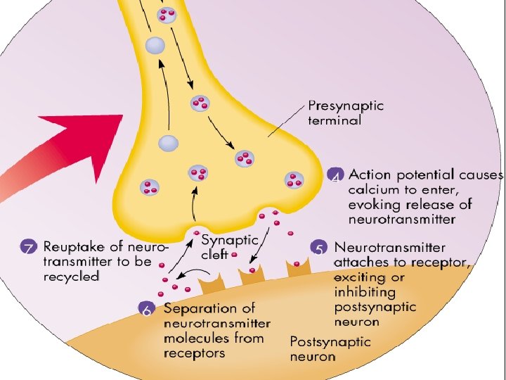
Continuation of the Nerve Impulse between Neurons · Impulses are able to cross the synapse to another nerve · Neurotransmitter is released from a nerve’s axon terminal · The dendrite of the next neuron has receptors that are stimulated by the neurotransmitter · An action potential is started in the dendrite Copyright © 2003 Pearson Education, Inc. publishing as Benjamin Cummings Slide 7. 21

How Neurons Communicate at Synapses Figure 7. 10 Copyright © 2003 Pearson Education, Inc. publishing as Benjamin Cummings Slide 7. 22

Diagram pg 403 • Talk through each event

Synapse

Transmission Across the Synapse Source: Gray

Major Neurotransmitters in the Body Neurotransmitter Role in the Body Acetylcholine Dopamine GABA (gamma-aminobutyric acid) Glutamate Glycine Norepinephrine Serotonin NIH Publication No. 00 -4871

Put It all Together - Stimulus- temperature/pressure on skin light- sound- eyes and ears correct shape molecule taste buds /nose - Action Potential propagates along axon - Neurotransmitter released into synapse - New action potential begins in adjoining nerve or muscle cell
- Slides: 38