Physiology of CNS Stretch Reflex By Dr Abdel


Physiology of CNS Stretch Reflex By Dr. Abdel Aziz M. Hussein Assist Prof. of Physiology

• The divergence function of interneurons is involved in : • • • a- temporal summation b- spatial summation c- reverberation d- irradiation

• Interneuron after-discharge circuits prolong the duration of : • • a- sensory input to the spinal motor centers b- synaptic delay in central synapses c- discharge of efferent neurons d- conscious perception of the evoked sensation

• The ability of stronger stimuli to produce wider range of reflex responses depends upon : • a- presence of reverberating circuits in reflex pathway • b- presence of parallel-chain circuits in reflex pathway • c- convergence of interneurons • d- divergence of interneurons

• Recruitment of a reflex response is due to : • a- difference in the amount of presynaptic inputs to the various efferent neurons initiating the reflex • b- difference in the conduction velocity of the various afferent neurons mediating the reflex • c- delay at the neuromuscular junction • d- presence of inhibitory interneurons in the reflex pathway

• After-discharge of reflex responses : • a- increase the magnitude of the reflex responses • b- delays the onset of fatigue of reflex responses • c- involves interneuron circuits • d- depends upon spatial summation

Stretch Reflex
 Reflex Def It is a reflex contraction of a muscle when it")
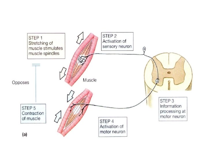
Stretch (Myotatic) Reflex Def It is a reflex contraction of a muscle when it is passively stretched Pathway 1. Stimulus: → passive stretch 2. Receptors: → ms spindles 3. Afferents: → fast-conducting Aα (Ia) and Aβ (II)nerve fibers 4. Centers: → alpha motor neurons in AHCs. 5. Efferents: → axons of alpha motor neurons. 6. Effector organ: → Extrafusal ms fibers 7. Response: → ms contraction.

Stretch Reflex

Ms spindle 1 ry and 2 ry endings AHCs Extrafusal ms fibers Alpha motor neurons

Muscle Spindle Site: • Fleshy parts of skeletal ms parallel to their fibres (extrafusal ms fibers). Shape: • It is capsulated fusiform stretch receptor. Structure: • Each spindle consists of several small ms fibres (412 fibers) called intrafusal fibres.

Muscle Spindle
Nuclear bag")
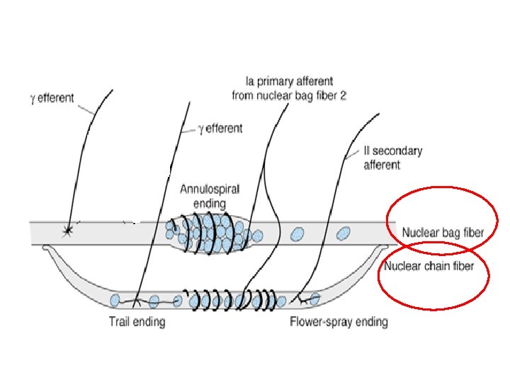
Muscle Spindle Types of intrafusal ms fibres: • There are 2 types; 1)Nuclear bag fibres; • Have a dilated central area filled with nuclei. • Are 2 of these fibres per spindle. 2)Nuclear chain fibres; • Have nuclei which are arranged as a chain in the receptor area. • Are 5 -8 of these fibres per spindle.
Central part: • It is")
Muscle Spindle Each ms fiber consists of 2 parts; a)Central part: • It is non-contractile part non-contractile • constitutes the receptor areas of the spindles receptor areas • It receives sensory innervation b)Peripheral part: • It is a contractile part • when contracts, it causes stretch of the central receptor area. • It receives motor innervation

Muscle Spindle

Muscle Spindle
 Afferent (sensory) innervations Anulospiral or 1 ry afferents Type")
Innervation of Muscle Spindle A) Afferent (sensory) innervations Anulospiral or 1 ry afferents Type Ia or A alpha (16 um) Supply nuclear bag and chain Rapidly responding and adapting Flower spray or 2 ry afferents Type II or A beta (8 um) Supply nuclear chain on sides of 1 ry endings Slowly Rapidly responding and adapting

1 ry endings 2 ry endings
 Efferent (motor) innervations Dynamic gamma motor neuron Static gamma")
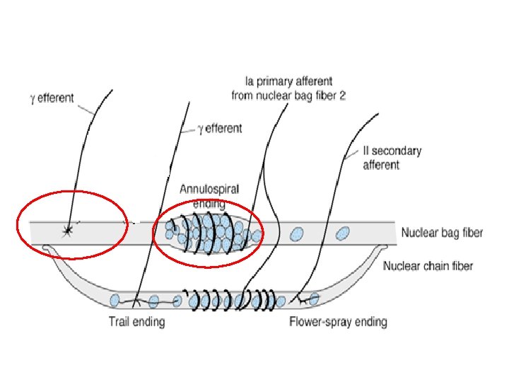
Innervation of Muscle Spindle B) Efferent (motor) innervations Dynamic gamma motor neuron Static gamma motor neuron A gamma (4 um) Supply nuclear bag Supply nuclear chain Enhance dynamic response Enhance static response

Efferent Innervation of Muscle Spindle

Stimulation of Muscle Spindle • The adequate stimulus that excites the ms spindles is stretching of their central part→ depolarizes it→ initiates an AP in 1 ry and 2 ry endings. • This can occur in 2 ways: • 1) Passive stretch of the whole ms: • It causes stretch of the ms spindle which lies parallel to ms fibers. Muscle spindle Whole Muscle Afferents
 Passive stretch of the whole ms:")
Stimulation of Muscle Spindle • 1) Passive stretch of the whole ms:
Activation of the γ-MNs: • By supraspinal centers or")
Stimulation of Muscle Spindle • 2)Activation of the γ-MNs: • By supraspinal centers or reflexely • It causes contraction of the peripheral part the intrafusal fibres→ stretch of receptor area
Activation of the γ-MNs:")
Stimulation of Muscle Spindle 2)Activation of the γ-MNs:
 Dynamic Response")
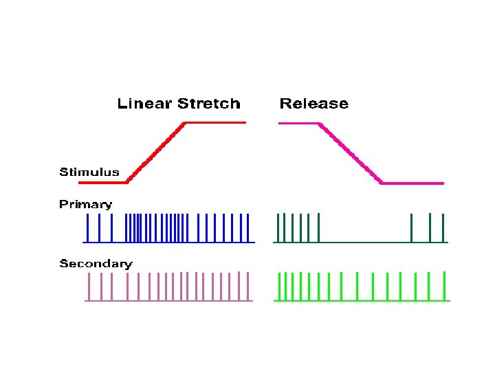
Types of Responses of Ms spindle to Stretch (Types of Stretch Reflex) Dynamic Response Stimulus Sudden stretch Static Response Maintained (steady) stretch Receptors nuclear bag nuclear chain Afferents 1 ry endings primary and secondary endings Center Alpha motor neurons Response Rapid contraction Maintained followed by rapid subtetanic relaxation contraction Examples e. g. tendon jerk e. g. muscle tone
 Dynamic response")
Types of Responses of Ms spindle to Stretch (Types of Stretch Reflex) Dynamic response Nuclear bag (1 ry) Basal dischar ge Nuclear chain (1 ry & 2 ry endings Static response

Dynamic stretch Reflex 1 ry endings Sudden stretch Nuclear bag AHCs Alpha MNs

Static stretch reflex AHCs 1 ry and 2 ry endings Alpha MNs Nuclear chain maintained stretch e. g. gravity

Functions of Stretch Reflex or Muscle Spindle

Functions of stretch reflex or Ms Spindle 1. Generation of ms tone 2. Smoothing of ms contraction or Damping function 3. Load Reflex: stabilize joint posture 4. Proprioceptive Functions

1. Skeletal Muscle Tone

Skeletal Ms Tone Def. • It is a state of Flexors of the UL. continuous (partial or Elevators of the lower jaw subtetanic) contraction of skeletal ms during rest. Back ms and back of neck Distribution: • It is present in all Anterior abdominal wall ms. skeletal ms but specially in the antigravity ms Extensors of LL

Skeletal Ms Tone Mechanism: • It is a static type of SR Continuous mild stretch of skeletal ms because ms length is shorter than distance ( ) origin and insertion Maintained stretch of nuclear chain fibers Continuous mild discharge along 2 ry endings (flower spray) Stimulate α-MNS of muscle Mild continuous (partial) contraction of skeletal ms

Muscle Tone AHCs 1 ry and 2 ry endings Alpha MNs Nuclear chain maintained stretch e. g. gravity

Skeletal Ms Tone Dynamic state of skeletal ms tone: • The ms tone is not static but dynamic • in upright standing the magnitude of the ms tone is greater in the antigravity ms especially extensors of lower limb and trunk • If the trunk is tilted backward the ms tone ↑es in ms of anterior abdominal wall and ↓es in extensors. • This by modulation of ɤ-MNs activity by the supraspinal centers which may facilitate or inhibit the muscle tone

Skeletal Ms Tone Regulation of skeletal ms tone: • By supraspinal centers (facilitatory or inhibitory) Inhibitory areas Facilitatory areas 1. Area 4 2. Neocerebellu m 3. Excitatory RF 4. Vestibular N. 1. Area 6 + - 2. Paleocerebllum 3. Inhibitory RF 4. Red N.
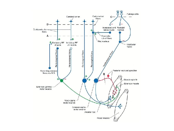
 Postural control: • Is the")
Skeletal Ms Tone Functions of skeletal ms tone: a) Postural control: • Is the basic mechanism for control of posture and equilibrium • By adjusting the magnitude of ms tone of different groups of ms. b) Help in heat production and maintain of body temperature c) It helps both the venous return & lymph flow: • Ms tone has a mild squeezing effect on the walls of veins and lymphatics of skeletal ms→ help venous return to the heart. d) Keeps viscera in position and prevents visceroptosis:

2. Damping Function or smoothing of ms contraction

Smoothing or Damping Function

Smoothing or Damping Function • Stretch reflex prevents oscillations or jerkiness of body movements • Motor signals from the motor areas are transmitted to the ms in an unsmooth form (↑ for few Sec and ↓ for another Sec) • This causes irregularities or oscillations of movements • The signals discharged from the ms spindles cause partial activity of αMNs of the ms • So, the motor signals find αMNs in state of partial activity, so they cause continuous activation of them → cause smooth ms contraction

Smoothing or Damping Function • When the signal decrease, produces weak direct contraction less shortening of the ms the stretch reflex signals will increase more contraction • The resultant contraction with be sum of both direct and reflex. • When the signal increase, produces strong direct contraction more shortening of the ms the stretch reflex signals will decrease less contraction. • The resultant contraction will be the sum of both direct and reflex
 Reflex contraction )")
Direct contraction ) Reflex contraction )

Smoothing or Damping Function • Different signal intensities produce equal, average contraction • This function of the stretch reflex is consequently termed signal averaging function • Interruption of the ms spindles sensory discharge cause the ms contraction to become unsmooth and jerky.

Smoothing or Damping Function

3. Load Reflex

Load Reflex • Def. , • It is a reflex responsible for keeping the hand or foot in position when a moderate loads are applied • E. g. when a person holding up a cup while someone is filling it with tea, as the load gets bigger and bigger, the force required to keep the hand in position must be continually increased.
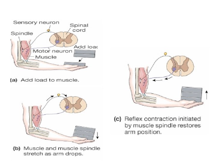
 • The higher motor centers")
Load Reflex • Mechanism (Coactivation of α-MNs and γ-MNs) • The higher motor centers activate α-MNs to initiate contraction of a ms and also activate γ-MNs discharge →coactivation of α-MNs and γ-MNs. • This increases the sensitivity of the ms spindles to any slight degree of stretch produced by increasing the load consequently

Load Reflex

4. Proprioceptive Functions

Proprioceptive Functions • Ms spindles provide proprioceptive information to the brain and cerebellum for keeping them continually informed about muscle length and changes in that length

Gamma Motor Neurons

Gamma motor Neurons Alpha motor neuron 70% Gamma motor neuron 30 %

Gamma MNs Site: • • γ-MNs are small motors neurons represent 30% of AHCs. The axons (about 4 u) supply the peripheral parts of intrafusal ms fibers. Types: • Are 2 types of γ-MNs 1. Dynamic or d- γ-MNs→ supply nuclear bag ms fibers. 2. Static or s- γ-MNs→ supply nuclear chain ms fibers.

Functions of Gamma MNs • They adjust ms spindle sensitivity • ↑ γ-MNs cause contraction of the peripheral parts of intrafusal fibers → stretch of central parts of ms spindle → ↑es the sensitivity of the ms spindle to stretch i. e. ms spindle needs a small amount of passive stretch to be stimulated • Vice versa.

Functions of Gamma MNs

Functions of Gamma MNs
 Stabilization of body")
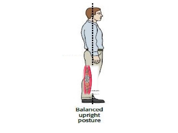
Functions of Gamma MNs • This is important in • A) Stabilization of body position and equilibrium (standing); • In upright posture , there is ↑ in the descending excitatory signals from the facilitatory RF in the brain stem to both and -MNs of the antigravity ms. • These excitatory signals ↑ the contraction of the extrafusal ms fibers and at the same time the ms spindle sensitivity increased.

Left Internal Capsule Midbrain L A Facilitatory Pontine RF Pons Medulla Alpha MN Gamma MN Spinal Cord
 Stabilization of body position and equilibrium (standing); •")
Functions of Gamma MNs • A) Stabilization of body position and equilibrium (standing); • Also deviation of the upright attitude toward any direction → will cause additional stretching of the postural ms →↑ ms tone of these ms → restores equilibrium before it is disturbed.

Functions of Gamma MNs B. Load reflex
 Load reflex (coactivation of alpha and gamma MNs)")
Functions of Gamma MNs • B) Load reflex (coactivation of alpha and gamma MNs)

Higher motor centers Left Internal Capsule Midbrain L A Pons Medulla Alpha MN Gamma MN Spinal Cord

Control of Gamma MNs Discharge

Supraspinal control of Stretch Reflex Facilitatory Areas Inhibitory Areas

Facilitatory Areas Inhibitory Areas Supraspinal facilitation is predominant +- ↓ ms tone and stretch reflex

Supraspinal control of Stretch Reflex Facilitatory Areas Inhibitory Areas

Facilitatory Areas Area 4 Facilitatory R. F. Neocerebellum Vestibular N. Reticulospinal T. Vestibulospinal T. Corticospinal T.

Inhibitory Areas Area 6 Basal ganglia Paleocerebellum Inhibitory R. F. Reticulospinal T.

Supraspinal Control of Stretch Reflex • At the cortical level, the net effect of area 4 & area At the cortical level, 6 & area 4 s on the stretch reflex & muscle tone is inhibitory • So, a lesion causing damage of area 4, 4 s & 6 (UMNL) leads to increase in muscle tone • In animals, separation ( ) cerebral cortex & brain stem → marked ↑ in ms tone due to the removal of the net inhibitory effect of the cerebral cortical areas & leaving the facilitatory areas centers in the brain stem (VN and facilitatory RF) to act. • ↑ed ms tone leads to a state known as Decerebrate Rigidity.

Properties of Stretch Reflex
 Has a short latent period: • has a very")
Properties of Stretch Reflex 1) Has a short latent period: • has a very short time between start of stretch and start of contraction. • It is due to ; a)It is monosynaptic. b)Its afferent and efferents are rapidly conducting nerve fibers. 2)High localization: • It is highly localized i. e. contraction occurs only in the stretched ms. • No divergence due to absence of interneurons

 It has no recruitment nor after discharge: • It")
Properties of Stretch Reflex 3) It has no recruitment nor after discharge: • It is due to lack of interneurons 4)Graded response: • The strength of ms contraction is directly proportional to the extent of stretch. 5) Reciprocal innervation: • In which stretch of a ms results in reflex contraction of the stretched ms and relaxation of the antagonistic ms.
Resist fatigue: • Sustained for a prolonged period without fatigue")
Properties of Stretch Reflex 6)Resist fatigue: • Sustained for a prolonged period without fatigue (e. g. in antigravity ms): • Delayed fatigue is due to; a)Alternation between motors units during stretch reflex i. e. not all units contracting at the same time. b)Antigravity ms are tonic (slow) ms which resist fatigue because; 1. Rich in blood supply 2. Rich in mitochondria 3. Its contraction is slow.

Inverse Stretch Reflex
 of a ms")
Inverse Stretch Reflex • It is a reflex relaxation (or lengthening) of a ms in response to excessive stretch or contraction of that ms.

Inverse Stretch Reflex

Inverse Stretch Reflex

Inverse Stretch Reflex

Inverse Stretch Reflex Neural pathway: • Stimulus: ↑ed ms tension by; 1. Overstretch or 2. Severe contraction • Receptors: Golgi tendon organs 1) Site: • tendons of skeletal ms in series with ms fibers 2) Structure: • Are encapsulated sensory receptor • 6 -20 elastic fibers 3) Innervations:
 Receptors: GTOs • Stimulated by ↑ed ms tension caused by")
Golgi Tendon Organs (GTOs) Receptors: GTOs • Stimulated by ↑ed ms tension caused by passive overstretch or active contraction of the ms Afferents: • A alpha or Ib Center : a)inhibitory interneurons→ inhibit the α -MNs supplying the same ms b)excitatory interneurons→ excite the αMNs supplying the antagonistic ms Response: • Relaxation of the same ms • Contraction of antagonistic group of
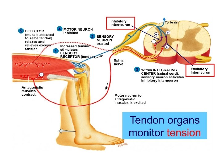
Physiological significance: • Protective reaction which prevent tearing of")
Inverse Stretch Reflex Significance GTR: a)Physiological significance: • Protective reaction which prevent tearing of the ms or avulsion of its tendon from its bony attachment when the ms is overstretched.
Clinical significance: (clasp knife effect) • Demonstrated clinically by")
Inverse Stretch Reflex Significance GTR: b)Clinical significance: (clasp knife effect) • Demonstrated clinically by passive flexion of a spastic limb (e. g. in upper motor neuron lesions) at its main joint. • As the limb is flexed, an initial resistance occurs due to contraction of this ms a result of the stretch reflex. • With persistent flexion, at a certain point, GTR is excited→ sudden disappearance of the initial resistance → the limb flexes easily, as occurs due closing-of a pocket knife→ clasp knife effect. • E. g. Flexion of knee and ankle

Tendon Jerk

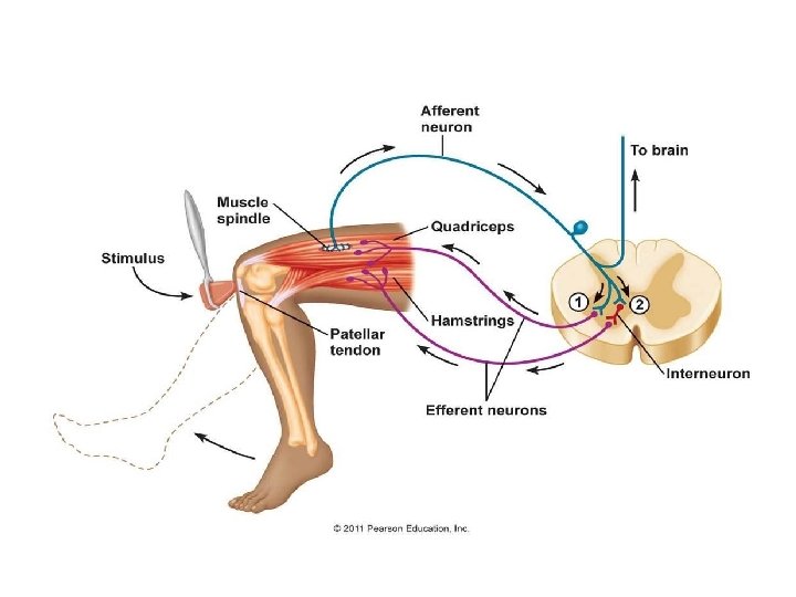
Tendon Jerks Def. , • Rapid contraction followed by relaxation of a ms due to sudden stretching of that ms by tapping on its tendon using a medical hammer Mechanism: • It is a dynamic type of the stretch reflex

The Tendon Reflex To brain 6 Primary afferent neuron stimulates inhibitory interneuron 4 Primary afferent neuron stimulates alpha motor neuron to extensor muscle 1 ry endings 3 Primary afferent neuron excited Nuclear bag Alpha MNs 7 Interneuron inhibits alpha motor neuron to flexor muscle 5 Alpha motor neuron stimulates extensor muscle to contract Ms contraction Sudden stretch 2 Muscle spindle stimulated 1 Extensor muscle stretched Flexor muscle 8 (antagonist) relaxes

Tendon Jerks
 Stoppage")
Tendon Jerks • Cause of relaxation after contraction in the tendon jerk: a) Stoppage of discharge from the ms spindles. b) Stimulation of the Golgi tendon organs. c) Stimulation of the Renshaw’s Cells.

Examples of Tendon Jerks Jerk Biceps jerk Triceps jerk Knee jerk Ankle jerk Jaw jerk Center Limb position Tendon Response C 5, 6 The elbow is Tapping on biceps Flexion of 120° tendon the forearm C 6, 7 The elbow is Tapping on triceps Extension of 90° tendon directly the forearm L 2, 3 & 4 knee is semi Tapping on Extension of flexed by patellar tendon the knee seating with the leg to be tested crossing over other S 1, 2 feet slightly Tapping dorsiflexed tendoachilles on Plantar flexion. Trigemin Mouth slightly Tapping on chin al nerve opened Closure mouth of

Biceps Jerk

Triceps Jerk

Knee Jerk

Ankle Jerk

Tendon Jerk Reinforcement of the tendon jerks • The response of the tendon jerks can be reinforced by facilitating the spinal centers. • This can be done by either; a) Jendrassik's maneuver → ask the patient to hook his fingers or to clench his teeth→ send signals from the contracted ms which stimulating γ-MNs. b) Distracting patient’s attention→ prevents any voluntary inhibition of the reflex.

Clinical Significance of TJ 1. Localization of spinal cord lesions: • Loss of TJ means the lesion in its center e. g. ankle jerk is lost in sacral region lesion. 2. Assessment of the ms tone : • In hyperreflexia (exaggerated tendon jerks) → hypertonia (↑ms tone). • In hyporeflexia (↓ed tendon jerks) → hypotonia (↓ms tone). • In areflexia (lost tendon jerks) → atonia (lost ms tone).

Clinical Significance of TJ 3. Assessment of the integrity of pathway of stretch reflex: so areflexia or absent tendon jerk may be due to; Site of lesion Condition • Afferent lesion Tabes dorsalis • Center (AHC) lesion Poliomyelitis • Efferent lesion Trauma or neuritis 4. Assessment of the state of Supraspinal centers: Hyperactive(exaggerated) Hypoactive (decreased) TJ TJ Physiological causes Anxiety and nervousness Sleep and anaesthesia Pathological causes UMNL Lesion in area 6 Tetany and hyperthyroidism Paleocerebellum lesion LMNL Lesion in area 4 Hypothyroidism Neocerebellar syndrome

Pendular Knee Jerk • Occurs in the neocerebellar syndrome and chorea. • Characterized by hyporeflexia & hypotonia • Knee jerk is weak than normal and during relaxation of the quadriceps ms, the leg falls like a dead weight(due to hypotonia) & swings for sometime like a pendulum before resting.

Clonus Def. • Alternating regular rhythmic contractions with incomplete relaxations of a ms (its MNs is in a state of facilitation) in response to sudden maintained stretch. Cause: • UMNL
 Ankle Clonus: • Produced by sudden maintained dorsiflexion of the foot")
Clonus Types 1) Ankle Clonus: • Produced by sudden maintained dorsiflexion of the foot leads to regular rhythmic planter flexions due to rhythmic contractions of soleus and gastrocnemius muscles. 2) Knee Clonus: • Produced by the sudden downward displacement of the patella rhythmic oscillations of the patella.

Clonus Mechanism of clonus: • Clonus is the result of a stretch reflex - inverse stretch reflex sequence, which occurs as follows : • Sudden stretch of the ms results in its contraction through the stretch reflex. • This is followed by relaxation due to; a) Stoppage of impulse discharge from the ms spindles. b) Initiation of an inverse stretch reflex due to stimulation of the GTOs. • As stretch is maintained, a new stretch reflex occurs (helped by the state of excessive spinal facilitation), and the cycle is repeated.

Test yourself The shortest reflex time is recorded with : a- a flexor withdrawal reflex b-an inverse stretch reflex c- a stretch reflex d- a scratch reflex A tendon jerk : a- is a dynamic stretch reflex b- is a static stretch reflex c- is evoked by gradually stretching the muscle d- is evoked by stimulation of tendon receptors

A reflex arc includes : a- at least two sets of sequential neurons b- at least two sequential sets of central synapses c- at least two types of sensory receptors d- at least two types of efferent neurons

Stretch reflex is characterized by the following except : a- disynaptic reflex b- high localization c- shows reciprocal innervations. d- it is of graded response

The nuclear-chain fibers of spindles are innervated by : a- Aα and Aδ nerve fibers b- Aδ and C nerve fibers c- Ia and II nerve fibers d- only type II nerve fibers

Inverse stretch reflex : a- increases the possibility of avulsion of the excessively stretched muscle from its bony attachments b- has no reciprocal innervation circuits c- is clinically manifested by lengthening reaction d- is clinically tested by examining the tendon jerks

THANKS
- Slides: 119