
Urinary System Models The Kidney Nephron the functional


- Slides: 38
Urinary System Models
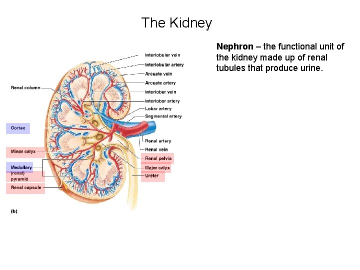
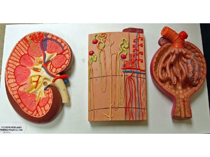
The Kidney Nephron – the functional unit of the kidney made up of renal tubules that produce urine.
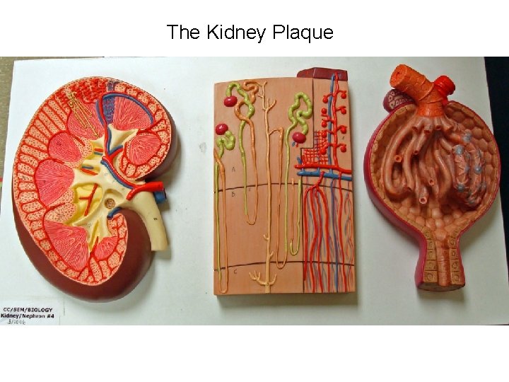
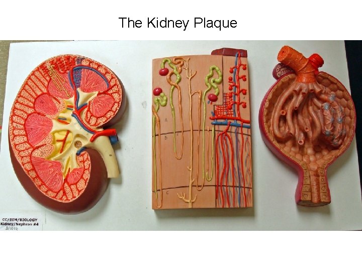
The Kidney Plaque
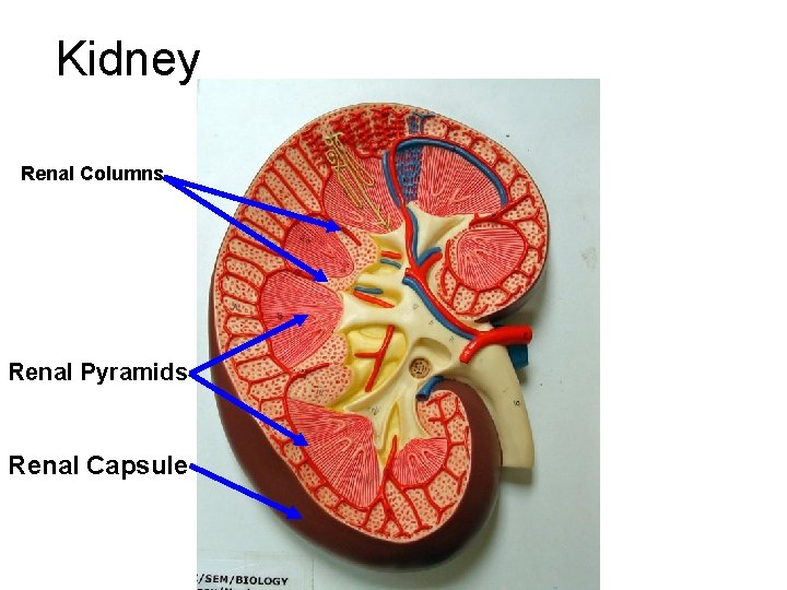
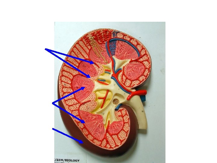
Kidney Renal Columns Renal Pyramids Renal Capsule
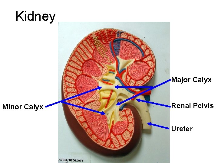
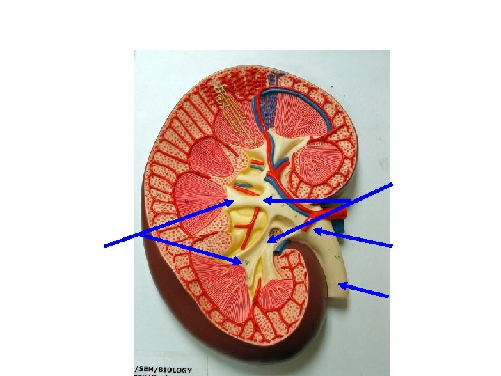
Kidney Major Calyx Minor Calyx Renal Pelvis Ureter
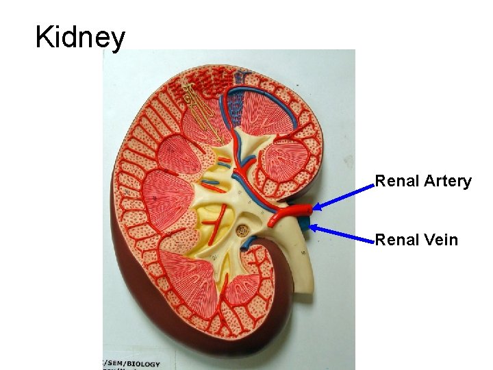
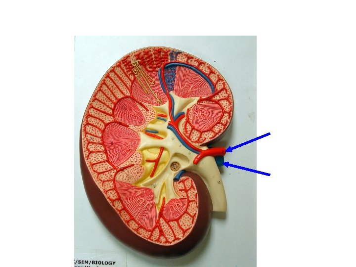
Kidney Renal Artery Renal Vein
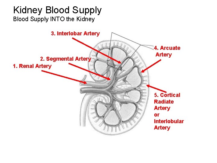
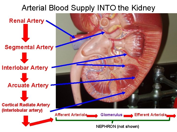
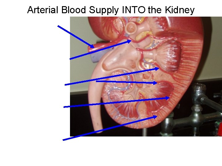
Kidney Blood Supply INTO the Kidney 3. Interlobar Artery 2. Segmental Artery 1. Renal Artery 4. Arcuate Artery 5. Cortical Radiate Artery or Interlobular Artery
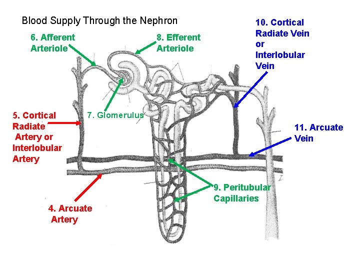
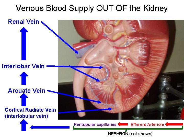
Blood Supply Through the Nephron 6. Afferent Arteriole 5. Cortical Radiate Artery or Interlobular Artery 8. Efferent Arteriole 10. Cortical Radiate Vein or Interlobular Vein 7. Glomerulus 11. Arcuate Vein 9. Peritubular Capillaries 4. Arcuate Artery
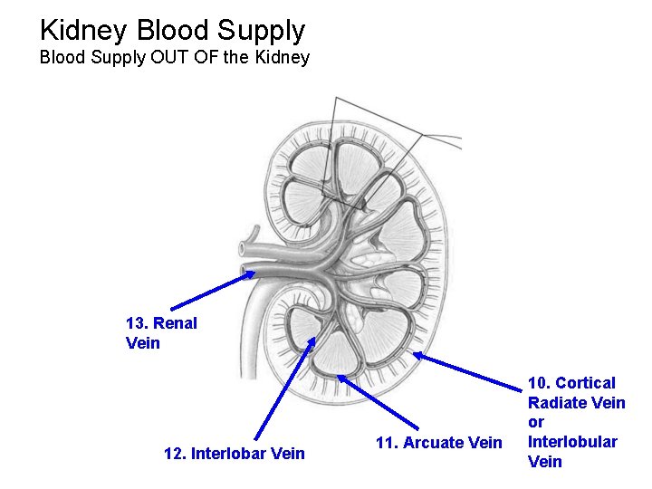
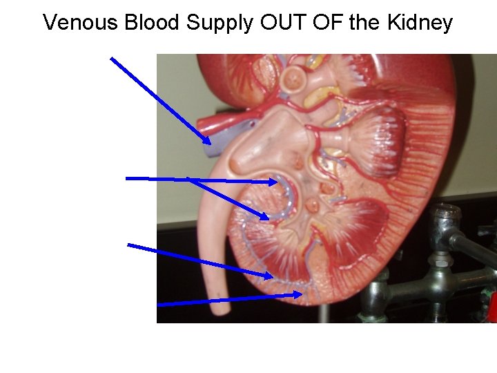
Kidney Blood Supply OUT OF the Kidney 13. Renal Vein 12. Interlobar Vein 11. Arcuate Vein 10. Cortical Radiate Vein or Interlobular Vein
Arterial Blood Supply INTO the Kidney Renal Artery Segmental Artery Interlobar Artery Arcuate Artery Cortical Radiate Artery (Interlobular artery) Afferent Arteriole Glomerulus Efferent Arteriole NEPHRON (not shown)
Venous Blood Supply OUT OF the Kidney Renal Vein Interlobar Vein Arcuate Vein Cortical Radiate Vein (interlobular vein) Peritubular capillaries Efferent Arteriole NEPHRON (not shown)
The Kidney Plaque
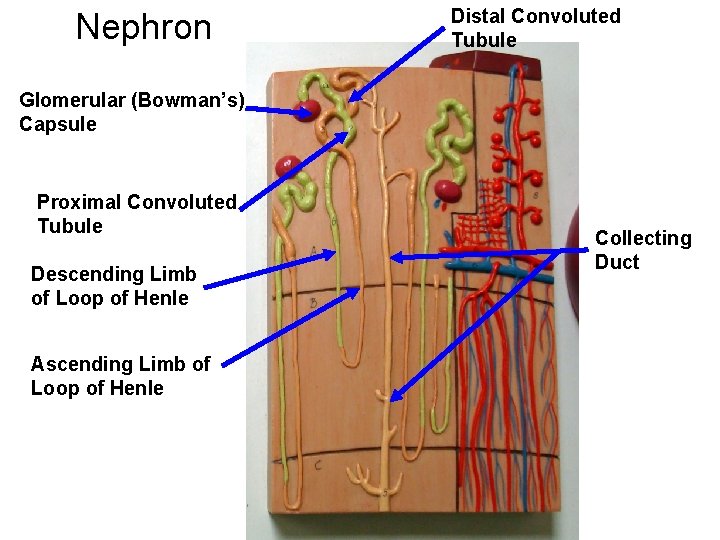
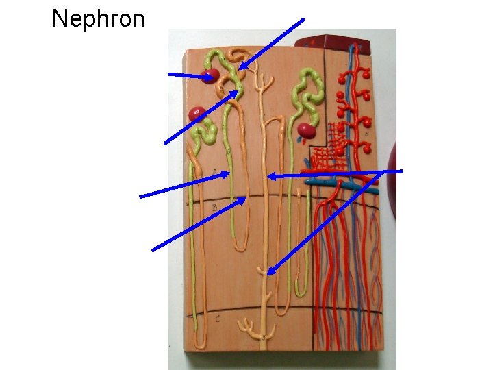
Nephron Distal Convoluted Tubule Glomerular (Bowman’s) Capsule Proximal Convoluted Tubule Descending Limb of Loop of Henle Ascending Limb of Loop of Henle Collecting Duct
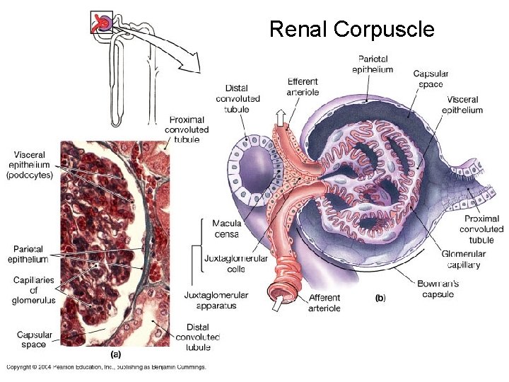
Renal Corpuscle
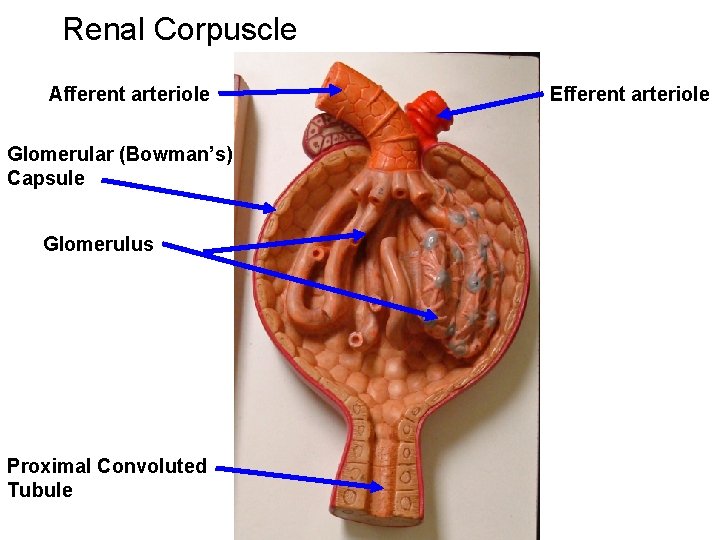
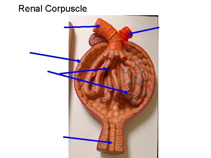
Renal Corpuscle Afferent arteriole Glomerular (Bowman’s) Capsule Glomerulus Proximal Convoluted Tubule Efferent arteriole
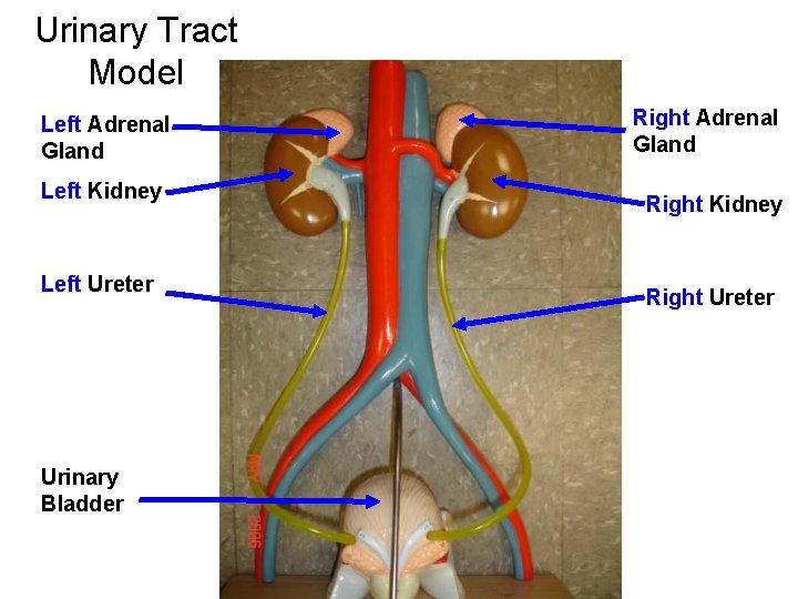
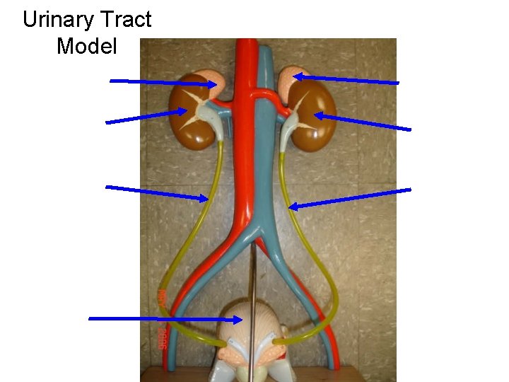
Urinary Tract Model Left Adrenal Gland Left Kidney Left Ureter Urinary Bladder Right Adrenal Gland Right Kidney Right Ureter
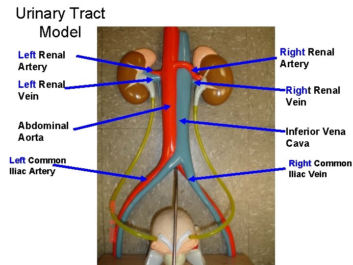
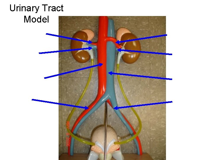
Urinary Tract Model Left Renal Artery Left Renal Vein Abdominal Aorta Left Common Iliac Artery Right Renal Vein Inferior Vena Cava Right Common Iliac Vein
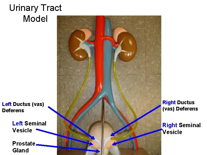
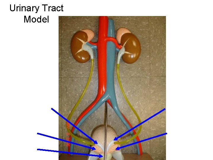
Urinary Tract Model Left Ductus (vas) Deferens Left Seminal Vesicle Prostate Gland Right Ductus (vas) Deferens Right Seminal Vesicle
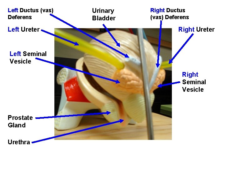
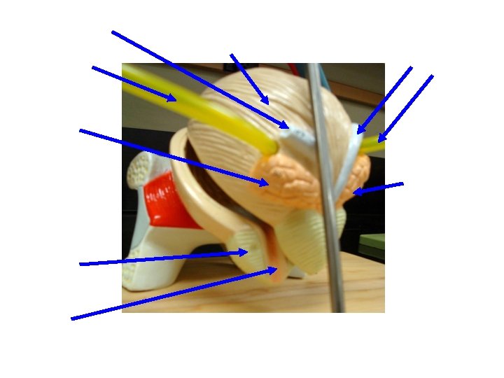
Left Ductus (vas) Deferens Left Ureter Urinary Bladder Right Ductus (vas) Deferens Right Ureter Left Seminal Vesicle Right Seminal Vesicle Prostate Gland Urethra
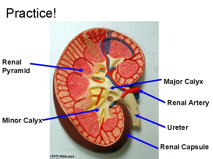
Practice! Renal Pyramid Major Calyx Renal Artery Minor Calyx Ureter Renal Capsule
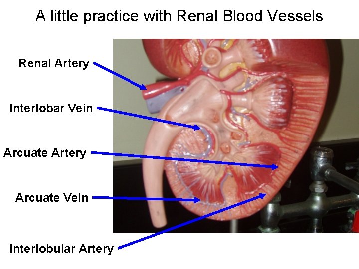
A little practice with Renal Blood Vessels Renal Artery Interlobar Vein Arcuate Artery Arcuate Vein Interlobular Artery
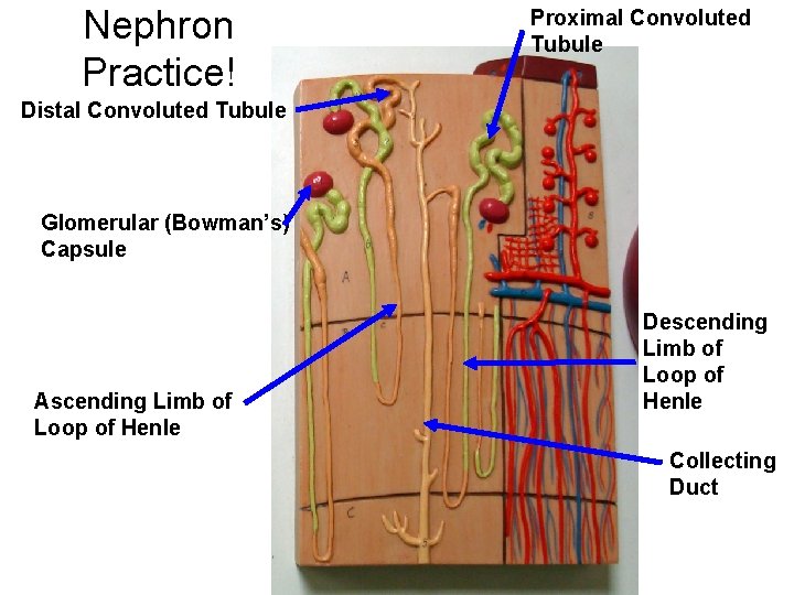
Nephron Practice! Proximal Convoluted Tubule Distal Convoluted Tubule Glomerular (Bowman’s) Capsule Ascending Limb of Loop of Henle Descending Limb of Loop of Henle Collecting Duct
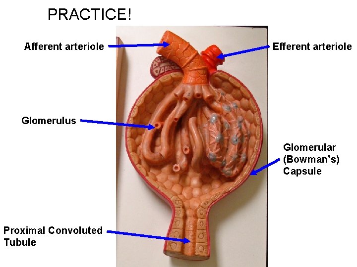
PRACTICE! Afferent arteriole Efferent arteriole Glomerulus Glomerular (Bowman’s) Capsule Proximal Convoluted Tubule
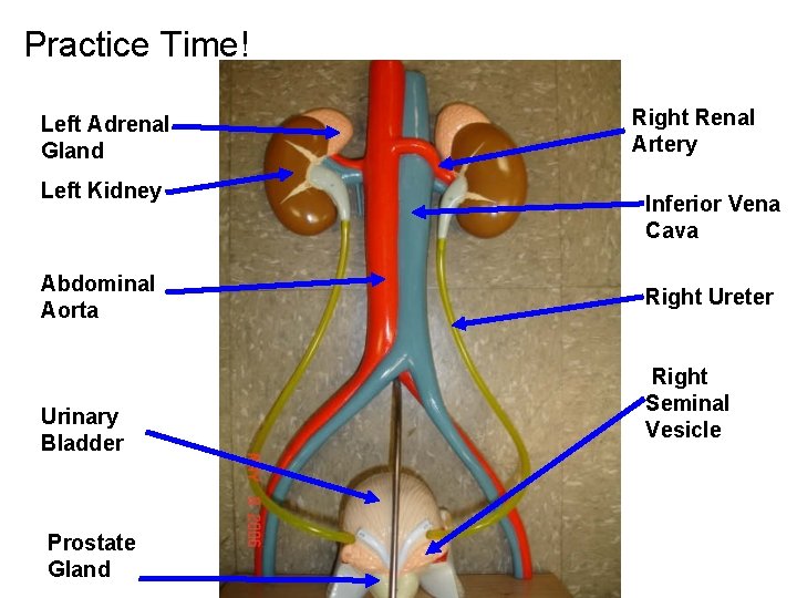
Practice Time! Left Adrenal Gland Left Kidney Abdominal Aorta Urinary Bladder Prostate Gland Right Renal Artery Inferior Vena Cava Right Ureter Right Seminal Vesicle
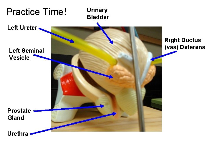
Practice Time! Urinary Bladder Left Ureter Left Seminal Vesicle Prostate Gland Urethra Right Ductus (vas) Deferens
Practice Slides for Labeling
Arterial Blood Supply INTO the Kidney
Venous Blood Supply OUT OF the Kidney
Nephron
Renal Corpuscle
Urinary Tract Model
Urinary Tract Model
Urinary Tract Model