Nervous Tissue Dr Michael P Gillespie Structures of


 w Sensory neurons. w Motor neurons located in skeletal muscles.")
 w Sensory neurons from the autonomic sensory receptors in the")

 w “The brain of the gut”. w Functions independently of")

. w")


- Slides: 78
Nervous Tissue Dr. Michael P. Gillespie
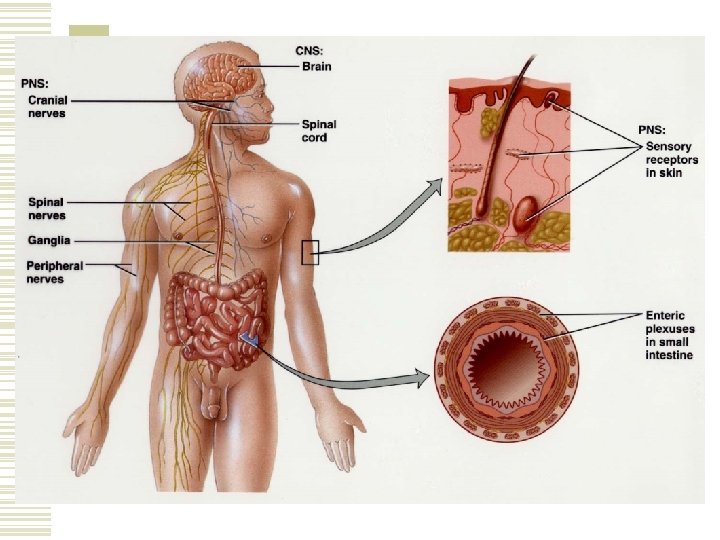
Structures of the Nervous System w Brain w Spinal cord w Nerves w Cranial nerves w Ganglia w Sensory receptors
Functions of the Nervous System w Sensory function – afferent neurons w Integrative function - interneurons w Motor function – efferent neurons n The cells contacted by these neurons are called effectors
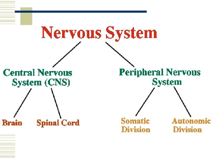
Organization of the Nervous System w Central nervous system n n Brain Spinal cord
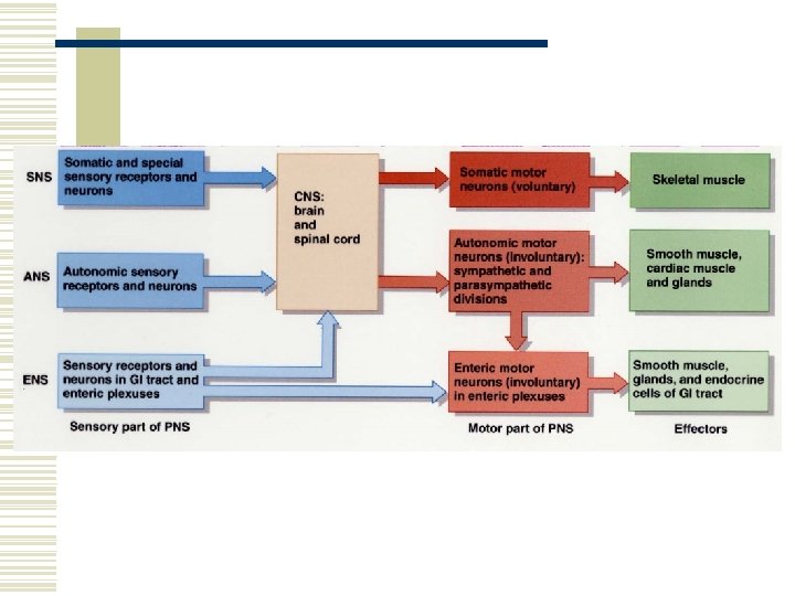
Organization of the Nervous System w Peripheral nervous system n n n n Cranial nerves and their branches Spinal nerves and their branches Ganglia Sensory receptors Somatic nervous system Autonomic nervous system Enteric nervous system
Somatic Nervous System (SNS) w Sensory neurons. w Motor neurons located in skeletal muscles. w The motor responses can be voluntarily controlled; therefore this part of the PNS is voluntary.
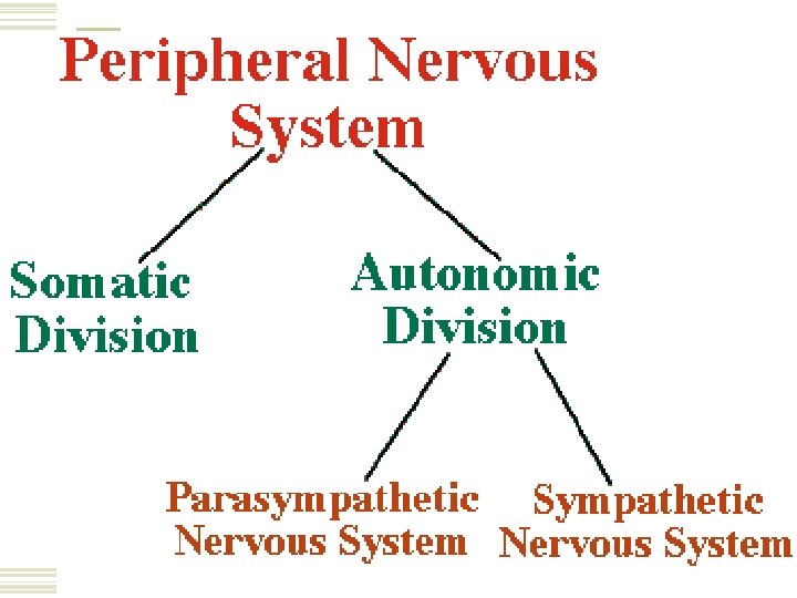
Autonomic Nervous System (ANS) w Sensory neurons from the autonomic sensory receptors in the viscera. w Motor neurons located in smooth muscle, cardiac muscle and glands. w These motor responses are NOT under conscious control; Therefore this part of the PNS is involuntary.
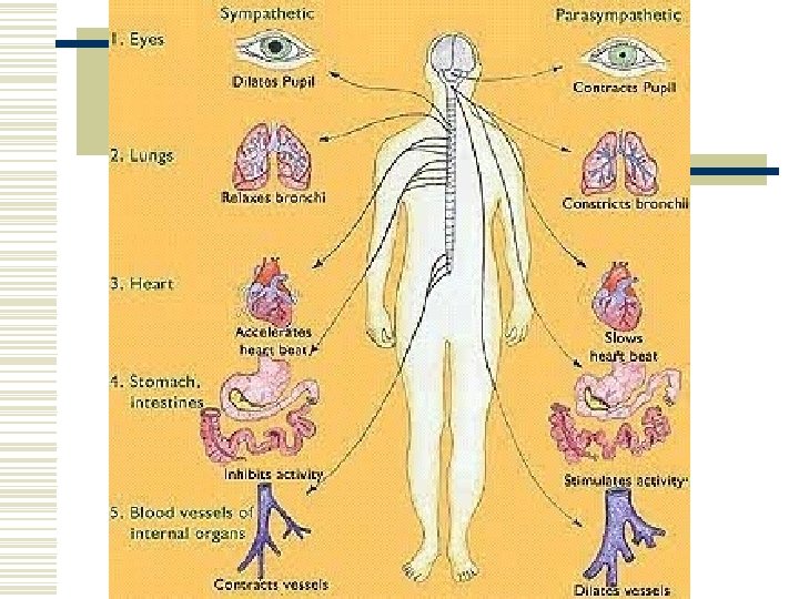
ANS Continued… w The motor portion of the ANS consists of sympathetic and parasympathetic divisions. w Both divisions typically have opposing actions.
Enteric Nervous System (ENS) w “The brain of the gut”. w Functions independently of the ANS and CNS, but communicates with it as well. w Enteric motor units govern contraction of the GI tract. w Involuntary.
Nervous Tissue w Neurons. n n n Sensing. Thinking. Remembering. Controlling muscular activity. Regulating glandular secretions. w Neuroglia. n Support, nourish, and protect neurons.
Neurons w Have the ability to produce action potentials or impulses (electrical excitability). w Action potentials propagate from one point to the next along the plasma membrane.
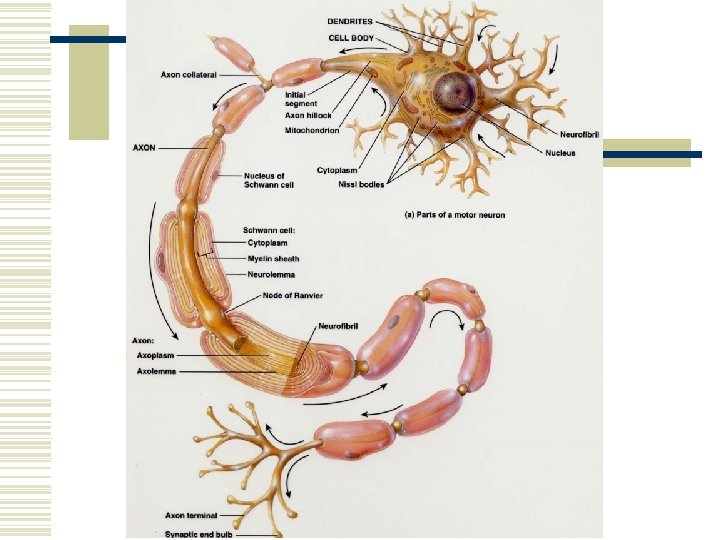
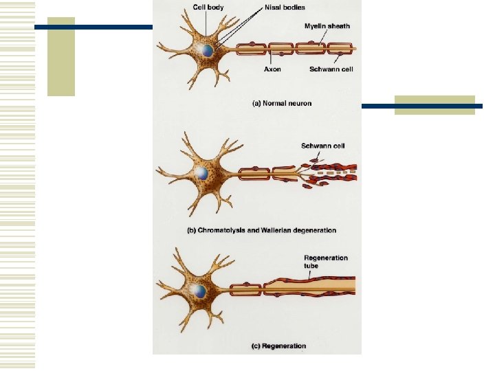
Parts of a Neuron w Cell body. n Contains the nucleus surrounded by cytoplasm which contains the organelles. w Dendrites (= little trees). n The receiving (input) portion of a neuron. w Axon. n n Each nerve contains a single axon. The axon propagates impulses toward another neuron, muscle fiber, or gland cell.
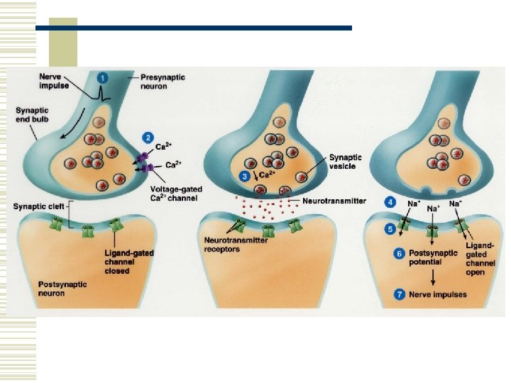
Synapse w The site of communication between two neurons or between a neuron and an effector cell. w Synaptic end bulbs and varicosities contain synaptic vesicles that store a chemical neurotransmitter.
Axonal Transport w Slow axonal transport. n n 1 -5 mm per day. Travels in one direction only – from cell body toward axon terminals. w Fast axonal transport. n n n 200 – 400 mm per day. Uses proteins to move materials. Travels in both directions.
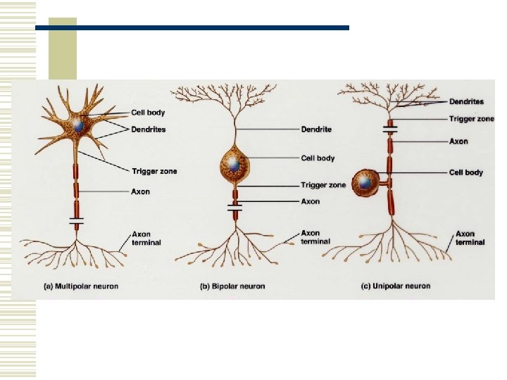
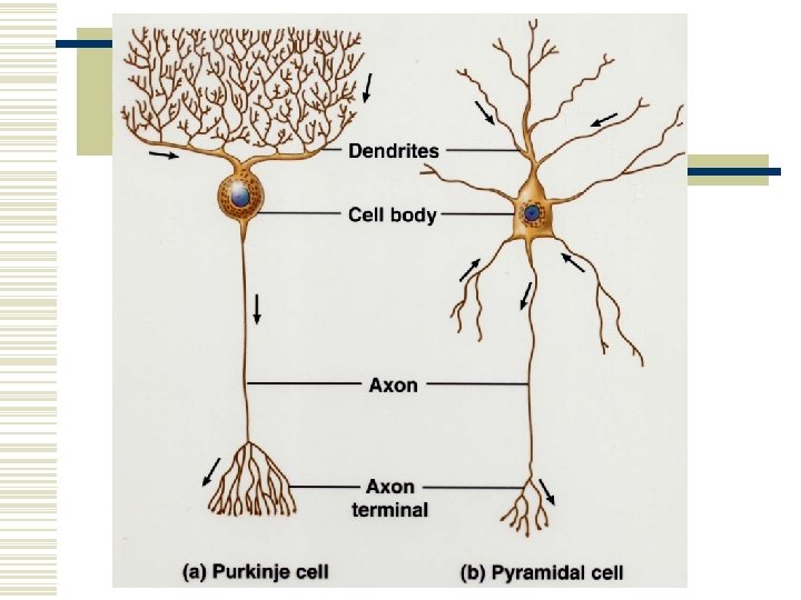
Structural Classifications of Neurons w Multipolar neurons. n n One axon and several dendrites. Most neurons of the brain and spinal cord.
Structural Classifications of Neurons w Bipolar neurons. n n One axon and one main dendrite. Retina of the eye, inner ear, and the olfactory area of the brain. w Unipolar neurons. n n The axon and the dendrite fuse into a single process that divides into two branches. The dendrites monitor a sensory stimulus such as touch or stretching.
Neuroglia w Half the volume of the CNS. w Generally, they are smaller than neurons, but 5 to 50 times more numerous. w They can multiply and divide. w Gliomas – brain tumors derived from glia.
Types of Neuroglia w CNS n n Astrocytes Oligodendrocytes Microglia Ependymal cells w PNS n n Schwann cells Satellite cells
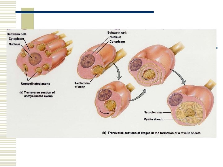
Myelination w The myelin sheath is a lipid and protein covering. It is produced by the neuroglia. w The sheath electrically insulates the axon of a neuron. w The sheath increases the speed of nerve impulse conduction. w Axons without a covering are unmyelinated. Axons with a covering are myelinated.
Myelination Continued… w Two types of neuroglial cells produce myelination. n n Schwann cells – located in the PNS. Oligodendrocytes – located in the CNS.
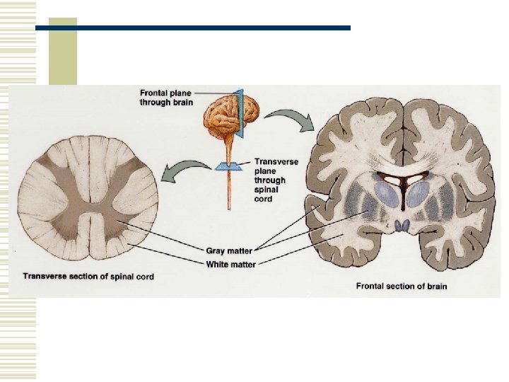
Gray and White Matter w The white matter consists of aggregations of myelinated and unmyelinated axons. w The gray matter consists of neuronal cell bodies, dendrites, unmyelinated axons, axon terminals, and neuroglia.
Electrical Signals in Neurons w Neurons are electrically excitable and communicate with one another using 2 types of electrical signals. n n Action potentials. Graded potentials. w The plasma membrane exhibits a membrane potential. The membrane potential is an electrical voltage difference across the membrane.
Electrical Signals in Neurons w The voltage is termed the resting membrane potential. w The flow of ions produces the electrical current.
Ion Channels w The plasma membrane contains many different kinds of ion channels. w The lipid bilayer of the plasma membrane is a good electrical insulator.
Ion Channels w The main paths for flow of current across the membrane are ion channels.
Ion Channels w When ion channels are open, they allow specific ions to move across the plasma membrane down their electrochemical gradient. n n n Ions move from greater areas of concentration to lesser areas of concentration. Positively charged cations move towards negatively charged area and negatively charged anions move towards a positively charged area. As they move, they change the membrane potential.
Ion Channel “Gates” w Ion channels open and close due to the presence of “gates”. w The gate is part of a channel protein that can seal the channel pore shut or move aside to open the pore.
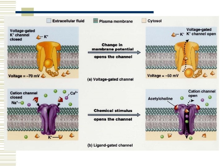
Types of Ion Channels w There are 4 types of ion channels. n n Leakage channels – gates randomly alternate between open and closed positions. Voltage-gated channels – opens in response to c change in membrane potential (voltage). Ligand-gated channels – opens and closes in response to a specific chemical stimulus. Mechanically gated channels – opens or closes in response to mechanical stimulation.
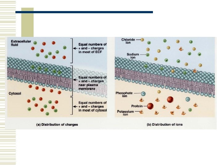
Resting Membrane Potential w The resting membrane potential occurs due to a buildup of negative ions in the cytosol along the inside of the membrane and positive ions in the extracellular fluid along the outside of the membrane. w The potential energy is measured in millivolts (m. V).
Resting Membrane Potential w In neurons, the resting membrane potential ranges from – 40 to – 90 m. V. Typically – 70 m. V. n The minus sign indicates that the inside of the cell is negative compared to the outside. w A cell that exhibits a membrane potential is polarized.
Electrochemical Gradient w An electrical difference and a concentration difference across the membrane.
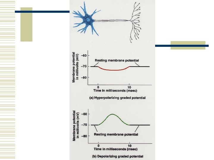
Graded Potentials w A graded potential is a small deviation from the resting membrane potential. w It makes the membrane either more polarized (more negative inside) or less polarized (less negative inside). w Most graded potentials occur in the dendrites or cell body.
Graded Potentials w Hyperpolarizing graded potential. w Depolarizing graded potential. w Graded potentials occur when ligand-gated or mechanically gated channels open or close. n n Mehcanically gated channels are present in sensory neurons. Ligand-gated channels are present in interneurons and motor neurons.
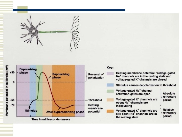
Action Potentials w An action potential is known as an impulse. w Depolarizing phase – the resting membrane potential decreased towards zero. w Repolarizing phase – restores the resting membrane potential.
Action Potentials w Threshold – depolarization reaches a certain level (about – 55 m. V), voltage gated channels open. w Action potentials arise according to an all or none principal.
Comparison of Graded Potentials and Action Potentials w See table 12. 2 p. 404
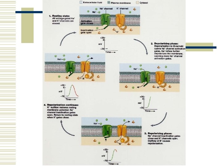
Depolarizing Phase w A depolarizing graded potential or some other stimulus causes the membrane to reach threshold. w Voltage-gated ion channels open rapidly. w The inflow of positive Na+ ions changes the membrane potential from – 55 mv to +30 m. V. w About 20, 000 Na+ enter through the gates. Millions are present in the surrounding fluid. w Na-k pumps bail them out.
Repolarizing Phase w While Na+ channels are opening during depolarization, K+ channels are opening, although slowly. w K+ channels allow outflow of K+ ions. w The closing of Na+ channels and the slow opening of K+ channels allows for repolarization.
Refractory Period w The period of time after an action potential begins during which an excitable cell cannot generate another action potential. n n Absolute refractory period – a second action potential cannot be initiated, even with a very strong stimulus. Relative refractory period – an action potential can be initiated, but only with a larger than normal stimulus.
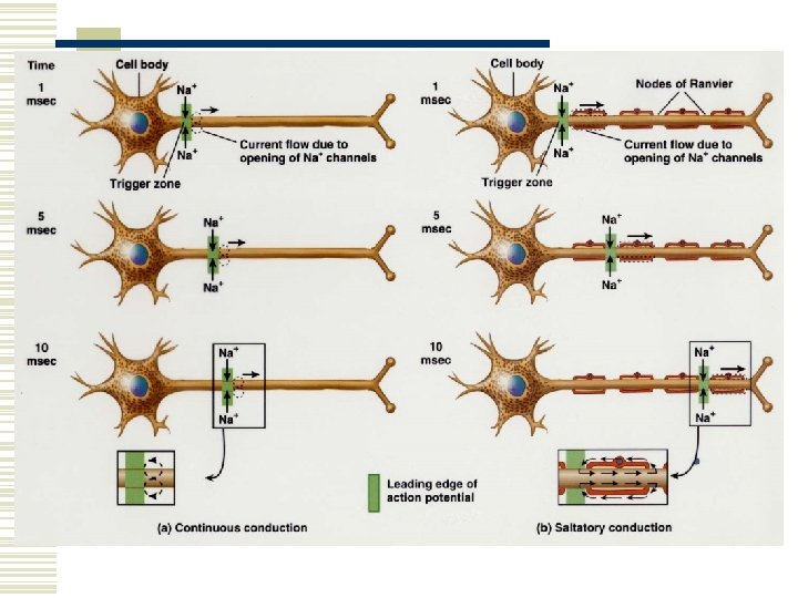
Propagation of Nerve Impulses w The impulse must travel from the trigger zone to the axon terminals. w This process is known as propagation or conduction. w As Na+ ions flow in, they trigger depolarization which opens Na+ channels in adjacent segments of the membrane.
Neurotoxins & Local Anesthetics w Neurotoxins produce poisonous effects upon the nervous system. w Local anesthetics are drugs that block pain and other somatic sensations. w These both act by blocking the opening of voltage-gated Na+ channels and preventing propagation of nerve impulses.
Continuous and Saltatory Conduction w Continuous conduction – step-by-step depolarization and repolarization of adjacent segments of the plasma membrane. w Saltatory conduction – a special mode of impulse propagation along myelinated axons.
Continuous and Saltatory Conduction w Few ion channels are present where there is myelin. w Nodes of Ranvier – areas where there is no myelin – contain many ion channels. w The impulse “jumps” from node to node. n n This speeds up the propagation of the impulse. This is a more energy efficient mode of conduction.
Effect of Axon Diameter & Myelination w Larger diameter axons propagate impulses faster than smaller ones. w Myelinated axons conduct impulses faster than unmyelinated ones.
Effect of Axon Diameter & Myelination w A fibers. n n n Largest diameter. Myelinated. Convey touch, pressure, position, thermal sensation.
Effect of Axon Diameter & Myelination w B fibers. n n n Smaller diameter than A fibers. Myelinated. Conduct impulses from the viscera to the brain and spinal cord (part of the ANS).
Effect of Axon Diameter & Myelination w C fibers. n n Smallest diameter. Unmyelinated. Conduct some sensory impulses and pain impulses from the viscera. Stimulate the heart, smooth muscle, and glands (part of ANS).
Encoding Intensity of a Stimulus w A light touch feels different than a firmer touch because of the frequency of impulses. w The number of sensory neurons recruited (activated) also determines the intensity of the stimulus.
Signal Transmission at Synapses w Presynaptic neuron – the neuron sending the signal. w Postsynaptic neuron – the neuron receiving the message. w Axodendritic – from axon to dendrite. w Axosomatic – from axon to soma. w Axoaxonic – from axon to axon.
Types of Synapses w Electrical synapse w Chemical synapse
Electrical Synapses w Action potentials conduct directly between adjacent cells through gap junctions.
Electrical Synapses w Tubular connexons act as tunnels to connect the cytosol of the two cells. w Advantages. n n Faster communication than a chemical synapse. Synchronization – they can synchronize the activity of a group of neurons or muscle fibers. In the heart and visceral smooth muscle this results in coordinated contraction of these muscle fibers.
Chemical Synapses w The plasma membranes of a presynaptic and postsynaptic neuron in a chemical synapse do not touch one another directly. w The space between the neurons is called a synaptic cleft which is filled with interstitial fluid. w A neurotransmitter must diffuse through the interstitial fluid in the cleft and bind to receptors on the postsynaptic neuron. w The synaptic delay is about 0. 5 msec.
Removal of Neurotransmitter w Diffusion. w Enzymatic degradation. w Uptake by cells. n n Into the cells that released them (reuptake). Into neighboring glial cells (uptake).
Spatial and Temporal Summation of Postsynaptic Potentials w A typical neuron in the CNS receives input from 1000 to 10, 000 synapses. w Integration of these inputs is known as summation.
Spatial and Temporal Summation of Postsynaptic Potentials w Spatial summation – summation results from buildup of neurotransmitter released by several presynaptic end bulbs. w Temporal summation – summation results from buildup of neurotransmitter released by a single presynaptic end bulb 2 or more times in rapid succession.
Summary of Neuronal Structure and Function w Table 12. 3 w P. 408
Neural Circuits
Neurogenesis in the CNS w Birth of new neurons. w From undifferentiated stem cells. w Epidermal growth factor stimulates growth of neurons and astrocytes. w Minimal new growth occurs in the CNS. n n Inhibition from glial cells. Myelin in the CNS.
Damage and Repair in the PNS w Axons and dendrites may undergo repair if the cell body is intact, if the Schwann cells are functional, and if scar tissue does not form too quickly. w Wallerian degeneration. w Schwann cells adjacent to the site of injury grow torwards one another and form a regeneration tube.