Knee Ligament Injuries The ligaments around the knee


- Slides: 63

Knee Ligament Injuries

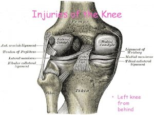
ØThe ligaments around the knee are strong. However, sometimes they can become injured. Ligaments injury Sprained Majority tend to stretched and quickly settle down Ruptured Partial Knee injuries by Lakeesha Perera Complete

ØThere a number of different things that can cause injury to the ligaments in your knee: ØYou may have a direct blow to your knee or knock into something with your knee. ØYour knee may be moved outside of its usual range of movement. For example, this can happen during a fall, if you land awkwardly during sport, or after a sudden movement. Knee injuries by Lakeesha Perera

Sprains and partial tears • Intact fibers splint the torn ones and so spontaneous healing will occur • Adhesions may result, so active exercise is prescribed • Aspirating the haemarthrosis and applying ice packs intermittently relieves pain • Weight-bearing is allowed • Knee is protected from rotation or angulation strains by a heavily padded bandage or a functional brace Knee injuries by Lakeesha Perera

Complete tears • Isolated MCL or LCL treated as above • Isolated tears of ACL may be treated by early operative reconstruction if the individual is a professional sportsman • Cast-brace is worn until symptoms subside, thereafter movement and muscle-strengthening exercise. This is sufficient in about half of the patients as they regain good function and need no further treatment. • Remainder will have varying instability, late assessment will identify those who will benefit from ligament reconstruction. • Isolated tears of the PCL are usually treated conservatively Knee injuries by Lakeesha Perera

Combined injuries • In ACL and collateral ligament injury treatment starts with joint bracing and physiotherapy to restore a good range of movements before ACL reconstruction • Combined injuries involving the PCL the same approach is used however all damaged structures need to be repaired Knee injuries by Lakeesha Perera

Complications � Adhesions • If the knee with a partial ligament tear is not actively exercised, torn fibers will stick to intact fibers and bone. • The knee gives way with catches of pain, localized tenderness and pain on lateral or medial rotation occur • Confusion with a torn meniscus can be resolved by the grinding test or arthroscopy � Instability • The knee continues to give way and tends to get worse predisposing to osteoarthritis. Reconstruction before degeneration is wise. Knee injuries by Lakeesha Perera

Grading Ligament Injuries Knee injuries by Lakeesha Perera

Anterior cruciate ligament injury ØACL injury most often occurs during sports such as football, basketball, skiing and tennis. ØThe injury often happens if you land on your leg and then quickly pivot or twist your knee in the opposite direction. ØAbout half of people with an ACL injury also have injury to their meniscus or another ligament in the same knee. ØWoman > men Knee injuries by Lakeesha Perera

Types of ACL Tears Knee injuries by Lakeesha Perera

Physical Exam of the Knee • Inspection • Palpation • Range of Motion • Special tests • Neurovascular assessment Knee injuries by Lakeesha Perera

ACL: HISTORY • Contact vs noncontact • Immediate effusion (first 4 -12 hr) • Unable to continue • Mechanism = pivot, hyperextension Knee injuries by Lakeesha Perera

ACL Special Tests • Anterior drawer • Lachman test • Pivot shift test • Valgus stress test at full extension! Knee injuries by Lakeesha Perera

ACL: PHYSICAL EXAM • Decreased ROM • Effusion-hemarthrosis, immediate • + Instability tests • Lachman: most accurate • Pivot shift • Anterior drawer • + MCL and meniscus tests Knee injuries by Lakeesha Perera

“Partial” ACL tear • > 40% ACL substance • + Lachman, - pivot shift • Clinically • Most behave functionally as full tears • Continued shifting ↑’s risk of meniscus damage • Rx as full tear Knee injuries by Lakeesha Perera

ACL TREATMENT • Grade 3 - Nonsurgical • modify activity • splint & crutches, Closed chain WB to strengthen • PRICES • Hamstrings, gastroc! • Functional bracing • 100% @ 9 -12 months Knee injuries by Lakeesha Perera

ACL TREATMENT • Grade 3 Injuries- Surgery • Indications • Most active people will require surgery to restore adequate function and decrease instability • Recurrent instability • Inability to modify activity • Associated injuries: meniscus • Age • Wait three weeks due to arthrofibrosis risk • 100% @ 6 -12 months Knee injuries by Lakeesha Perera

Posterior cruciate ligament injury ØNot as common as an ACL injury. Because the PCL is wider and stronger than the ACL. ØPCL sprains usually occur because the ligament was pulled or stretched too far, anterior force to the knee, or a simple misstep. ØPCL injuries disrupt knee joint stability because the tibia can sag posteriorly. Knee injuries by Lakeesha Perera

ØThe ends of the femur and tibia rub directly against each other, causing wear and tear to the thin, smooth articular cartilage. ØThis abrasion may lead to arthritis in the knee ØThere a number of ways that the PCL can become injured. For example, üIt may be injured during a car accident if the front of your bent knee hits the dashboard. Knee injuries by Lakeesha Perera

ü It may also be injured from falling on to your bent knee. üYour PCL can also be injured if your knee is hit from the front whilst your leg is stretched out in front of you with your foot on the ground - for example, during a game of football. Knee injuries by Lakeesha Perera

ØAt first, some people with a PCL injury may not have much in the way of symptoms. ØIt may take a while for you to realize that there is a problem. For example, you may later notice pain that comes on when going up and down stairs or when starting a run; or, your knee may feel unstable when walking on uneven ground. Knee injuries by Lakeesha Perera

PCL INJURIES PHYSICAL EXAM • + Effusion • + Posterior drawer test • + Posterior sag sign • False positive Lachman test • Common to have isolated injuries Knee injuries by Lakeesha Perera

PCL INJURIES TREATMENT • • PRICES Functional bracing (early) Rehab Surgery if continued instability, effusions Non-operative • • • Aggressive rehab Focus quadriceps No support for bracing closed kinetic chain Open kinetic chain extension avoided 90% quads strength prior to normal athletics Knee injuries by Lakeesha Perera

Medial collateral ligament injury ØInjuries to the MCL can happen in almost any sport and can affect people of all age groups. ØThey often happen when your leg is stretched out in front of you and the outer side of your knee is knocked at the same time - for example, during a rugby or football tackle. Knee injuries by Lakeesha Perera

MCL INJURIES PHYSICAL EXAM • Tender to palpation along MCL • Pain + instability with valgus stress • 30 o flexion = MCL • 90 o flexion = associated ACL • Pain with Apley’s distraction test • COMPARE SIDES

MCL INJURIES Treatment Of Grade 1 &2 • Early mobilization • Weight-bearing as tolerated • Hinged knee brace • PRICES • Recovery 4 -6 weeks Knee injuries by Lakeesha Perera

MCL INJURIES Treatment of Grade 3 (full tears) • Isolated = nonsurgical management • Combined = surgery consistent with associated injuries • Natural Hx = lack of long-term degenerative changes seen with ACL, meniscus Knee injuries by Lakeesha Perera

Lateral collateral ligament injury ØInjury to the LCL is less common than injury to the MCL. This is because your other leg usually protects against injury to the inner side of your knee. Ø(It is usually a direct blow to the inner side of your knee that causes an LCL injury. ) ØBut, this ligament injury can sometimes happen if one leg is stretched out in front of you and doesn't have the other leg for protection - for example , during a rugby or football tackle. Knee injuries by Lakeesha Perera

What are the symptoms of a knee ligament injury? If you have injured one or more of the ligaments in your knee, the symptoms are likely to be similar regardless of the ligament that is injured. The severity of the symptoms depends on the degree of the injury to the ligament. For example, a ligament that is completely torn may produce more in the way of symptoms than a ligament that is just sprained (stretched). Knee injuries by Lakeesha Perera

Symptoms can include: 1. A popping sound, or a popping or snapping feeling Can hear at the time of injury if ligament completely torn 2. Swelling of your knee. Bleeding inside from the damaged ligament It leads to swelling Completely torn ligament Minor ligament sprains Knee injuries by Lakeesha Perera

3. Pain in your knee. depend on the severity of the knee injury. 4. Tenderness around your knee on touching. This may be minor sprains ----mild tenderness over the actual ligament torn -----more generalised and severe tenderness 5. Not being able to use or move your knee normally. complete ligament tears--- severely reduce minor sprains----relatively good Knee injuries by Lakeesha Perera

6. A feeling that your knee is unstable or perhaps giving way if you try to stand on it. This may cause you to limp. Again, this depends on how severe the ligament injury is. You may be able to stand if you only have a minor sprain. 7. Bruising around your knee can sometimes appear, although not always. It may take some time for bruising to develop. Knee injuries by Lakeesha Perera

Historical Clues to Knee Injury Diagnoses Noncontact injury with “pop” ACL tear Contact injury with “pop” MCL or LCL tear, meniscus tear, fracture Acute swelling ACL tear, PCL tear, fracture, knee dislocation, patellar dislocation Lateral blow to the knee MCL tear Medial blow to the knee LCL tear Knee “gave out” or “buckled” ACL tear, patellar dislocation Fall onto a flexed knee PCL tear Knee injuries by Lakeesha Perera

Special tests for ligaments Anterior Cruciate • Posterior Cruciate Assess stability of 4 knee ligaments via applied stresses* Medial Collateral Lateral Collateral Knee injuries by Lakeesha Perera

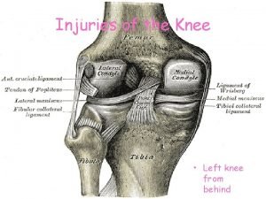
The stabilizing roles of each ligament include: MCL prevents the knee from buckling inwards (valgus injury) LCL prevents the knee from buckling outwards (varus injury) ACL prevents the tibia from sliding forward under the femur PCL prevents the tibial from sliding backward under the femur Knee injuries by Lakeesha Perera

Stress Testing of Ligaments Use a standard exam routine q Direct, gentle pressure q No sudden forces Abnormal test q Excessive motion = laxity q Soft/mushy end point** Knee injuries by Lakeesha Perera

Normal Stability Medial and Lateral collateral ligaments • Normal test is no motion with varus and/or valgus stress with knee in neutral and 30 degrees of flexion Anterior and Posterior Cruciate Ligaments' control anterior/posterior motion • Lachman’s test assesses Anterior Cruciate Ligament: • Normal test is <5 mm of forward movement of tibia on femur with knee at 30 degrees of flexion Anterior and posterior drawer testing assesses ACL and PCL With knee in 90 degrees of flexion and foot stabilized, normal test will have <5 mm of anterior motion (assessing ACL) or <5 mm of posterior motion (assessing PCL) Knee injuries by Lakeesha Perera

Normal end point of ligament that examiner feels with applied stress is FIRM. A soft or mushy end point implies ligament damage (stretching or complete tear). Knee injuries by Lakeesha Perera

Collateral Ligament Assessment Patient and Examiner Position* Knee injuries by Lakeesha Perera

*Position patient supine on table with thigh resting on edge of exam table and foot supported by examiner • Knee in 30 degrees of flexion – WHY? Increased laxity of medial side of knee in extension may indicate additional damage to posterior structures (posterior joint capsule & PCL) Knee injuries by Lakeesha Perera

Valgus Stress Test for MCL* Note Direction Of Forces Knee injuries by Lakeesha Perera

*VALGUS (MCL) stress • Proximal hand on lateral aspect of knee holds and stabilizes thigh • Distal hand directs ankle laterally • Attempt to open knee joint on medial side • Estimate the medial joint space and evaluate the stiffness of motion. • Positive test = Significant gap in medial aspect of knee with valgus stress = MCL injury. Knee injuries by Lakeesha Perera

Varus Stress Test for LCL* Note direction of forces Knee injuries by Lakeesha Perera

*VARUS (LCL) Stress • Supine position, with knee at 20 to 30 degrees of flexion and thigh supported. • Stabilize medial aspect of knee and push ankle medially, trying to open knee joint on lateral side • Disruption of LCL is indicated by difference in degree of lateral knee tautness with varus stress. Compare affected knee to uninjured side Knee injuries by Lakeesha Perera

Lachman Test* • Patient Position • Physician hand placement 45 Knee injuries by Lakeesha Perera

*Lachman Maneuver more sensitive and specific for ligamentous tears than drawer sign. • Patient is supine • Knee flexed to 20 -30 degrees • Hand placement: • Grasp and stabilize patient’s thigh just proximal to patella • With opposite hand, try to move proximal tibia forward on femur • POSITIVE TEST = Excessive forward motion of tibia (>5 mm) without firm endpoint indicates ACL damage Knee injuries by Lakeesha Perera

• Modification for patient with large thighs: • Thigh placed over knee of examiner • Push downward on femur with hand while other hand grasps proximal tibia, attempting to move it anteriorly Knee injuries by Lakeesha Perera

Lachman Test • View from lateral aspect* Note direction of forces 48 Knee injuries by Lakeesha Perera

Anterior Drawer Test for ACL • Physician Position & Movements* • Patient Position Note direction of forces Knee injuries by Lakeesha Perera

*Patient Position • Supine • Flex hip of affected knee to 45 degrees • Bend knee to 90 degrees • Patient's foot planted firmly on examination table Physician position: Sitting on dorsum of foot, place both hands behind knee Once hamstrings relaxed, try to displace proximal leg anteriorly Anterior drawer test is LESS SENSITIVE for ACL damage than Lachman’s Maneuver Knee injuries by Lakeesha Perera

Posterior Drawer Testing- PCL* Note direction of forces Knee injuries by Lakeesha Perera

*Patient Position • Supine • Affected knee at 90 degrees of flexion • Determine ‘neutral’ position by comparing resting position with unaffected knee Physician Position & Movements • Patient's foot placed between examiner's legs while the palms of the hands are used to push the tibia posteriorly. • Tester directs pressure backward upon proximal tibia, similar to Anterior Drawer Testing Interpretation of test: • Posterior instability - PCL injury indicated by increased posterior tibial translation • Confusion - trying to distinguish abnormal translation of tibia on femur - from excessive ACL or PCL laxity Knee injuries by Lakeesha Perera

• Commonly injured part Meniscal Tears Occur during twisting motions with the knee flexed Signs • tenderness • possible clicking • Symptoms • Pain • catching • buckling Can occur combine with other ligament – ACL mostly Knee injuries by Lakeesha Perera

• Older people can injure the meniscus without any trauma as the cartilage weakens and wears thin over time, setting the stage for a degenerative tear. • Medial Menisci: more prone to injury because of its restricted anatomy due to attachment to the joint capsule and to the tibial collateral ligament make it less mobile. Knee injuries by Lakeesha Perera

Meniscus Tears Pattern of tear Mechanism traumatic bucket handle horizontal degenerative The split is vertical, along the circumference of the meniscus leaving anterior and posterior segments attached loosely. Sometimes the torn part displaces towards the center, causing “locking” (extension block). Usually degenerative in origin or due to repetitive minor trauma, or with association with meniscal cysts. Generally speaking, most of the meniscus is avascular, except the outer third-from capsule-, due to this spontaneous repair doesn’t occur. *The loose part act as a mechanical irritant causing recurrent synovial effusion, and in severe cases secondary osteoarthritis. Knee injuries by Lakeesha Perera

Knee injuries by Lakeesha Perera

Menisci Tears Clinical Features: Patients may complain of pain at the joint line area, locking, clicking, giving way, and swelling with activity. In ptn >40 yrs the main complaint is recurrent giving way or locking. Physical exam: • Joint line tenderness (Mostly medial). • Joint held slightly flexed. • Joint effusion may be present. • In late cases quadriceps are wasted. • Flexion is full , extension limited. Knee injuries by Lakeesha Perera

Assess Meniscus – Knee Flexion • Most sensitive test is full flexion* • Examiner passively flexes the knee or has patient perform a full two-legged squat to test for meniscal injury • Joint line tenderness** • Flexion of the knee enhances palpation of the anterior half of each meniscus 58 Knee injuries by Lakeesha Perera

Joint line tenderness: the most imp and specific test _ Apley’s grind test: • Isolates meniscii • Prone with knee flexed, axial load and rotation. - Mc. Murray’s test • Flex/ext with varus / valgus and int/ext rotation. • Goal is to get torn piece to pop in and out of place. • Positive if pop or reproduction of pain. Knee injuries by Lakeesha Perera

Menisci Tears Imaging X-ray – Normal MRI – most useful may reveal tears missed by arthroscopy Arthroscopy : Diagnostic and therapeutic. You have to be certain that the lesion you can see is the one causing the patient’s symptoms. Knee injuries by Lakeesha Perera

Menisci Tears Treatment � Conservative treatment of meniscal injuries begins with RICE (Rest, Ice, Compression, and Elevation). � Arthroscopy is the preferred method. � peripheral tears – surgery. � The displaced portion should be excised. � Postoperative physiotherapy. � Surgical treatment of symptomatic meniscal tears is recommended because untreated tears may increase in size and may abrade articular cartilage, resulting in arthritis. Knee injuries by Lakeesha Perera

Knee injuries by Lakeesha Perera

 Biomechanics of throwing a football
 Knee joint line
 Muscle around knee
 Martin luther king of hinduism
 What goes around comes around example
 Vitreous humor cow eye
 Ligaments of female reproductive system
 Spermatic cord
 What type of connective tissue are tendons and ligaments
 Gluteal ligaments
 Pintos ligament
 Splenic ligaments
 Arch of foot muscles
 Rétrécissement foraminal l5-s1 traitement
 Where is peritoneal cavity located
 Arches of the foot anatomy
 Texture of sheep eye lens
 Voice box in respiratory system
 Hepar facies diaphragmatica
 Lower triangular space
 Hepatic ligaments
 Ligament falciforme
 Structure of the nail
 Supravesical fossa
 Structure pierced during lumbar puncture
 Abdominal part of esophagus
 Forearm tendonitis
 Muscles of pelvic girdle
 Ligamentum teres
 Semons law
 Normal shoulder extension
 Adipose tissue drawing labeled
 Thyroidoctomy
 Characteristics of ligaments
 Suspensory ligaments eye
 Periodontal ligament
 Leg tendons and ligaments
 Sacciform recess
 Chapter 11 assessment and evaluation of sports injuries
 Chapter 21 caring for head and spine injuries
 Yorkhill minor injuries
 Sports injuries angus, on
 How did rue die hunger games
 How to make fake skin with gelatin
 Chapter 4 preventing injuries through fitness
 Climatic injuries
 Jones and bartlett learning
 Clinton tendonitis
 Hunger games chapter questions
 Injuries first aid
 Discoid miniscus
 Kristen wilson injuries
 Two-person drag
 17:7 providing first aid for heat exposure
 How are sports injuries classified and managed
 Intentional fallacy
 Climatic injury
 Chapter 13 worksheet recognizing different sports injuries
 Chapter 13:2 preventing accidents and injuries
 Sentinel injuries in infants are
 How did mayella get rid of the children
 Characters of firearm injuries
 Preventing hand injuries
 Uc davis web scheduler
