
Introduction to urology Dr Malik Ayyad 1 Anatomy












- Slides: 46
Introduction to urology Dr. Malik Ayyad 1
Anatomy 2
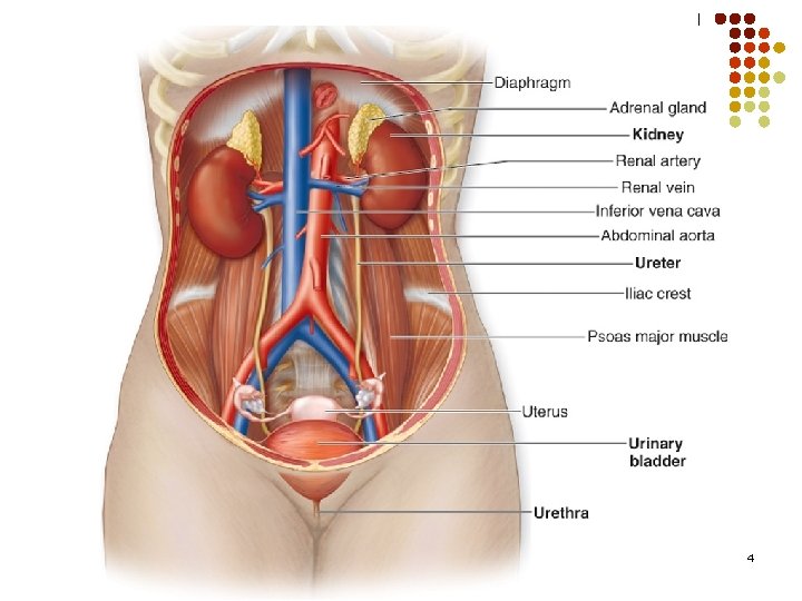
Renal Anatomy l The kidneys are retroperitoneal organs on each side of the vertebral column (T 12 -L 3), at about the level of the twelfth rib. l The left kidney is lightly higher in the abdomen than the right. l On an average, each kidney weighs 150 g. l The kidneys have the following coverings: Fibrous capsule Perirenal fat Renal fascia Pararenal fat 1. 2. 3. 4. 3
4
Ureteral Anatomy l l l The ureters are retroperitoneal tubes measuring each about 25 cm long and about 3 mm in diameter. They descend with an inclination medially on the posterior abdominal wall opposite the tips of the lumbar transverse processes. They continue following the contour of the pelvis. They pass obliquely in the wall of the bladder of 1 inch before they open at the sides of the trigone. 5
Bladder Anatomy l The adult bladder is located in the anterior pelvis and is enveloped by extraperitoneal fat and connective tissue. It is separated from the pubic symphysis by an anterior prevesical space known as the space of Retzius. The dome of the bladder is covered by peritoneum, and the bladder neck is fixed to neighboring structures by reflections of the pelvic fascia and by true ligaments of the pelvis. 6
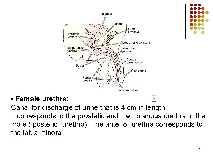
Urethral Anatomy Male urethra: Canal for the discharge of urine and seminal fluid. It is 18 -20 cm in length. The male urethra may be divided into 2 portions. The posterior urethra includes the prostatic urethra and the membranous urethra, The anterior urethra includes 3 segments. The bulbar urethra courses through the proximal corpus spongiosum and ischial cavernosus-bulbospongiosus muscles to reach the penile urethra. The penile urethra then extends through the pendulous portion of the penis to the final segment, the fossa navicularis. The fossa navicularis is invested by the spongy tissue of the glans penis. l 7
• Female urethra: Canal for discharge of urine that is 4 cm in length. It corresponds to the prostatic and membranous urethra in the male ( posterior urethra). The anterior urethra corresponds to the labia minora 8
Signs and Symptoms of GUT
volume and composition ♣ Polyuria : abnormally large volume of urine, and is most commonly due to excessive fluid intake. ♣ Oliguria : reduction in urine volume to <800 ml/day. - Acute renal failure ♣ Anuria : total absent of urine production ♣ Pneumaturia : passing gas bubbles in the urine
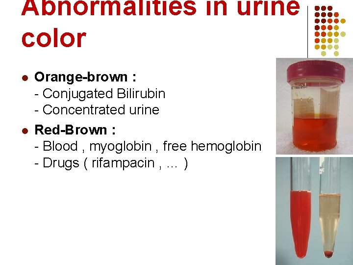
Abnormalities in urine color l l Orange-brown : - Conjugated Bilirubin - Concentrated urine Red-Brown : - Blood , myoglobin , free hemoglobin - Drugs ( rifampacin , … )
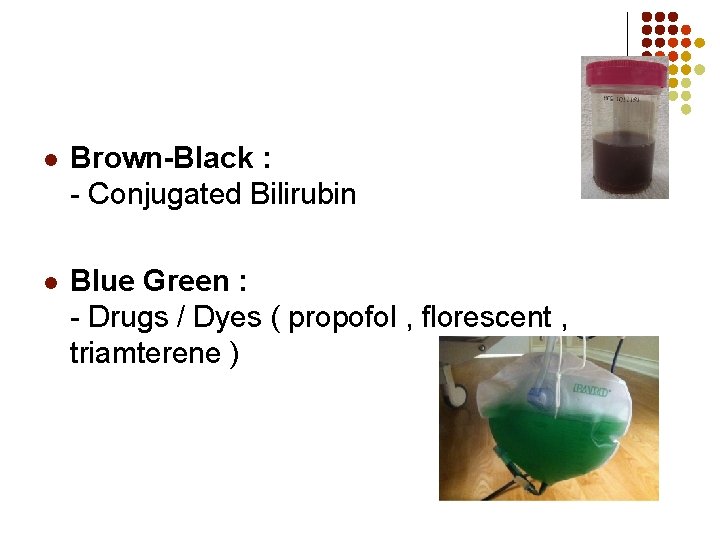
l Brown-Black : - Conjugated Bilirubin l Blue Green : - Drugs / Dyes ( propofol , florescent , triamterene )
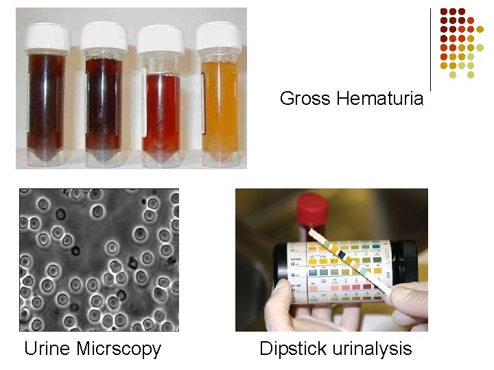
Hematuria * It is the presence of RBCs in the urine. - Macroscopic (gross) hematuria is visible to the naked eye (overtly bloody, smoky or blood clots) - Microscopic or dipstick hematuria is when blood is identified by urine microscopy or dipstick testing >3 RBCs/HPF on urinalysis of at least two separate samples
Gross Hematuria Urine Micrscopy Dipstick urinalysis
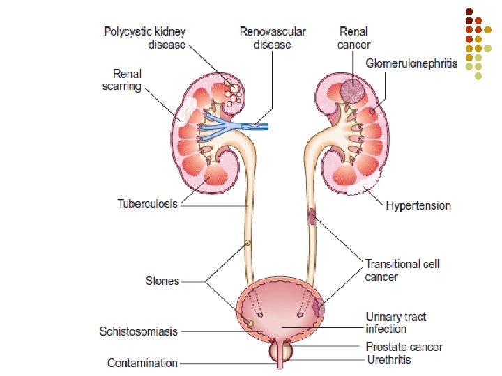
- Recent Urological Interventions : Bladder Catheterization , urethral stent , Prostate biopsy ) - Trauma - Radiotherapy , Chemotherapy - Coagulopathies ** Pseudohematuria : Mensis , Drugs (Rifampacin) , Dyes , Food (Beets) , porphyria
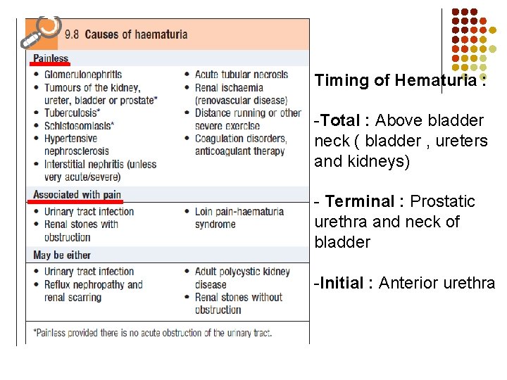
Timing of Hematuria : -Total : Above bladder neck ( bladder , ureters and kidneys) - Terminal : Prostatic urethra and neck of bladder -Initial : Anterior urethra
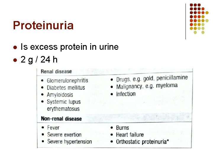
Proteinuria l l Is excess protein in urine 2 g / 24 h
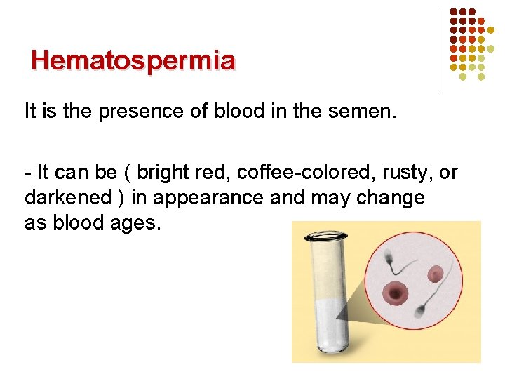
Hematospermia It is the presence of blood in the semen. - It can be ( bright red, coffee-colored, rusty, or darkened ) in appearance and may change as blood ages.
Nocturia and nocturnal polyuria l l l Nocturia is the frequent need to get up and urinate at night. Enuresis (bed wetting) in that the person does not wake and the bladder empties. Nocturnal polyuria (NP) refers to a condition in which the rate of urine output is excessive only at night and total 24 -hour output is within normal limits. Awakening 2 or more times at night considered as nocturia
Lower Urinary Tract Symptoms • two phases of lower urinary tract function : 1. Storage phase (bladder filling and urine storage) �accommodation and compliance �no involuntary contraction 2. Voiding phase (bladder emptying) �coordinated detrusor contraction �synchronous relaxation of outlet sphincters �no anatomic obstruction
Filling (Storage) or irritative symptoms : - Frequency : micturating more often with no increase in the total urine output. - Urgency : a sudden strong need to pass urine - Dysuria : painful urination - Nocturia Voiding or obstructive symptoms : - Poor stream (unimproved by straining) - Hesitancy (worsened if bladder is very full) ”difficulty in beginning the flow of urine “ - Terminal dribbling - Incomplete voiding - Urinary retention
Urinary Incontinence Definition • involuntary leakage of urine Types : 1 - Urgency Urinary Incontinence (UUI) : Involuntary leakage accompanied by or immediately preceded by urgency
2 - Stress Urinary Incontinence (SUI) : Urine leakage associated with increased abdominal pressure from laughing, sneezing, coughing, climbing stairs, or other physical stressors on the abdominal cavity and, thus, the bladder - Common in women , seen in men after prostate cancer surgery l Mechanism : - Urethral Hypermobility : weakness in pelvic floor - Intrinsic sphincter deficiency (ISD)
3 - Mixed incontinence : A combination of stress and urge incontinence, 4 - Overflow incontinence: increased residual or chronic urinary retention leads to urinary leakage from bladder overdistention. 5 - Functional incontinence : loss of urine related to deficits of cognition and mobility. (eg, delirium, psychiatric disorders, urinary infection, impaired mobility) 6 - Continuous incontinence : associated with fistulas ( vesicovaginal , urethrovaginal ) and ectopic ureter inserted distal to external sphincter
Urinary Retention Definition : It is an inability to completely empty the bladder. Etiology : - Outflow obstruction ( Stones , BPH , stricture ) - Bladder innervation (Nerve damage , DM , . . ) - Pharmacologic ( Anticholinergic , Narcotics , …) - Infection ( UTI , prostatitis , … )
PAIN PRESENTATIONS ( renal, ureteric, vesical, urethral ) - Renal angle pain – dull ache between 12 th rib and erector spine muscle on the side of the affected kidney – pyelonephritis. (Refer renal angle tenderness) - Renal colic – due to ureteric obstruction – a severe pain – lumbar region; radiates to abdomen, groin, testes, thigh – due to stone or tumour - Ureteric colic – spasmodic, severe , radiation path of renal colic; may be associated with vomiting, sweating. - Suprapubic pain from bladder / urethra - is referred to lower abdomen, perineum and glans penis in males
Urological Investigations and imaging studies
Urological Investigations : 1 -Dipstick testing 2 -Urine microscopy 3 -Urine cytology Imaging studies : 1 -Ultrasound 2 -plain abdominal radiography (KUB) 3 -Intravenous pyelography (IVP)/(IVU) 4 -Voiding cystourethrography [ VCUG ] 5 - Cystography 6 -Urethrography 7 -Retrograde pyelography 8 -CT 9 - MRI
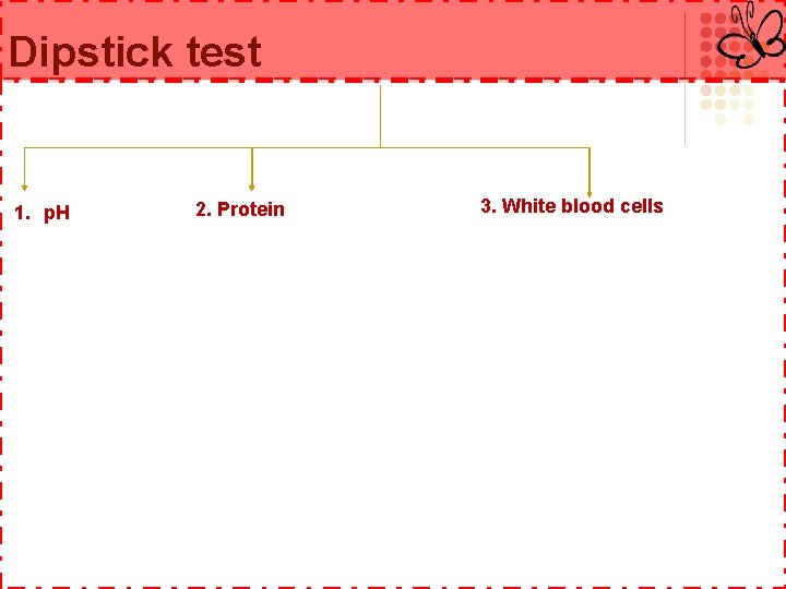
Dipstick test A urine dipstick test ( test strip ) is a basic diagnostic tool used to determine pathological changes in a patient’s urine in standard urinalysis.
Dipstick test 1. p. H 2. Protein 3. White blood cells
Dipstick test 4. Blood 5. Nitrite testing Cloudy urine that is positive for WBCs and positive for nitrite is very likely to be infected.
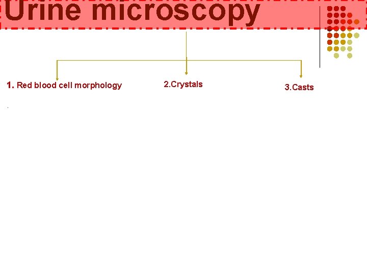
Urine microscopy 1. Red blood cell morphology. 2. Crystals 3. Casts
Urine cytology is a test to look for abnormal cells in the urine. It is used along with other tests and procedures to diagnose urinary tract cancers. It is most often used to diagnose bladder cancer, though it may also detect cancers of the kidney, prostate, ureter and urethra. It can best detect larger and more-aggressive urinary tract cancers , and might not detect small urinary tract cancers that grow more slowly.
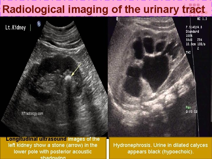
Radiological imaging of the urinary tract -Ultrasound : • Non – invasive method. • provides good images of the kidneys and bladder. • poor anatomical detail of the ureter , and the mid-ureter cannot be imaged at all by ultrasound because of overlying bowel gas.
Radiological imaging of the urinary tract Longitudinal ultrasound images of the left kidney show a stone (arrow) in the lower pole with posterior acoustic Hydronephrosis. Urine in dilated calyces appears black (hypoechoic).
Radiological imaging of the urinary tract - Plain abdominal radiography (KUB) : • Needs praparation • detection of stones. [ size & location ]. • not useful for following ureteric stones that are radiolucent (e. g. , uric acid) or small (generally a stone must be 3– 4 mm to be visible on plain X-ray), or when the stones pass through the ureter as it lies over the sacrum. • The ability of KUB X-ray to visualize stones is dependent on the amount of overlying bowel gas.
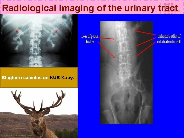
Radiological imaging of the urinary tract Staghorn calculus on KUB X-ray.
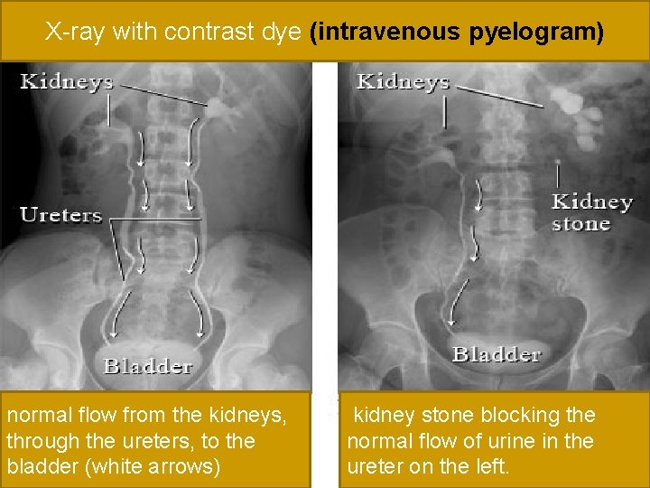
Radiological imaging of the urinary tract -Intravenous pyelography (IVP)/(IVU) : • A control film is obtained before contrast is given. • Intravascular contrast is administered followed by a series of X-rays of the kidneys, ureters, and bladder over the following 30 minutes or so, to image their anatomy and pathology, and to give some indication of renal function. *Films and “phases” of IVP : • Plain film : This is used to look for calcification overlying the region of the kidneys, ureters, and bladder. • Nephrogram phase : This is the first phase of IVP; film is taken immediately following intravenous administration of contrast. Pyelogram phase : As the contrast passes along the renal tubule (into the distal tubule) it is concentrated , As a consequence, the contrast medium is concentrated in the pelvicalyceal system, thus this pyelogram phase is much denser than the nephrogram phase.
X-ray with contrast dye (intravenous pyelogram) normal flow from the kidneys, through the ureters, to the bladder (white arrows) kidney stone blocking the normal flow of urine in the ureter on the left.
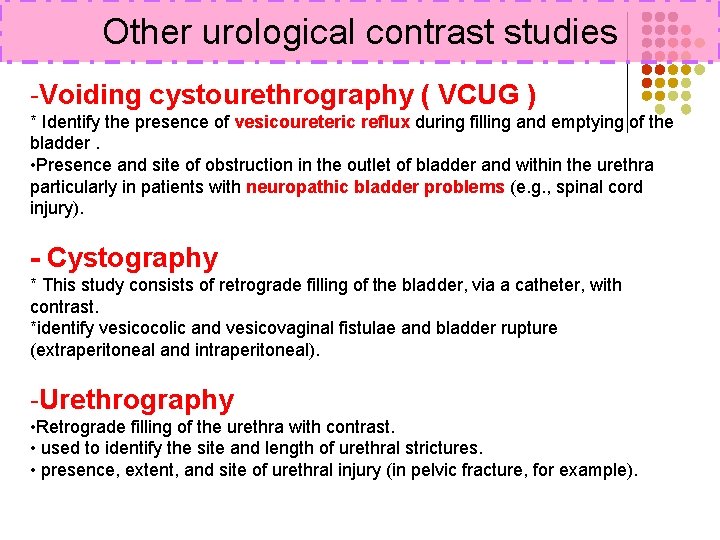
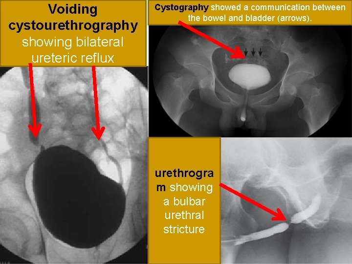
Other urological contrast studies -Voiding cystourethrography ( VCUG ) * Identify the presence of vesicoureteric reflux during filling and emptying of the bladder. • Presence and site of obstruction in the outlet of bladder and within the urethra particularly in patients with neuropathic bladder problems (e. g. , spinal cord injury). - Cystography * This study consists of retrograde filling of the bladder, via a catheter, with contrast. *identify vesicocolic and vesicovaginal fistulae and bladder rupture (extraperitoneal and intraperitoneal). -Urethrography • Retrograde filling of the urethra with contrast. • used to identify the site and length of urethral strictures. • presence, extent, and site of urethral injury (in pelvic fracture, for example).
Voiding cystourethrography showing bilateral ureteric reflux Cystography showed a communication between the bowel and bladder (arrows). urethrogra m showing a bulbar urethral stricture
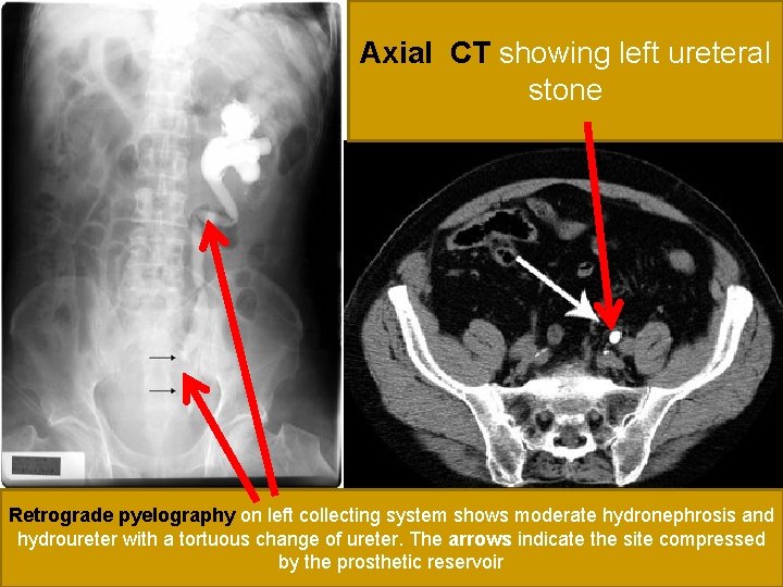
Other urological contrast studies -CT : Uses of CT 1 -Renal • Investigation of renal masses • Staging of renal cancer (establishes local, nodal, and distant spread) • Assessment of stone size and location • Detection and localization of site of intrarenal and perirenal collections of pus (pyonephrosis, perinephric abscess) • Staging (grading) of renal trauma • Determination of cause of hydronephrosis 2 -Ureters : >> Locates and measures size of ureteric stones 3 -Bladder : >> Bladder cancer staging (establishes local, nodal, and distant spread)
Axial CT showing left ureteral stone Retrograde pyelography on left collecting system shows moderate hydronephrosis and hydroureter with a tortuous change of ureter. The arrows indicate the site compressed by the prosthetic reservoir
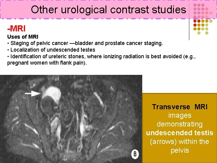
Other urological contrast studies -MRI Uses of MRI • Staging of pelvic cancer —bladder and prostate cancer staging. • Localization of undescended testes • Identification of ureteric stones, where ionizing radiation is best avoided (e. g. , pregnant women with flank pain). Transverse MRI images demonstrating undescended testis (arrows) within the pelvis
Thank you 46