
HEMOGLOBINOPATHIES BIOCHEMISTRY DR AQSA MALIK HEMOGLOBINOPATHIES Mutations in
HEMOGLOBINOPATHIES BIOCHEMISTRY DR AQSA MALIK

HEMOGLOBINOPATHIES � Mutations in the genes for the hemoglobin protein in a species result in hemoglobin variants, some of these mutant forms of hemoglobin cause a group of hereditary diseases termed the hemoglobinopathies

�A large number of mutations have been described in the globin genes. These mutations can be divided into two distinct types: �Those that cause qualitative abnormalities (e. g. sickle cell anemia) and those that cause quantitative abnormalities (the thalassemias). �Taken together these disorders are referred to as the hemoglobinopathies.
�The best known is sickle-cell disease, which was the first human disease whose mechanism was understood at the molecular level. �A separate set of diseases called thalassemias involves underproduction of normal and sometimes abnormal hemoglobins, through problems and mutations in globin gene regulation. All these diseases produce anemia.
Methemoglobinemias �The iron ion may either be in the Fe 2+ or (ferric) Fe 3+ state, but methemoglobin (Fe 3+) cannot bind oxygen �In binding, oxygen temporarily oxidizes (Fe 2+) to (Fe 3+), so iron must exist in the +2 oxidation state to bind oxygen. �The enzyme methemoglobin reductase reactivates hemoglobin found in the inactive (Fe 3+) state by reducing the iron center
�They are therefore particularly susceptible to the effects of methemoglobin-producing compounds. �The methemoglobinemias are characterized by chocolate cyanosis (a brownish-blue coloration of the skin and membranes) and chocolate-colored blood, as a result of the dark- colored methemoglobin.
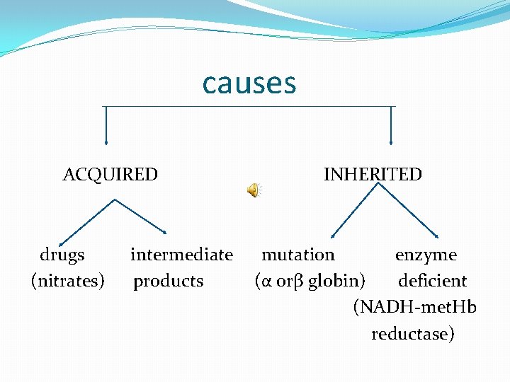
causes ACQUIRED drugs (nitrates) intermediate products INHERITED mutation enzyme (α orβ globin) deficient (NADH-met. Hb reductase)
�Symptoms are related to tissue hypoxia, and include anxiety, headache, and dyspnea. � In rare cases, coma and death can occur � Treatment is with methylene blue, which is oxidized as Fe+3 is reduced to ferrus.
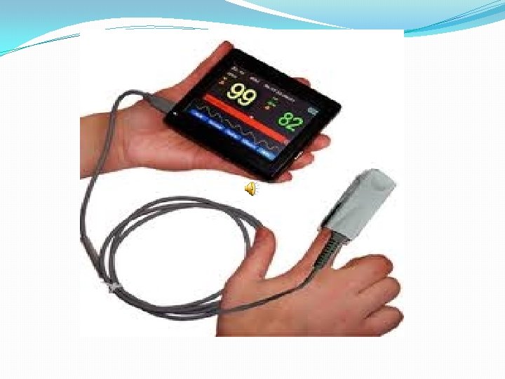
�The absorption spectra of oxyhemoglobin and deoxyhemoglobin differ. The oxyhemoglobin has significantly lower absorption of the 660 nm wavelength than deoxyhemoglobin, while at 940 nm its absorption is slightly higher. �This accounts for hemoglobin's red color and deoxyhemoglobin's blue color. This difference is used for measurement of the amount of oxygen in patient's blood by an instrument called pulse oximeter.
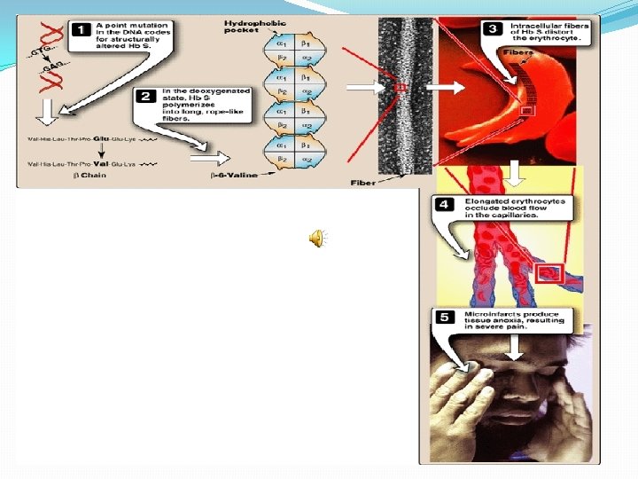
Sickle Cell disease �The mutation causing sickle cell anemia is a single nucleotide substitution (A to T) in the codon for amino acid 6 �The change converts a glutamic acid codon (GAG) to a valine codon (GTG). �The form of hemoglobin in persons with sickle cell anemia is referred to as Hb. S
�Sickle cell anemia is an autosomal recessive disorder �However, individuals who are heterozygous have sickle cell trait
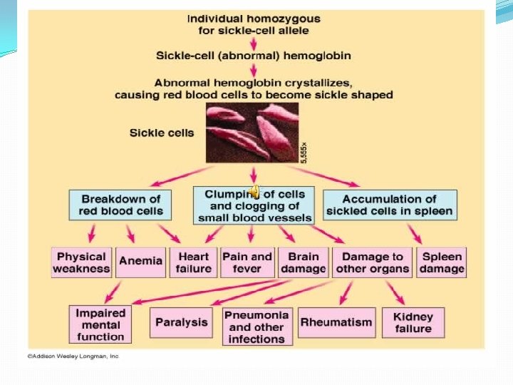
�The underlying problem in sickle cell anemia is that the valine for glutamic acid substitution results in hemoglobin tetramers that aggregate into arrays upon deoxygenation in the tissues. �This aggregation leads to deformation of the red blood cell into a sickle-like shape making it relatively inflexible and block the flow of blood in the narrow capillaries.
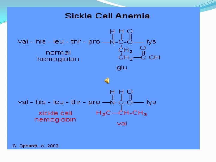
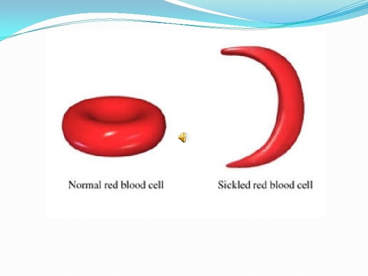
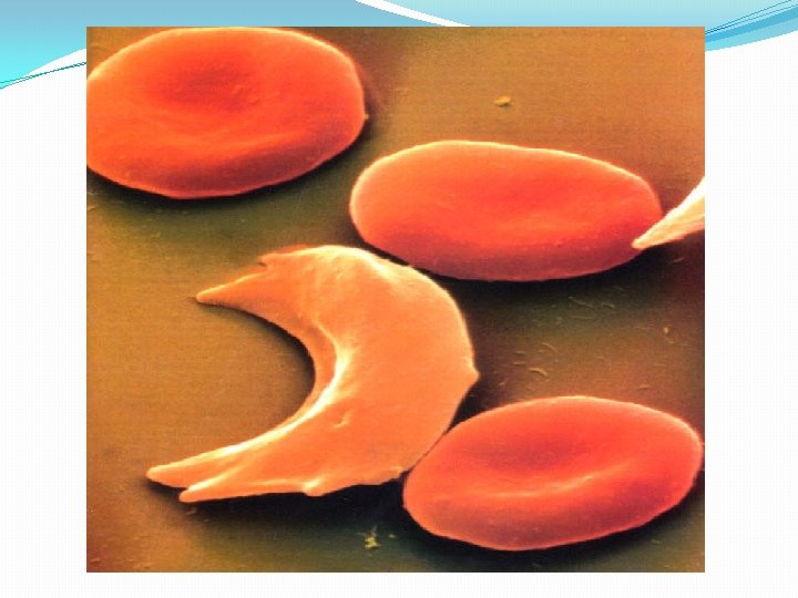
�Valine residue forms a protrusion on the beta chain that fits into the complementary site on the alpha chain of another hemoglobin molecule �At low oxygen tension deoxy. Hb S polymerizes inside the cell, that distort the cell
�Hb S contains two normal alpha globin chains and two mutant beta globin chains (Bs ) �Altered mobility on electrophoresis is due to less negative charges.
�Sickling caused by: 1. High altitude 2. Increased p. CO 2 3. Decreased p. H 4. Dehydration 5. Increased concentration of 23 BPG.
Pathophysiology of Sickle Cell Anemia �Sickle cell anemia is characterized by persistent episodes of hemolytic anemia and the occurrence of acute episodes referred to as sickling crises �The sickling red cells result in clogging of the fine capillary beds
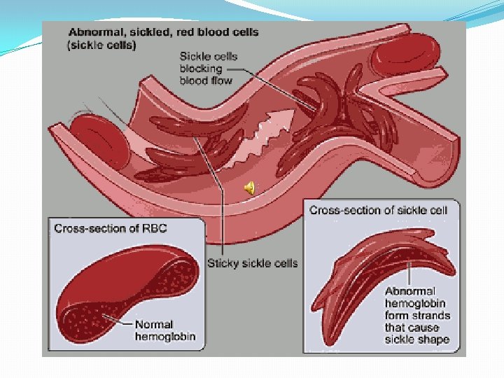
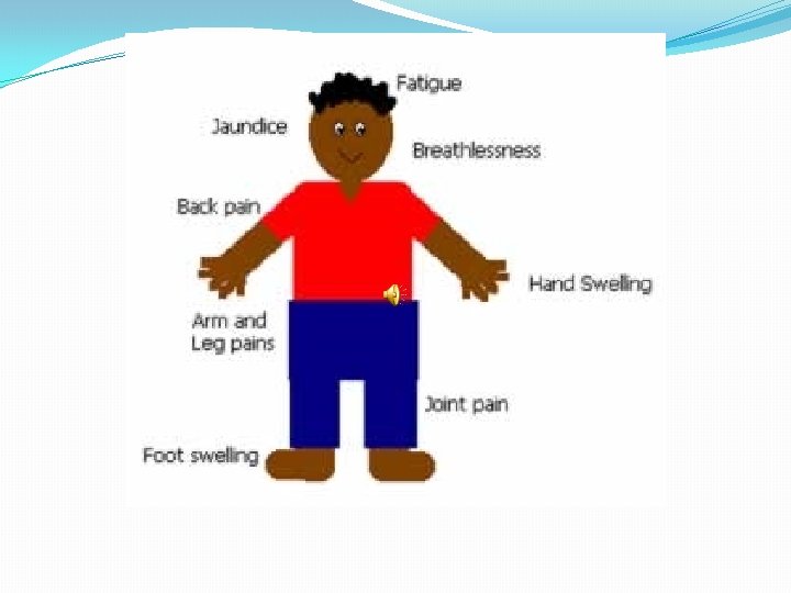
�In addition, due to these recurrent vasculo-occlusive episodes there a series of complications: 1. Because bones are particularly affected by the reduced blood flow, frequent and severe bone pain results. This is the typical symptom during a sickle cell crisis.
2. Long term, the recurrent clogging of the capillary beds leads to damage to the internal organs, in particular the kidneys, heart and lungs 3. The continual destruction of the sickled red blood cells leads to chronic anemia and episodes of hyperbilirubinemia
Treatment of sickle cell disease �Hydration �Analgesics �Antibiotics �Transfusions (risks- hemosiderosis) �Hydroxyurea
HEMOGLOBIN C DISEASE �Glutamate is replaced by lysine at position 6. �Hb C will move more slowly towards the anode than Hb S �Patient have generally mild , chronic anemia. �Patients don't suffer infarctive crises �No specific therapy is required.
HEMOGLOBIN SC DISEASE �Some β chains have the sickle cell mutation whereas other β globin chains carry the mutation found in Hb C. �Hb level tends to be high in Hb. SC than in sickle cell anemia. �These patient will remain well until they suffer an infarctive crises.
�Hemoglobin AS - A heterozygous form causing Sickle cell trait with one adult gene and one sickle cell disease gene �Hemoglobin SC disease - Another heterozygous form with one sickle gene and another encoding Hemoglobin C.
advantage �Selective advantage for heterozygotes. �Less susceptible to malaria-(plasmodium falciparum).

- Slides: 32