Introduction liver is the largest internal organ 2


![CUSA [Cavitron Ultrasonic Surgical Aspirator] • well-defined transection plane • low blood loss +](https://slidetodoc.com/presentation_image_h2/84018e5dc69455e2ad67e6cbe0391690/image-26.jpg "CUSA [Cavitron Ultrasonic Surgical Aspirator] • well-defined transection plane • low blood loss +")


- Slides: 36
Introduction • liver is the largest internal organ • 2 -3% of total BW • fixation of liver – IVC – Rt + Lt triangular ligament – coronary ligament – falciform ligament
Liver surgery • Dr. Luis, 1886 – the first liver surgery – Pt died 6 hours later due to bleeding • Dr. Langenbuch, 1888 – the first successful liver resection – Re-open for bleeding • Kousnetzoff & Pensky, 1896 – suture fracture technique
Liver surgery • Dr. Cantlie, 1897 – further understanding of liver anatomy – better bleeding control • Dr. Pringle, 1908 – compression of portal inflow • Improvement in morbidity and mortality – – subcostal incision with better exposure anesthesia technique technological advance peri-operative care
Hepatocellular Carcinoma • HCC – the most common primary hepatic carcinoma • 3 rd leading cause of cancer deaths worldwide – Asia & sub-Saharan Africa • HBV, HCV, alcohol • Liver cirrhosis
HCC survival to treatment
before Surgery…. • understanding hepatic anatomy is crucial
Liver anatomy • Based on arterial blood supply, portal vein, biliary drainage, and hepatic venous drainage
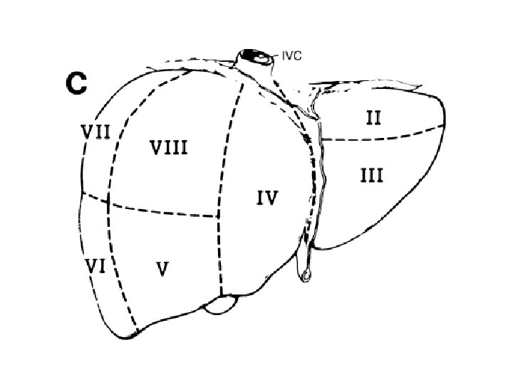
Liver anatomy • Couinaud, 1954 – 8 segments
Hepatic artery 25 -30% 17%
Lt hepatic artery
Portal vein
Bile duct
Hepatic Vein
CT interpretation of liver Lt medial Lt lateral Rt anterior Rt posterior
CT interpretation of liver anterior branch Rt portal vein posterior branch
CT interpretation of liver I
HCC management BCLC staging 2011 < 3 yr 16 M 8 M 24 M < 3 M
Pre-OP evaluation • • • CBC, PT/a. PTT liver function test α-FP Abd CT – Tri-phasic Angiography ± CT (CTAP ) ICG test (15 min <20%)
Resectability • Tumor size, number, location, relation to vessels • Liver resection / hepatic failure • Liver condition – cirrhosis, post-chemotherapy • portal vein embolization • Anatomical resection • Resection margin
Types of major resection • • • Rt lobectomy Lt lateral segmentectomy extended Rt lobectomy extended Lt lobecomy
Abd wall Incision
Hepatic Resection Techniques Vascular control Blood loss is the most important factor to post -op outcome • Pringle’s maneuver • Total hepatic vascular exclusion • Intra-hepatic pedicle ligation
Liver parenchymal transection • • • finger or clamp-fracturing the tissue ultrasonic or radiofrequency energy water-jet tissue-sealing device surgical stapler
Crush-Clamp Technique • crushing parenchyma to expose small vessels and bile duct • Pringle maneuver • non-cirrhotic liver
CUSA [Cavitron Ultrasonic Surgical Aspirator] • well-defined transection plane • low blood loss + low risk of bile leak
Harmonic scalpel • 55, 500 Hz vibration cut and seal vessel up to 3 mm • protein denaturization, not heat • increase risk of bile leak
Liga. Sure Vessel Sealing System • simultaneous parenchymal division and vessel hemostasis • bipolar vessel sealing device, up to 7 mm
Tissue. Link • radiofrequency energy • blunt parenchymal dissection and hemostasis
Radiofrequency-assisted liver resection • Radiofrequency energy to thermocoagulate liver parenchyma • higher complication rate – abscess, biliary fistula or stenosis – infection
Water-jet dissection • high-pressure water jet to break apart the liver tissue • No thermal damage
Vascular Stapler • Division of major vessel and liver parenchyma
Laparoscopic Liver Resection • segment II, IVb, V, VI – wedge resection – Lt lateral segmentectomy
da Vinci Robot • 3 D visualization + Wristed instruments – more flexibility to perform fine movements not possible with laparoscopy
Liver Transplantation • Milan criteria – solitary ≤ 5 cm or if multiple, a maximum of 3 nodules ≤ 3 cm – without vascular invasion or extrahepatic spread – the 5 -year survival > 70% – recurrence ranging from 5% to 15%
Thank You for Your Attention!!