The Integumentary System Ya gotta have skin to


 • Active person (1 L/ hr) In")
")


 • A")


 • This bacterium")
 • Structurally they are the same. The skin has responded")


 of dermal blood vessels • always present at")
 is a lack of pigmentation")


- Slides: 74
The Integumentary System Ya gotta have skin to keep your insides in!!! Chapter 5 pages 142 -159
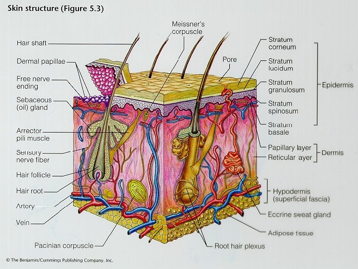
Skin Factoids Skin is part of the Integumentary System. -Largest organ of the body. (21. 5 ft 2)( 12 -15% of body weight). -Thickness varies: . 5 cm on upper back. 05 cm on eyelids Hair and sweat glands are not located in all areas! - No hair on the palms - No sweat glands on the eye lids.
Skin is always being replaced from within. Most of the dust in your house is dead skin. By the time you are 70 you will have shed 70 lbs. of skin. • Skin is important in helping the body to maintain homeostasis (“steady state) of such things as body temperature, fluid balance and Skin Trivia
General Skin Structure • 2 main layers - epidermis and dermis • Epidermis • 4 to 5 layers of epithelium • 3 main cell types – Keratinocytes – Melanocytes – Langerhans cells (like the one pictured here) are free ranging white blood cells that are found in the stratum spinosum. They phagocytize foreign material and bacteria.
• • The skin supports a wide variety of microorganisms. Species of Bacteria and yeasts inhabit the surface of the skin and help to protect us from more dangerous microbes. We are all hosts of tiny mites which feed on our dead skin. They are most common in the eyebrows and eyelashes. All houses have dust mites that eat the dead skin that accumulates in pillows and cushions. These mites can become very numerous and some people can become allergic to their droppings. It is suspected that this may be the explanation for some cases of asthma. The skin as an ecosystem
This picture shows a group of follicle mites nested in the follicle of an eyebrow.
Dust Mite
• Dust Mites
Perspiration • Inactive person (3 m. L/hr) • Active person (1 L/ hr) In hot climates this can increase to 2 to 3 L/hr in 6 weeks. • In addition to cooling the body sweat provides nutrients for certain bacteria and fungi that lie on the skins surface. • These bacteria produce lactic acid which prevents many kinds of harmful bacteria from growing on the skin! • Sweat is produced in the sweat glands and exits through the sweat pore. • It is mostly water. There is some urea, salts, proteins and oils that are also secreted.
Sweat Gland, cross section (430 X)
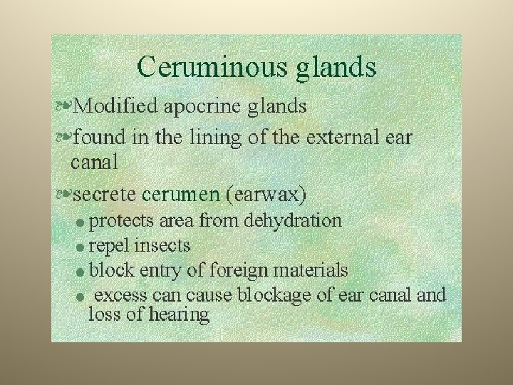
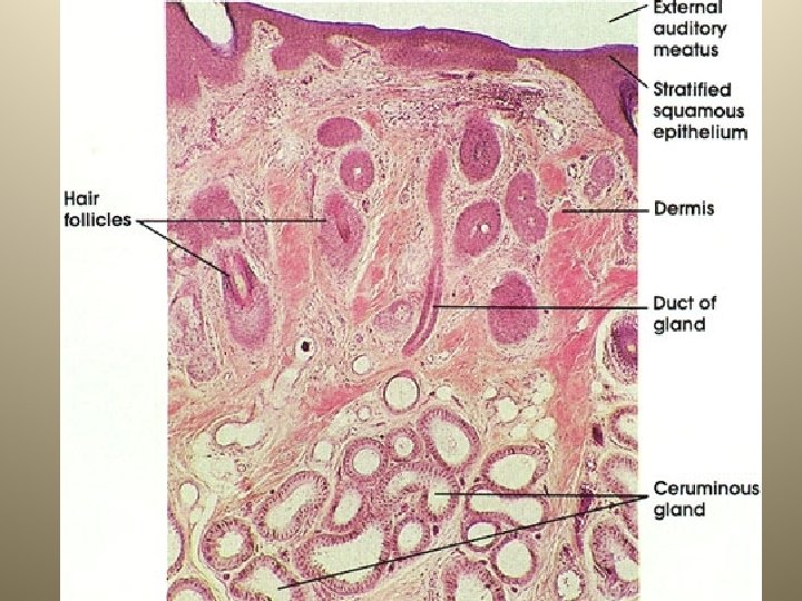
Sebaceous gland Secreting oil to help lubricate and protect the hair shaft.
Apocrine sweat glands are formed from the same structure as the hair follicle and sebaceous glands. • They produce a thicker, viscous liquid that produces a noticeable odor. • The apocrine glands become very active with the onset of puberty. They are found in the armpit and the genital area. • The milk glands in the breasts are modified apocrine sweat glands. • Body odor is produced by microorganisms that grow where the apocrine glands exist. • The bacteria produce an odor as they digest this sebum. • Since they can only do this if water is present many deodorants are antiperspirants as well. Apocrine glands
• The skin provides protection against mechanical injury. • The skin helps to prevent the penetration of many kinds of harmful chemicals. • This photograph shows a typical case of poison ivy. Poison ivy is caused by an allergic reaction between the body and an oil, urushiol, that is produced by this plant. • 85% of the people in the US are sensitive to this oil. Functions of Skin
Functions of Skin • The skin, with its continuous waterproof covering, helps to prevent microbes from getting inside the body. • The skin is host to a community of microbes that assists in the prevention of disease.
Functions of Skin • Prevention of dehydration. • Burn patients who lose more than 50% of their skin are likely to become severely dehydrated. • The skin provides protection of the internal sea that bathes all of our cells. • This slide is human skin magnified 1000 X by a scanning electron microscope.
• The skin helps to maintain a constant body temperature. • Capillary networks and sweat glands help to radiate excess heat and to cool the body through evaporation of water. Functions of Skin
Functions of Skin • Excretion of Wastes. • Water, urea and some salt are excreted when people sweat. • Recently people discovered a new compound in sweat, a protein called dermicidin. • This protein seems to play a role in inhibiting bacterial growth.
Functions of Skin • • • The skin serves as a large sensory receptor organ of the body. Many nerve endings end in the skin. Sensations include touch, pressure, heat, cold, and pain. • • • Meissner’s corpuscle (fine touch), free nerve endings (touch, pressure and pain, (tickle? ). Merkel’s discs (light touch and pressure), Krause’s end bulbs (cold? ), Pacinian corpuscle (heavy pressure and vibration)
Skin nerve receptors • Pacinian corpuscle • Sensitive to heavy pressure and vibration • Abundant near joints and skeletal muscles, palms and soles
• • Meissner’s corpuscle Fine touch receptors Found in high concentrations in fingertips Important in two point discrimination
Skin nerve receptors • Pain, cold and warmth nerve receptors are free nerve endings located very close to the epidemis • Cornea of the eye contains only these nerve receptors
Cold and Warmth • Detected by specific free dendrites • 3 to 10 times more cold than warm receptors • Extremes in temp. (<100 C and >450 C) involve pain receptors
Functions of Skin • Vitamin D synthesis • Skin that is exposed to the sun will produce Vitamin D. • Vitamin D seems to play a role in preventing certain types of cancer.
Hair Structure and function • Hair protects the head, eyes and ears. • For creatures in cold climates often there are two layers. • Most hair structure is the same. • Hair grows at different rates.
Hair continued • Composed of keratin • Grows in cycles • Grows faster in the morning than the evening. • Curly hair is oval in shape • Grey hair lacks pigment
Hair Transplants • • • The first step in the hair transplant process is the design of the hairline. Next the donor area is prepared. Hair in the back is lifted and a narrow strip of hair is trimmed to about 1/16 th of an inch.
• • Both the top and the back of the scalp are then anesthetized. Patients are given the option for nitrous oxide (laughing gas) while the local anesthesia is applied. Narrow strips of hair bearing skin are removed from the donor area and the hair from above and below is then brought back together with a running stitch. The stitches are covered by hair and removed in ten days, usually leaving a thin scar line that is concealed by your own hair. Hair Transplant Procedure
Hair Transplant procedure • From the donor strip tiny grafts are prepared. • Grafts for the hairline are usually between 1 to 3 hairs and are dissected under a microscope to preserve every follicle possible.
Hair Transplant procedure • • The hair transplant surgeon next creates the sites where the grafts will be placed. Tiny slit incisions are made at the hairline for the smallest single hair grafts.
Hair Transplants cont. • • • The surgeon will gradually make larger incisions (approximately 1/32" wide by 1/8" long). After the procedure most patients are given a baseball cap or bandana. There is usually no bandage and the hair can be washed in two days. Tiny crusts will form where the transplants have been placed and usually shed in 4 to 7 days. The small hairs in the newly transplanted grafts normally shed within 2 to 4 weeks after the procedure. Permanent hair growth usually begins in 8 to 12 weeks. Significant hair growth within 6 to 8 months and full hair growth within 9 to 12 months.
• Hair Transplant surgery
Caribou hair is hollow and is rooted in a deep layer of fat. So, even though it is shorter than other species of deer hair it does a better job of insulating the animal from the cold.
Timber wolf fur, like many northern mammals is in two layers. There is an inner short dense layer and a longer, oily, water proof outer layer. The temperature of the outer fur is often no warmer than the environment!
Nails • Horny plates composed of keratin • Growth occurs in nail matrix. • Cells grow and fill with keratin • . 1 mm/day average growth rate
African Black Rhinoceros • The horns of the Black Rhinoceros is made up of the same protein as found in your nails and the stratum corneum. • The Black Rhinoceros’ thick skin is really a very thick layer of keratin filled cells.
Skin pigmentation • Oxyhemoglobin, carotene and melanin determine skin color • Melanin produced by melanocytes in stratum basale. • Melanocytes thought to originate from nervous tissue. • Melanin is produced in reaction to UV radiation.
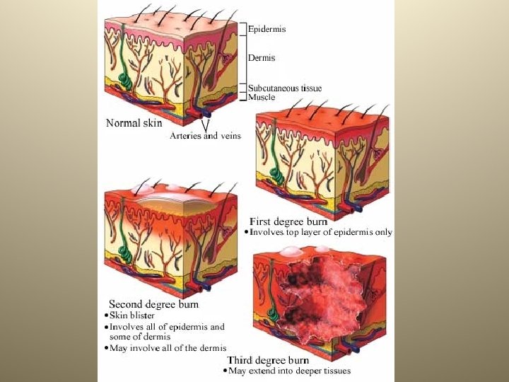
• Classified by degree • 1 st degree - redness, swelling • 2 nd degree - blisters • 3 rd degree - destroyed skin tissue. • 2 nd and 3 rd degree burns need medical attention. • Burns to the head, neck and groin are the most serious. • When the skin is burned, it can’t protect the body against bacteria, prevent the loss of body fluid, or keep the body at a normal temperature. • 2 nd and/or 3 rd degree burns over 30% of the body are usually fatal. Burns
• Skin Transplant Surger video
Burn Care • The patient is placed in a special cart that allows for cleansing of the burned area. • Cleansing is initially done with soap and water. • Medication is given to relieve pain and antibiotics and bandages are used. • Surgery is performed to remove dead and dying tissue. (debridement) • Skin grafting is done using skin from healthy areas of the patient. • If the area burned is large it may be necessary to do more than one grafting operation.
Skin Grafting • Dead skin is removed from the damaged area (debridement) • A section of skin larger than the injury is placed over the burn and covered with bulky, wet bandages that have been soaked in an antibiotic. • The graft begins to grow and adhere in 48 hours and usually is totally attached in 4 to 5 days. • The patient will need to protect this area for up to two weeks before it is totally healed
• The typical cover for extensive burn wounds was cadaver skin, known as allografts. • The skin grafts did not always have the desired effects. They sometimes introduce disease to the burn victim or were rejected by the body. • In the 1980’s artificial skin was successfully developed. • Fibroblasts from neonatal foreskin samples are enmeshed in a collagen gel. • After several weeks keratinocytes are added and they form an epidermal layer. Artificial Skin
• An artificial skin graft eliminates the need for tissue typing. • Artificial skin can be made in large quantities and frozen for storage and shipping, making it available as needed. • Each culture is screened for pathogens, reducing the chance of infection. • Because artificial skin does not contain immunogenic cells such as dendritic cells and capillary endothelial cells, it is not rejected by the body. • Finally, rehabilitation time is significantly reduced. Artificial Skin
http: //gizmodo. com/5749968/the-skin-gunthat-sprays-new-skin-on burn-victims-isreal
Skin Cancer • 3 kinds basal, squamos and melanoma • Basal cell carcinoma involves cells in the stratum basale which become cancerous • Usually noticed as persistant skin ulcers. • metastasis is uncommon
Skin Cancer • Squamous cell carcinoma • Begins in stratum spinosum • Metastasis may occur so these growths are operated on as soon as they are diagnosed.
Skin Cancer • Malignant Melanoma is cause by cancerous melanocytes • Metastasis is common and early treatment is essential • Increased 600% in last 15 years • 6/7 of all skin cancers • Most common cancer in women aged 25 -29. • Often starts as a mole that suddenly changes in size and texture.
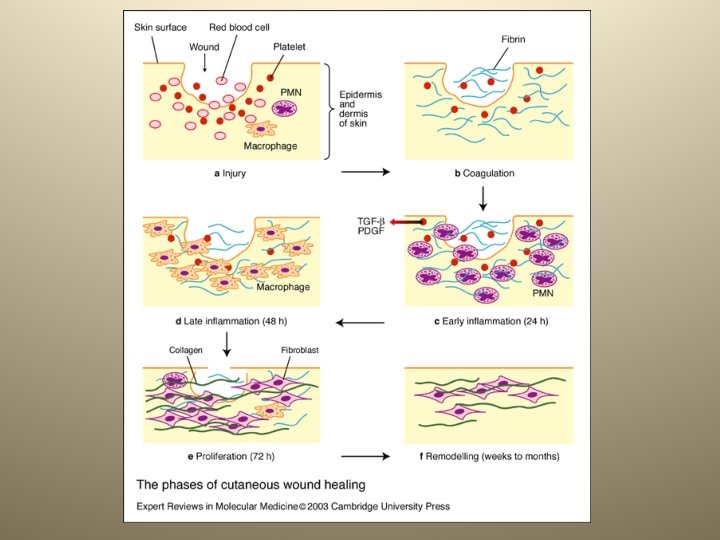
Wounds • Injured cells release histamine which causes swelling and dialation of blood vessels. • White blood cells released from bone marrow. • Clot forms • A decrease in chalones causes increased cell division. • The drying scab helps to pull the injured layers together. • Fibroblasts in the dermis produce collagen which fills in the wound below the scab.
• • • Chinese first used fingerprints thousands of years ago as signatures on documents. The formal name for fingerprinting in dactyloscopy. Fingerprints are caused by the pattern of ridges on the dermal papilla. They are also found in monkeys, gorillas, orangutans and some kinds of birds as well as humans. There also toe prints, palm prints, soleprints (footprints) and lip prints. 1893 Sir Frances Galton proved that no two prints were alike. The following year the first identification of a criminal was made Fingerprints
The Henry System of Fingerprint classification
Fingerprint residue • Twins, triplets and quadruplets all have different prints. • Fingerprints are composed mostly of sweat or moisture with tiny traces of urea, proteins and sebum. • Latent prints can be dusted and lifted several months after the impressions is made and up to 10 years if a laser is used. • Fingerprints can not be copied or forged. • The F. B. I. identifies over 2. 700 fugitives a month through fingerprints, and they have nearly 200 million fingerprints on file.
• For reasons not completely understood, follicles or pores often become blocked. • Sebum (oil) which normally drains to the surface is trapped and bacteria start growing. • Both whiteheads and blackheads start out as microcomedones as the one indicated in this picture. • Microcomedones becomedones which are either whiteheads or blackheads Acne
Trapped bacteria under the surface of the skin cause a small infection in the area of the follicle
Blackheads occur when the trapped sebum and bacteria partially open to the surface. They turn black because of the skin pigment melanin.
Acne Treatments • Acne is caused by the bacterium (propionibacterium acnes) • This bacterium can be killed by the compound benzoyl peroxide. (pictured to the right) • Treatments can include antibiotics, but the bacteria can develop a tolerance for antibiotics. • Scrubbing and irritating the skin can make matters worse.
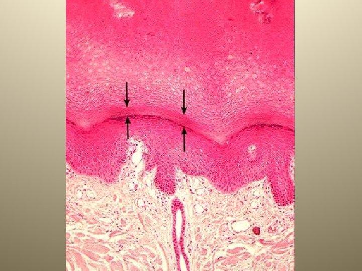
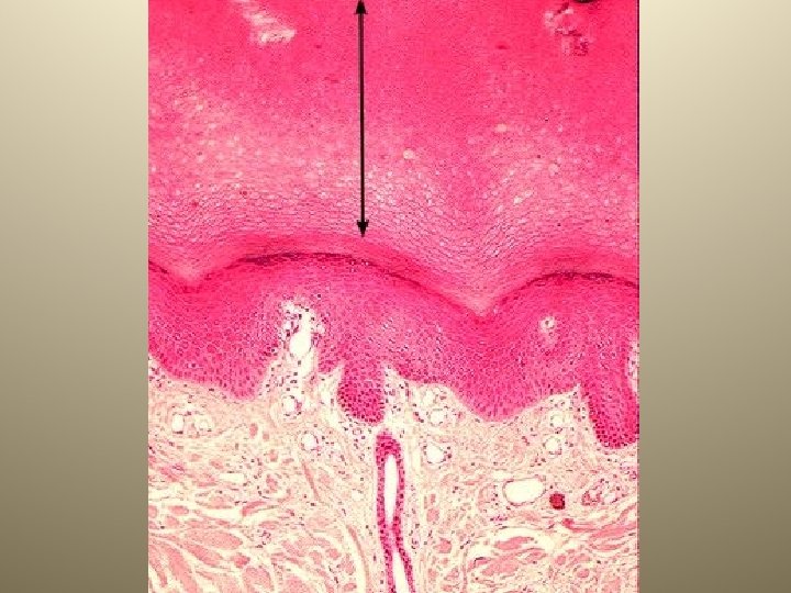
Corns and Calluses (Hyperkeratosis) • Structurally they are the same. The skin has responded to friction and pressure with increased epidermal growth. • Most of the time this is a natural and beneficial response that protects the skin and the tissues underneath the skin. • They tend to form on the foot and the hands. • Corns are the result of a callus on the foot continuing to grow to the point that it becomes painful. They are a horny thickening of the skin, dome shaped, with the dome pointing down into the skin.
• • • Freckles are flat, circular spots that typically are the size of the head of a nail. The spots develop randomly on the skin, especially after repeated exposure to sunlight and particularly in persons of fair complexion. Freckles vary in color -- they may be red, yellow, tan, light-brown, or black -- but they are always darker than the skin around them since they are due to deposits of the dark pigment called melanin. There are two types of freckles -- ephelides and lentigines: Ephelides (singular: ephelis, the Greek word for freckle): This term refers to flat spots that are red or light-brown and typically appear during the sunny months and fade in the winter. Lentigines (singular: lentigo, from the Latin word for lentil): Children may develop a small tan, brown, or black spot which tends to be darker than an ephelis-type freckle and which does not fade in the winter. Freckles
• • • Plantars Warts are the most common skin viral infection. The virus (human papilloma virus (HPV))invades the skin cells and causes them to grow abnormally. People commonly get the virus from public swimming pools or showers. They occur mostly on the plantar or the soles of the feet. They appear as raised, skincolored bumps with a rough surface. The surface is made up of dozens of tiny finger-like projections. They vary in size from the tip of a pencil to as big as a dime. In time the wart does go away. Many people will treat these warts because of the pain they can cause when walking or running. After a while a person becomes immune to these viruses. Plantars Warts
• • • Molluscum contagiosum warts These are another type of wart caused by a different virus from the classic wart. They have a different appearance and usually occur on the chest, abdomen, upper thighs, and occasionally the face. They are skin-colored bumps, can vary in size from the tip of a ballpoint pen to about half the size of the eraser end of a pencil, are smooth surfaced, often have a dimple in the center, and usually occur in clusters. They can itch. They are more contagious than classic warts, and are transmitted by repeated close physical contact with the warts. These often do not require any treatment, and will go away on their own usually within 1 year. They are more difficult to treat because there can be several dozen at any time. Molluscum warts
• keloids extend beyond a healed wound with clawlike extensions. • keloids may arise without history of injury. • more common in black people than white people. • earlobes, shoulders, upper back, chest • hypertrophic scars tend to regress in time • keloids may expand for decades • histology: densely packed, bright pink collagen bundles in a haphazard array; increased number of fibroblasts and blood vessels in early lesions Keloids
• vascular malformation (not neoplasm) of dermal blood vessels • always present at birth • never spontaneously disappears • macular in infancy; become papular and darker with age • histology: increased number of blood vessels, with larger lumens, in upper dermis Port Wine Stain
• • Albinism (from Latin albus, meaning "white") is a lack of pigmentation in the eyes, skin and hair. It results from the combination of recessive alleles passed from both parents of an individual. This condition is known to affect mammals, fish, birds, reptiles, and amphibians. The gene which results in albinism prevents the body from making the usual amounts of a pigment called melanin. The lack of pigment in the skin leads to increased incidence of burning and skin cancer. The lack of pigment in the iris of the eyes leads to increased sensitivity of the eyes to light. People with albinism are not less healthy and lead normal lives of normal length. Albinism
A girl with albinism and her mother.
• Same basic structure as human skin. • Tends to have a thicker epidermis overall. • The outer epidermis heals quickly and can lock bacteria inside the body creating abscesses. • Abscesses, if severe, may need to be operated on to remove the infection. • In cat species that live in colder weathere will be an inner dense layer of fur as in dogs. • Note the lack of sweat glands. Cat Skin cross section
• The outermost fat layer, the blubber, provides insulation for the body; the thickness varies from 5 to 50 centimetres, depending on the species. • The limited transportation of blood to the outer layers of the body also reduces heat loss. • The insulating capability of whale blubber is so effective, that the temperature of a stranded whale rises rapidly from 37 0 C to 50 - 60 0 C and the carcass begins to decompose immediately Whale Blubber