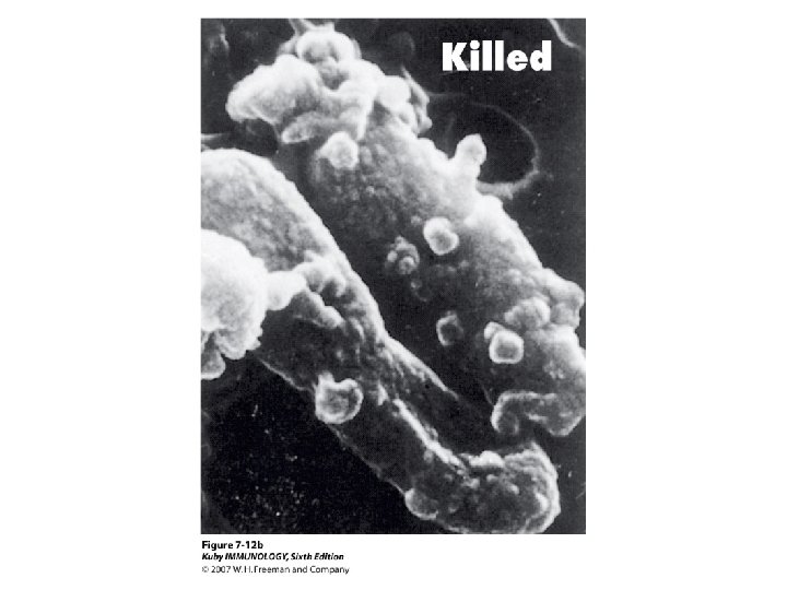
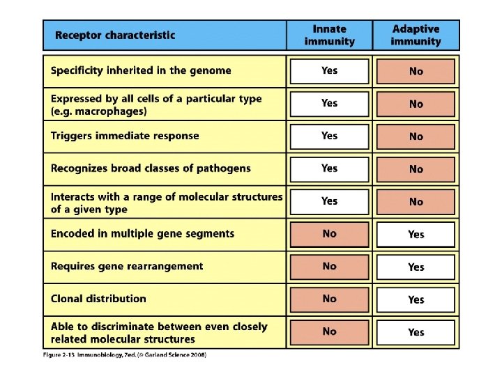
Innate and adaptive immunity The mechanisms of innate


 were acquired by")


 MBL Reconstitution Does Not Rescue C 4 Deposition Patient+")


 Functional Analysis of the Mutated and Wild Type Recombinant")


- Slides: 61
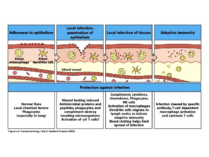
Innate and adaptive immunity. The mechanisms of innate immunity provide the initial defense against infections. Adaptive immune responses develop later and consist of activation of lymphocytes. The kinetics of the innate and adaptive immune responses are approximations and may vary in different infections.
Plants or non-vertebrate organisms have molecules and systems that have innate immunity properties.
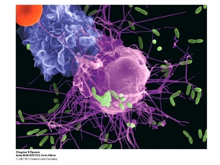
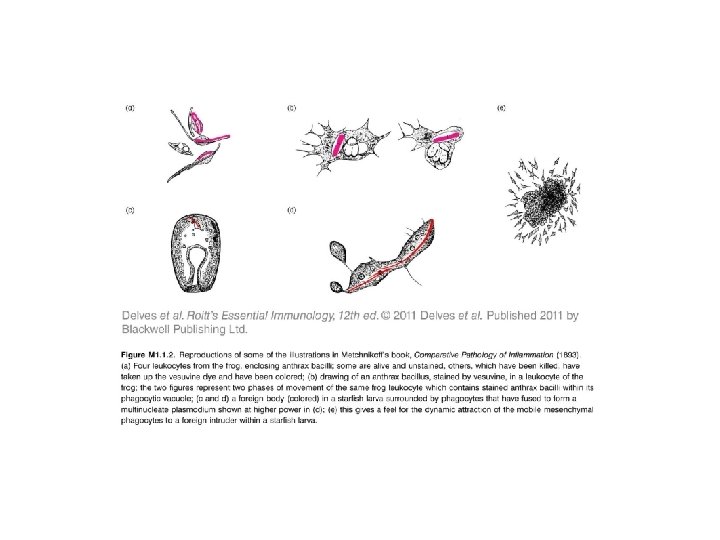
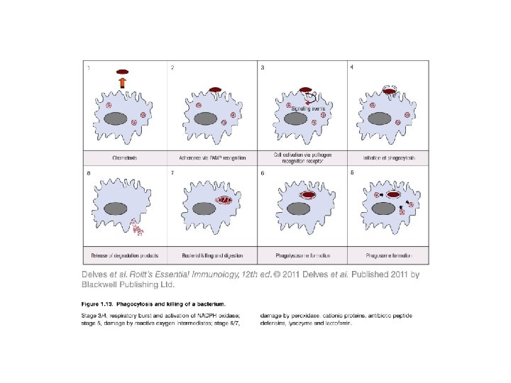
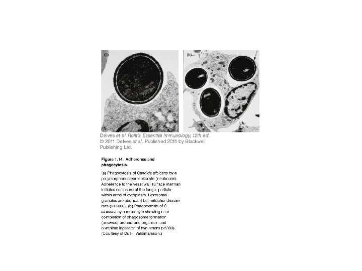
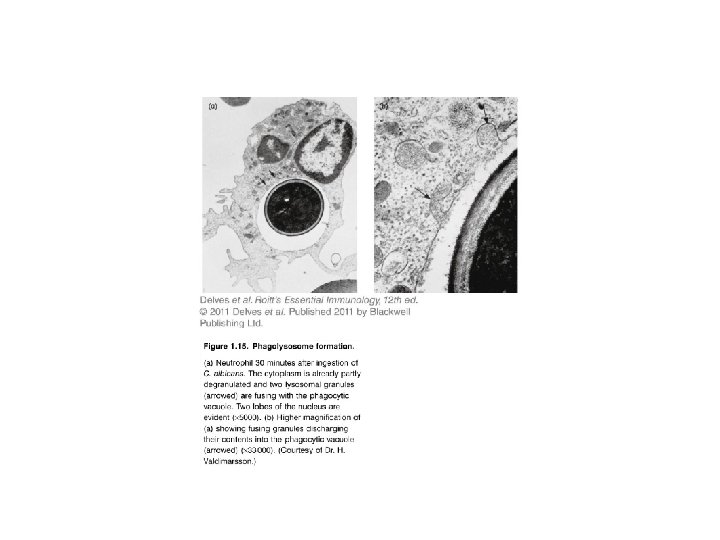
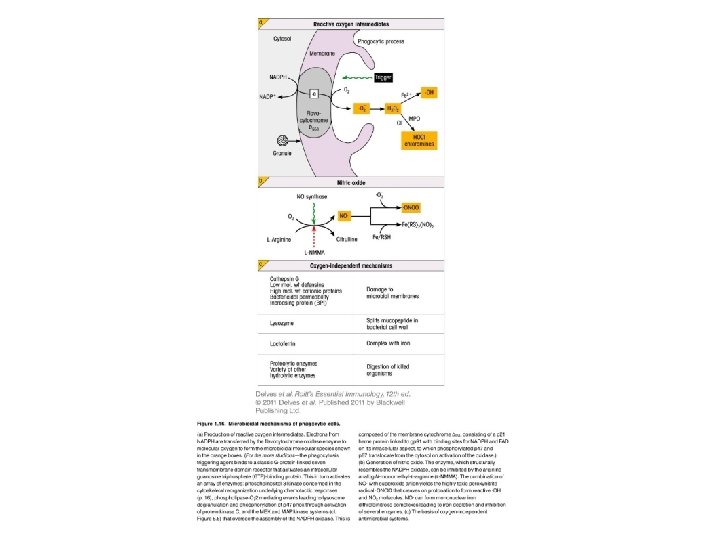
Phagocytes
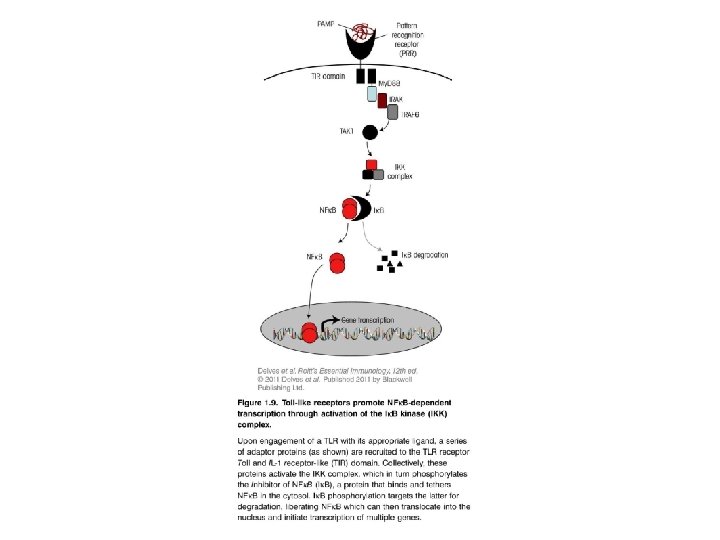
Mammalian Toll like receptors are activated by many different pathogenassociated molecular patterns
TLR 4 recognizes LPS in association with MD 2
Intracellular NOD proteins sense the presence of bacteria, recognizing bacterial peptidoglycans and activating NFk. B to induce the expression of pro-inflammatory genes.
Structural mechanisms of inflammasome assembly FEBS Journal Volume 282, Issue 3, pages 435 -444, 21 NOV 2014 DOI: 10. 1111/febs. 13133 http: //onlinelibrary. wiley. com/doi/10. 1111/febs. 13133/full#febs 13133 -fig-0001
Structural mechanisms of inflammasome assembly FEBS Journal Volume 282, Issue 3, pages 435 -444, 21 NOV 2014 DOI: 10. 1111/febs. 13133 http: //onlinelibrary. wiley. com/doi/10. 1111/febs. 13133/full#febs 13133 -fig-0004
Important “take home” concepts: 1. Cells of the innate immune system like neutrophils, macrophages rapidly eliminate microbes through phagocytosis. 2. Pattern recognition receptors like TLRs initiate intracllular cascade of signaling that triggers transcription of proinflammatory mediators. 3. NOD receptors act as intracellular sensors for bacterial infection 4. NALP proteins initiate processing of proinflammatory mediators of the IL-1 family by activation of inflammasome
NK cells
Activating and inhibitory receptors of NK cells. A. Activating receptors of NK cells recognize ligands on target cells and activate protein tyrosine kinase (PTK), whose activity is inhibited by inhibitory receptors that recognize class I MHC molecules and activate protein tyrosine phosphatase (PTP). NK cells do not efficiently kill class I MHC-expressing healthy cells. B. If a virus infection or other stress inhibits class I MHC expression on infected cells, and induces expression of additional activating ligands, the NK cell inhibitory receptor is not engaged and the activating receptor functions unopposed to trigger responses of NK cells, such as killing of target cells and cytokine secretion.
Functions of NK cells. A. NK cells recognize ligands on infected cells or cells undergoing other types of stress, and kill the host cells. In this way, NK cells eliminate reservoirs of infection as well as dysfunctional cells. B. NK cells respond to IL-12 produced by macrophages and secrete IFN-γ , which activates the macrophages to kill phagocytosed microbes.
NK cell behavior under homeostatic conditions. Images of NK cells (green) were acquired by twophoton microscopy. As a reference for velocity, images were acquired on LN that had also received B cells (red). The image sequence was acquired 18 h after NK cell adoptive transfer. (Scale bar: 15 mm. ) Time indicated as min/sec. Movie is shown at 15 frames per sec.
NK cells form stable conjugate pairs with allogeneic B cells. The image sequence was acquired 20 h after NK cell (green) adoptive transfer and shows a single NK-B cell (red) conjugate. (Scale bar: 5 mm. ) Time indicated as min/sec. Movie is shown at 15 frames per sec.
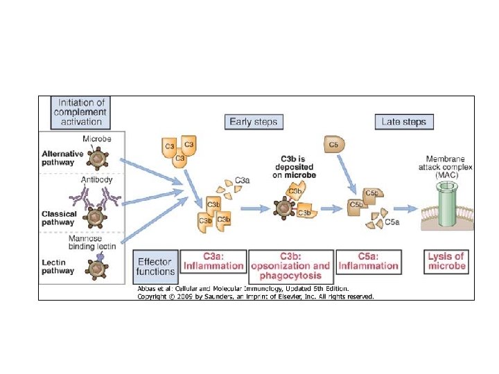
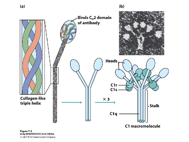
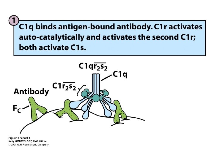
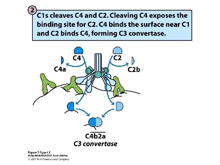
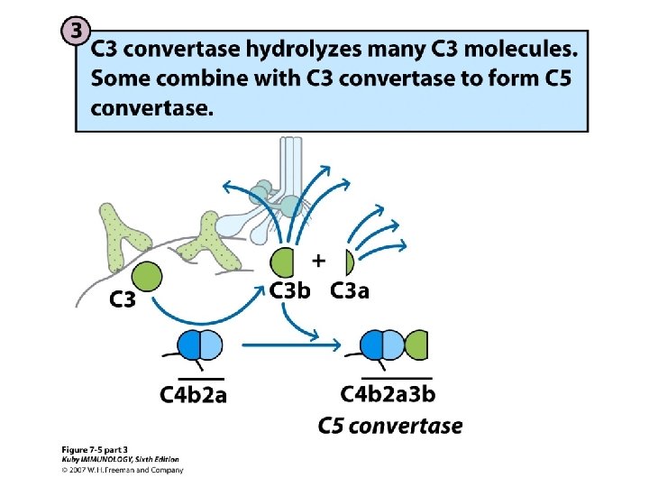
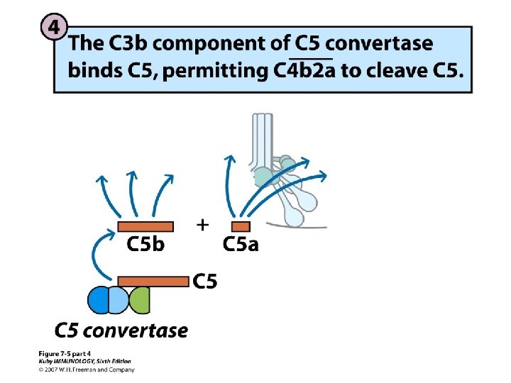
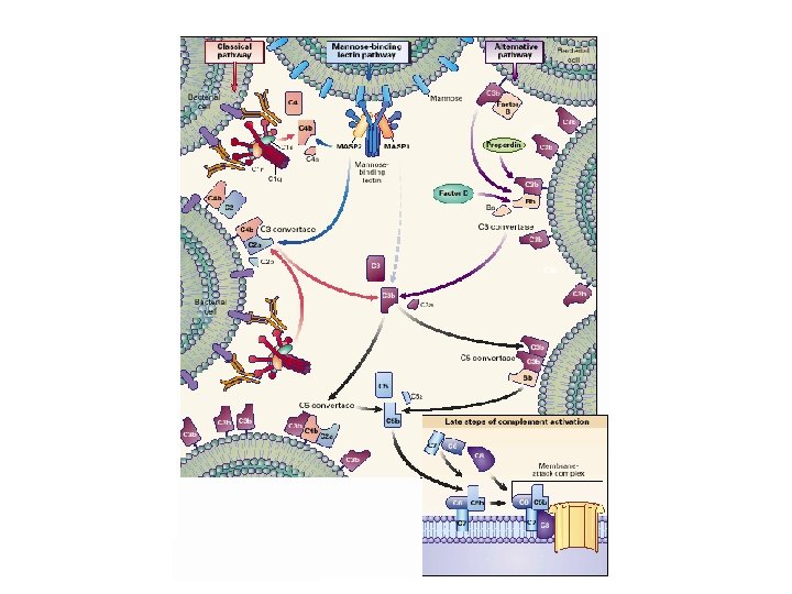
C 3 convertase activates C 3 for covalent bonding to microbial surfaces by cleaving it into C 3 a and C 3 b and exposing a highly reactive thioester bond in C 3 b.
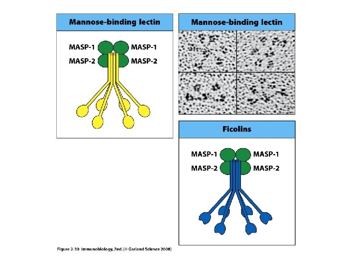
Mannan-Binding Lectin C-type lectin combining a collagen – like tail with sugar binding domain. Three gene products combine to form the basic subunit, which can form different oligomers The oligomerization increases the avidity of MBL – carbohydrate interaction. Distinct functions can be attributed to the different oligomers
Mannan-Binding Lectin C-type lectin combining a collagen – like tail with sugar binding domain. Three gene products combine to form the basic subunit, which can form different oligomers The oligomerization increases the avidity of MBL – carbohydrate interaction. Distinct functions can be attributed to the different oligomers
Why so promiscuous? Micropattern recognition: N-acetyl glucosamine, N-acetylmannoseamine, L-fucose and glucose
MBL as an Example for Pattern Recognition Parasites • Leishmania • Trypanosomes Viruses • Influenza • RSV • HIV Bacteria • gram negative • gram positive • Mycobacteria • Clamydia • Yeast/fungi • Candida • Aspergillus
MBL Levels in Circulation MBL plasma 1, 000 10 1 1000 ng/ml 40% 100 ng/ml 10%
MBL Haplotypes OH OH OHOHOH OH PGINGFPGKDGRDDTKGEKGEPGQ OH PGINGFPGKDGRDGTKGEKGEPGQ OH OH Haplotype B (G 34 D) OH Haplotype A (native) PGINGFPGKDGRDGTKQEKGEPGQ OH OH OH Haplotype C (G 37 Q) PGINGFPGKDGCDGTKGEKGEPGQ OH OH Haplotype D (R 32 C) OH OH OH
Low MBL plasma levels are caused by mutations in the promoter and/or coding region in the MBL gene locus A/A MBL plasma 1, 000 A/O 1000 ng/ml 40% 100 10 1 O/O 100 ng/ml 10%
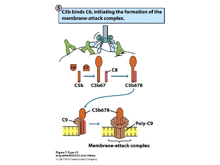
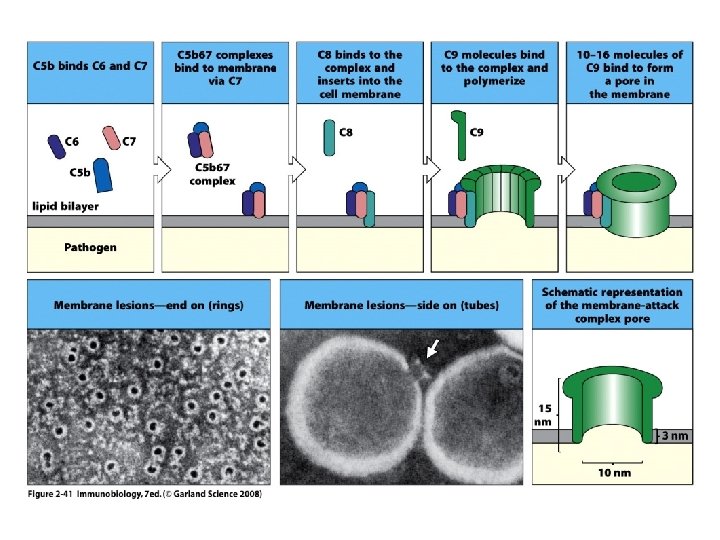
C 3 3 C 2 b C 4 2 C 4 b C 3 b 1 M Co AS mp P lex 4 MAC C 5 -9 C 5 a MBL-activating surface C 4 a C 3 a C 2 a
MBL is a Disease Modifier of Considerable Importance in the Context of Secondary Immune Deficiencies Plasma MBL activates complement by binding to carbohydrate structures presented by microorganisms. MBL levels were measured in leukemia patients who were scheduled to undergo chemotherapy and MBL was related to the results of the chemo. Low MBL concentrations are associated with serious infections related to chemotherapy (p<0. 0001). CSI- clinically significant infections Peterslund et. al. , Lancet, 2001, vol 358, p 637
Complement activation Recombinant MBL Functionally Equivalent to p. MBL ng/m Jensenius et. al. , Bioch Soc Transactions, 2003, 31: 763
Functional Analysis of the MBL/MASP-2 Complex
Correlation between MBL concentration in plasma and activity of MBL-MASP complexes C 4 deposition (m. U/ml) 10000 A/A A/B A/D A/C 0/0 Linear regression 1000 10 1 1 10 10000 MBL concentration (ng/ml)
C 4 deposition (m. U/ml) MBL Reconstitution Does Not Rescue C 4 Deposition Patient+ r. MBL Sera Healthy control sera + r. MBL deficient MBL concentration (ng/ml)
Patient Outline • Born 1967. • No medical history until 1980 when diagnosed with ulcerative colitis - treated with prednisolone. • 1996: erythema multiforme bullosum responding to prednisolone. Suspect SLE - joint symptoms, myalgia, weakly positive anti-nuclear antibodies. • 1995 -1997 Several courses of severe pneumococcal pneumonia. • Progressive lung fibrosis without vasculitis, alveolitis or granulomas.
Mutation in MASP-2/MAp 19 Gene in CUB 1 domain
Functional Analysis of the Mutated and Wild Type Recombinant MASP-2
MASP-2 (counts x 10 -3/sec) Functional Analysis of the Mutated and Wild Type Recombinant MASP 2 30 A. MBL B. L-ficolin C. H-ficolin 50 20 45 18 25 40 16 35 20 14 30 12 15 25 10 20 8 10 15 6 4 10 5 2 5 0 0 0 25 50 75 100 125 0 25 50 75 100125 00 25 50 75 100125 ng MASP-2/ml.
Normal CUB 1 ACC TTC GCG TCC GAC TCC AAC GAG AAG CCG TTC T F R S D Y S N E K P F Mutant CUB 1 105 G ACC TTC GCG TCC GGC TAC TCC AAC GAG AAG CCG TTC
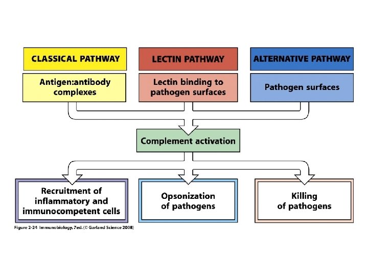
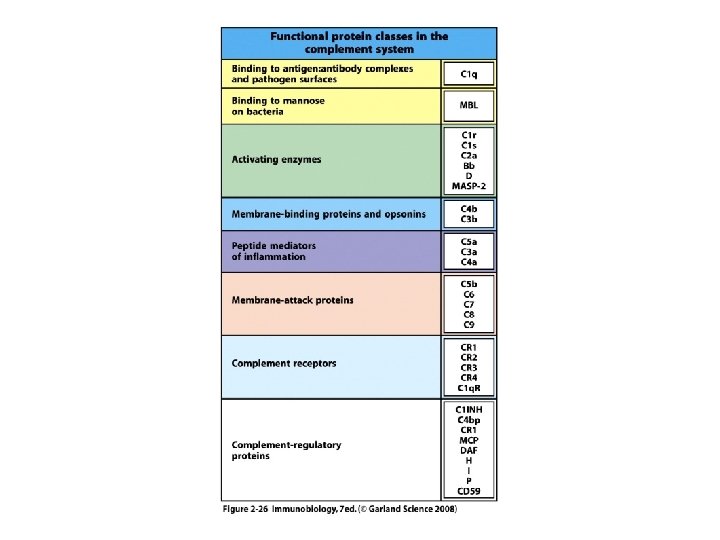
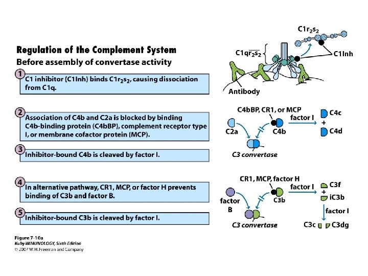
Conclusions • The complement system comprises a group of serum proteins. • The three pathways converge in a common sequence of events. • Activation of the alternative and lectin pathways is antibody independent. • In addition to cell lysis complement mediates opsonization, activation of inflammation, and clearance of the immune complexes. • Complement system can control the adaptive immunity • Because of its ability to cause damage to the host organism, the complement system requires a complex passive and active regulatory mechanisms.