Immune tolerance autoimmune diseases Immune tolerance Central negative


 • Systemic autoimmune disease affecting various tissues and organs •")


 Indirect immunofluorescence on Hep 2")
 • • • SLE: 95 - 100 %")


 thyroiditis Hyperthyroidism (Graves’ disease; thyrotoxicosis) Type I")


- Slides: 31
Immune tolerance, autoimmune diseases
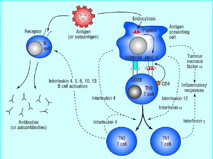
Immune tolerance • Central: – negative selection during thymic education – deletion of autoreactive B-lymphocytes in bone marrow
Positive selection in the thymus
Negative selection in the thymus
Tolerance ‘Central’
Immune tolerance • Peripheral: – Clonal deletion - elimination of autoreactive cells by apoptosis – Clonal anergy - costimulatory signals are lacking – Clonal ignorance - to low concentration of antigen does not stimulate immune response – Suppression - autoreactivity is blocked by regulatory cells
Tolerance ‘Peripheral’
Regulatory T cells • Treg cells – naturally occurring regulatory cells causing tolerance of autoantinegens. They cause active tolerance of autoantigens. Development in the thymus. Involved in inborn tolerance. Also the induction of these cells in periphery by foreign antigens seems to be possible. • TH 3 (Tr 1) cells: induced in periphery. They cause acquired tolerance.
Acquired immune tolerance • Low-zone tolerance: repeated injections of very low doses of antigen. Suppressor cells are stimulated. • High-zone tolerance: induced by highdoses of antigen. Clonal deletion is induced. • Oral tolerance
Mechanisms of breakage of immune tolerance • Visualization of „hidden antigens“ • Alteration of body antigens by chemical substances, burns, necrosis • Cross reactivity of antigens • Excessive stimulation of the immune system, abnormal expression of HLA-II antigens. • Defect in suppressor function of lymphocytes
Systemic autoimmune diseases Systemic lupus erythematosus Rheumatoid arthritis Sjogren’s syndrome Polymyositis Dermatomyositis Scleroderma (progressive systemic sclerosis)
SLE • A prototypic multi-system autoimmune and immune complex disease • Involvement of skin, kidneys, lungs, heart blood vessels • Immunoregulatory abnormalities • Many autoantibodies – ANA • ds DNA • ENA – Phospholipids
Systemic lupus etythematodes (SLE) • Systemic autoimmune disease affecting various tissues and organs • Many symptoms are caused by deposition of immune complexes (type-III immunopathological reaction) • Female : male ratio is 10: 1 • Usually begins in early adulthood
Systemic lupus erythematodes Clinical presentation • • General: fever, malaise, loss on weight Artralgia Skin: butterfly rash, urticaria Vascular: Raynaud´s phenomenon Neurological: vasculitis, seisures, neuritis Glomerulonephritis Haematological: leukopenia, thrombocytopenia anemia • Recurrent serositis
Systemic lupus etythematodes • Butterfly rash
Systemic lupus etythematodes
Autoantibodies in SLE - 1 Anti-nuclear anibody (anti-nuclear factor) Indirect immunofluorescence on Hep 2 cells Staining pattern may be clinically useful Interpretation depends on clinical story, titre and age Sensitive but not specific Good screening test for lupus (prevalence ~ 100%)
Positivity of antinuclear antibodies (ANA, ANF) • • • SLE: 95 - 100 % Rheumatoid arthritis: 15 - 30 % Systemic scleroderma: 75 -80 % Autoimmune hepatitis: 20 -60 % Healthy persons: 0 - 4 % Seniors: 10 - 20 %
ANA - homogenous type
ANA – granular type
Systemic lupus erythematosus
Livedo reticularis
Systemic lupus etythematodes
Organ-specific autoimmune diseases Endocrine system Autoimmune (Hasimoto’s) thyroiditis Hyperthyroidism (Graves’ disease; thyrotoxicosis) Type I diabetes mellitus (insulin-dependent or juvenile diabetes) Autoimmune adrenal insufficiency (Addison’s disease) Autoimmune oophritis Hermatopoietic system Autoimmune hemolytic anemia autoimmune thrombocytopenia Autoimmune neutropenia Neuromuscular system Myasthenia gravis Autoimmune polyneuritis Multiple sclerosis Skin Pemphigus and other bullous diseases Cardiopulmonary System Rheumatic carditis Postcardiotomy syndrome (Dressler’s syndrome) Gastrointestina tract Atrophic gastritis Crohn´s disease Ulcerous colitis Autoimmune hepatitis
Anti- parietal cells antibodies
Pernicious anemia • Antibodies against gastric parietal cells cause atrophic gastritis. • Decreased production of gastric juice results in dyspeptic problems. • Also production of intrinsic factor is decreased causing disturbed resorption of vitamin B 12. • Low serum levels of vitamin B 12 result in megaloblastic anemia.
Anti-receptor antibodies • Stimulatory – – Graves disease. Antibodies against TSHreceptors stimulate function of thyroid gland causing hypertyreosis. • Inhibitory – Myastenia gravis. Antibodies against acetylcholine receptor block activation of muscle in neuromuslular junction.
Treatment of autoimmune diseases • Substitution of function of the affected organ (insulin treatment, parenteral treatment by vitamin B 12…. ) • Anti-inflammatory drugs • Immunosuppressive treatment • Tolerance induction
Systemic Immunosuppression • • • High-dose steroids Purine antagonists: Azathioprin Alkylating agents: Cyclophosphamide Anti-pholates: Methotrexate Calcineurin antagonists: Cyclosporine A, Rapamycin, Tacrolymus • Block of purins synthesis: Mycophemolate • Antilymphocytic serum • Monoclonal antobodies: anti CD 3, anti CD 20, anti CD 54. . .
Imunostimulatory drugs • • Synthetic immunostimulators: inosiplex Cytokines: IL-2, interferons Thymic hormones Bacterial immunomodulators: Ribomunyl, Broncho-vaxom, Luivac, Imudon, Biostim. . .