Autoimmune diseases and tolerance Dr Eman Albataineh Assistant

Autoimmune diseases and tolerance Dr. Eman Albataineh, Assistant Prof. Immunology College of Medicine, Mu’tah university Immunology, 2 nd year students

Tolerance – – – Immunologic tolerance is defined as unresponsiveness to an antigen Antigens that induce tolerance are called tolerogens, or tolerogenic antigens, to distinguish them from immunogens, which generate immunity and failure of self-tolerance results in immune reactions against self (autologous) antigens. Such reactions are called autoimmunity

• Main contributor to tolerance is CD 4 T cells because – MHC relation to autoimmune diseases – T cell is the key regulator of immune response to proteins • Tolerance in CD 4+ helper T lymphocytes is an effective way of preventing both cell-mediated and humoral immune responses to auto protein antigens because helper T cells are necessary inducers of all such responses • Ways of tolerance • • Central tolerance; selection, T reg Peripheral tolerance= T cells regulation

v Mechanisms of central self tolerance v In T cell selection stage; Medullary thymic epithelial cells can express self antigens that related to many organs and this controlled by many genes one is called Aire (autoimmune regulatory) (Aire deficient cause polyendocrinopathy syndrome, addison, hypoparathyroid and chronic candidiasis. ) if they react to them they die or differentiated to T reg and non-self reacting mature T cell circulate

v Peripheral tolerance v Auto-reactive T cells may result because many self antigens are not presented in thymus or presented insufficiently (hidden). Or because infection with similar foreign antigen to self or genetic. • T cell regulation, • Absence of co-stimulatory signals (B 7) on APC-self antigen • expression of CTLA-4 after T cell activation • activation induced cell death by death receptors ( Fas-Fas. L) in the case of persistent activation, • or apoptosis or passive cell death in case of antigen elimination, • T cell anergy, presenting antigen by immature DCs,

B lymphocytes tolerance • Central – Editing, deletion and anergy • Peripheral – Mature B lymphocytes that recognize self antigens in peripheral tissues in the absence of specific helper T cells may be rendered functionally unresponsive or die by apoptosis – Expression of death protiens; Fas on B cell and Fas L on Tc, inhibitory receptor CD 22 and inhibitory Fc receptor (FcγRIIB). • polymorphism of this inhibitory Fc receptor (FcγRIIB). impairs inhibitory signaling and is associated with SLE in humans

Artificial induction of tolerance • Tolerance induction may also be useful for – preventing immune reactions to the products of newly expressed genes in gene therapy protocols, – for preventing reactions to injected proteins in patients with deficiencies of these proteins (e. g. , hemophiliacs treated with factor VIII), – for promoting acceptance of stem cell transplants, and graft transplantation – and in immunotherapy for allergy to foreign proteins.

Ways of induction tolerance • In general, protein antigens administered cutaneously with adjuvants favor immunity, whereas antigens administered without adjuvants tend to induce tolerance. • Immunosuppression by total body irradiation, drugs (cyclosporin and anti-lymphocytic antibodies as anti. CD 4, soluble CTLA-4, steroids • Oral administration of antigens ( lead to increase in IGA and IL-10 and TGF-beta)

Autoimmune diseases, etiology • multifactorial • After infection, trauma or surgery • Release of sequestrated antigens as a result of tissue injury. g; Post-trauma, exposed antigens of nucleus in SLE (systemic lupus erythromatosus) that immune system did not expose to before. • Exposure to microbial antigens that cross react with self antigens (molecular mimicry); strep-pyogenes and rheumatic fever. • Infection may also lead to enhanced expression of costimulators in tissues. Thus, the infection results in the activation of T cells that are not specific for the infectious pathogen; this type of response is called bystander activation.
; Rheumatoid arthritis (RA)in HLADR 4, thyroiditis in HLA DR")
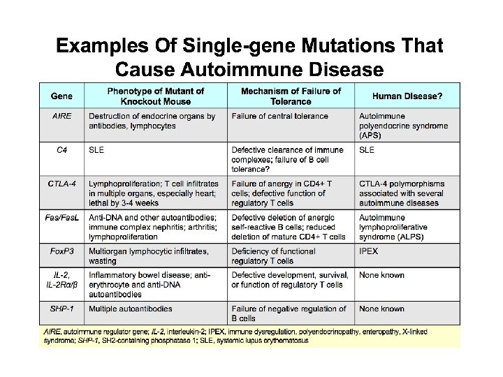
• Genetic pre-disposition (Polygenic); Rheumatoid arthritis (RA)in HLADR 4, thyroiditis in HLA DR 5, multiple sclerosis in HLA DR 2, systemic lupus erythromatosus (SLE) in HLA DR 3, Type 1 diabetes in HLA DR 3 and 4 • Other genetics (single gene), loss of fas, fas. L expression. AIRE gene, C 4 gene (SLE) and CTLA -4 gene mutations • Hormonal factors, RA and SLE more in females
 focus on one organ or")
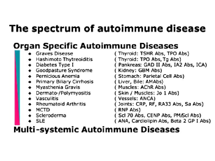
Classification of autoimmune v Organ-specific disorders (also called localized) focus on one organ or a specific type of tissue. Among those that can affect children are: • Addison’s disease (adrenal glands) • celiac disease (gastrointestinal tract) • Crohn’s disease (gastrointestinal tract) • multiple sclerosis (MS)(brain/spinal cord) • type 1 diabetes (pancreas ilets Beta cells) • ulcerative colitis (gastrointestinal tract) v Non-organ-specific disorders (also called systemic) cause problems more widely throughout the body. Among those that can affect children are: • Rheumatic fever (joints , skin and heart) • lupus (SLE) (joints, skin, kidneys, heart, brain and others) • Rheumatoid arthritis (RA) (joints, skin, muscles)

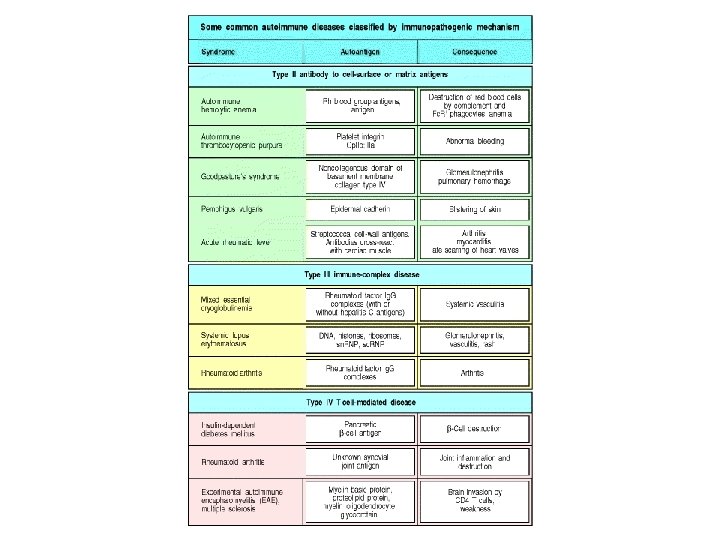
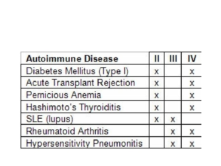
• Mechanisms of tissue damage – EFFECTOR MECHANISMS IN AUTOIMMUNE DISEASES: Both antibodies and effector T cells can be involved in the damage in autoimmune diseases – Bound self antigens (type 2 hypersensitivity) as hemolytic anemia, myasthenia gravis, thyroiditis, good Pasteur and rheumatic fever – Immune complex deposition; type 3 as SLE and RA – Cell mediate (Type 4), as Multiple sclerosis (MS), RA, type 1 diabetes and ulcerative colitis • Once autoimmune disease start it become chronic and progressive due to epitope spreading as a result of tissue damage • The symptoms is on and off, when it is on it is called flare up.

Lab. diagnosis • Elevated levels of immunoglobulins • High CRP, ESR • Auto-antibodies; anti-nuclear, anti-smooth muscle, anti-mitochondrial, rheumatoid factor • Complement levels may decreased • Biopsy; Antibody on the surface of the tissue or cells, immune complex or lymphocyte infiltration

management • Anti-inflammatory drugs; aspirin, corticosteroids • Immuno-suppressive drugs, azathioprine • plasmapheresis

Myasthenia gravis • MG is Ab against acetylcholine receptor of neuromuscular junction, block receptor and cause muscle weakness • Gravis disease – Antibodies against thyroid stimulating hormone receptor cause long lasting activation and hyperthyroidism • Idiopathic thrombocytic purpura (platelet antigen) low platelet count+bleeding • Good pasteur syndrome (renal and lung basement membrane collagen)lung and kidney bleeding; antiglomerular basement membrane (GBM) • Vitiligo (melanocytes) lead to depigmentation of skin
 • Red flush on face as wings of butterfly, disease")
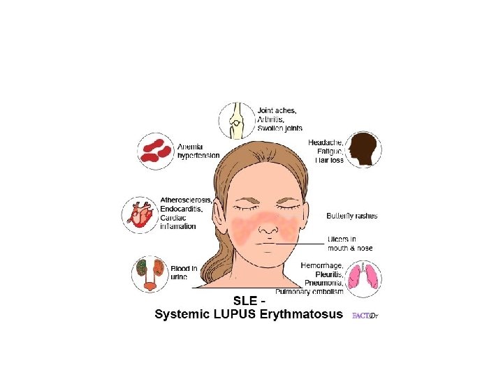
SLE (Systemic lupus erythematosus) • Red flush on face as wings of butterfly, disease attack many organs as CNS, heart and kidney • Mechanisms; 95% of patients have Abs to DNA and RNA (anti-nuclear Ab (ANA), and more specific anti-ds-DNA antibody (type 2 and 3 hypersensitivity) – as a result of tissue damage, exposed DNA attacked by Ab)
, basement membrane of")
SLE • Circulating immune complexes deposit in skin (vasculitis, skin rass), basement membrane of kidney (lampy bumpy deposits) lead to glumerolonephritis and proteinuria • Diagnosis, – Symptoms as skin rash, proteinuria and edema – Tissue biobsy; immunoflourescence microscope (granular appearance or linear)) to see ANA, – Blood levels of ANA and anti-DS DNA antibody – low complement levels

SLE

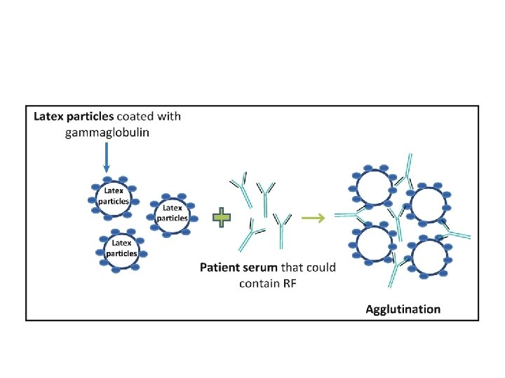
Rheumatoid arthritis • Synovium full with lymphocytes and immune complexes leading to destruction of bone and cartilage • Causes – rheumatic factor: Auto-antibodies mainly IGM but may be IGG, against Fc portion of self IGG and this factor present in 90% of patients – Tissue damage by Type 3 hypersensitivity reaction (Immune complexes), by anti-CCP antibody or RF and antigen complexes – Or Type 4 reaction; TH 1, CD 8 cells, IL-1, IL-6 and TNF alpha cytokines against antigens in synovial membrane of the joint – anti-nuclear Abs (ANA) in 50% of patients – Stiff painful joints, malformation in Joint x-ray – Diagnostic test, positive RF latex agglutination test. mixes the patient's serum with tiny latex beads covered with human antibodies (Ig. G). The latex beads clump or agglutinate if rheumatoid factor is present.

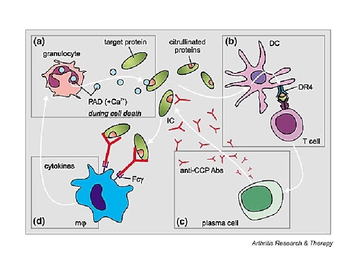
Anti-CCP • During inflammation, arginine amino acid residues can be enzymatically converted into citrulline residues in proteins such as vimentin, by a process called citrullination. That lead to generation of new antigenic epitopes • Anti-citrullinated protein antibodies (ACPAs) are autoantibodies (antibo dies to an individual’s own proteins) that are directed against peptides and proteins that are citrullinated. They are present in the majority of patients with rheumatoid arthritis (70%). Clinically, anti-cyclic citrullinated peptides (CCP) in patient serum or plasma are frequently used for diagnosis in very early stages(then referred to as anti– citrullinated peptide antibodies).

RA

RA disease modifying agents • Corticosteroids, pain killers, methotrexate (anti-folate that inhibit synthesis of DNA, and RNA) so prevent or stop immune cells growth • anti-inflammatory drug sulfasalazine

RA treatment

Rheumatic fever • Rheumatic fever is an inflammatory disease that occurs following a Streptococcus pyogenes infection, such as strep throat or scarlet fever. Believed to be caused by antibody cross-reactivity that can involve the heart, joints, skin, and brain the illness typically develops two to three weeks after a streptococcal infection. Acute rheumatic fever commonly appears in children between the ages of 6 and 15, with only 20% of first-time attacks occurring in adults. The illness is so named because of its similarity in presentation to rheumatism.
 is the antibody")
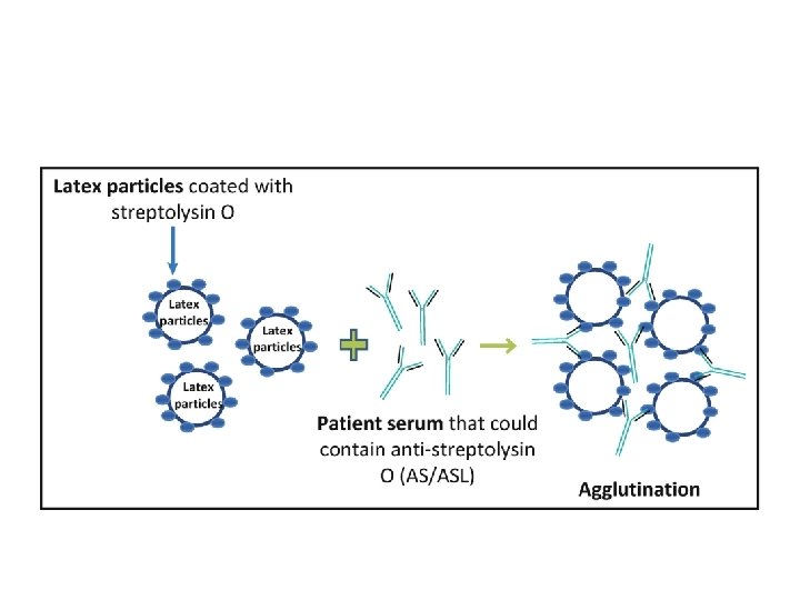
diagnosis • Elevated anti-ASO titer or Anti-streptolysin O (ASO or ASLO) is the antibody produced against an antigen produced by group A streptococci. The antigen is called streptolysin O, the titer varies being maximum 3 -5 weeks after infection. the presence of Ab indicate exposure to these bacteria. diagnosis depend also on clinical presentation as some people have this antibody but normal. • positive ASO latex agglutination test.
 (Type 2 hypersensitivity) • Also")
Multiple sclerosis • Antibodies against myelin basic protein (MBP) (Type 2 hypersensitivity) • Also cell infiltration with TH 1 and TH 17 and cytokine as TNF alpha (type 4) • Demyelination, perivascular inflammation, paralysis and ocular lesions • No certain treatment, disease modifying agents as interferon beta, anti-CD 20 to deplete B cells, injection of MBP to induce tolerance Hashimoto thyroiditis • Antibodies against thyroglobulin and/or thyro-pyroxidase (TPO) antigens (type 2 hypersensitivity) • hypothyroidism, and hard and large gland due to lymphocytic infiltrate (type 4 hypersensitivity) • Treatment, thyroid hormone replacement
 (type 2) •")
Type 1 diabetes • Antibodies against pancreatic beta cell protein (insulin) (type 2) • Or infiltration with cells TH 1 and CD 8 and (type 4) • cytokine effect (IL-1 and TNF alpha) • Lead to beta cell destruction and absence of insulin • Can be differentiated from type 2 DM by autoantibody testing • Symptoms polydepsia, polyphagis, polyuria • Treatment, insulin therapy, immune therapy by induce tolerance to diabetic antigen.

Causes of inflammatory bowel disease are mainly genetic and cellular infiltration mainly TH 1 and TH 17 (type 4)

Connective tissue diseases • Besides RA and SLE • Scleroderma – an activation of immune cells that produces scar tissue in the skin, internal organs, and small blood vessels. Lead to tight skin appear in fingers and chest • Sjögren's syndrome – also called Sjögren's disease, is a chronic, slowly progressing inability to secrete saliva and tears. • Mixed connective tissue disease – Mixed connective-tissue disease (MCTD) is a disorder in which features of various connective-tissue diseases (CTDs) such as systemic lupus erythematosus (SLE); scleroderma , MCTD is considered an intermediate stage of a disease that eventually becomes either SLE or Scleroderma. • Psoriasis in psoriasis is over growth of the skin epidermal layer, Th 17 and TH 1 cell attack (type 4) the skin epidermis and joint. (cluster plaques on the skin)

Immune-suppressive drugs • Initially, radiation and chemicals were used as nonselective immunosuppressive agents • Corticosteroids, inhibit immune response, it is a glucocorticoid-based medication that works principally to block T cell and APC derived cytokine. The major elements blocked are proinflammatory cytokines IL-1 and IL-6. • antiproliferative(azathioprin) inhibit T cell proliferation • Inhibitor of IL-2 – Calcineurine inhibitors that inhibits calcineurin. This inhibition ultimately inhibits the production and secretion of IL-2 and prevent T cell activation and growth. e. g. ; cyclosporin antibiotic and tacrolimus – sirolimus. It inhibits activation of T cells and B cells by reducing their sensitivity to interleukin-2 (IL-2). • antibodies – Anti-lymphocyte antibodies; anti-CD 3, Anti-CD 20 – Two antibodies that are IL-2 receptor antagonists (basiliximab and daclizumab) • Future immunosuppressive agents by induction of tolerance. Under trial. Administration of CTLA-4, high dose of soluble donor MHC. May be useful in chronic rejection
- Slides: 39