Forearm Fractures Derek J Donegan M D University

- Slides: 55
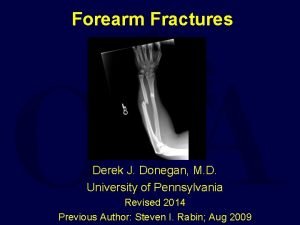
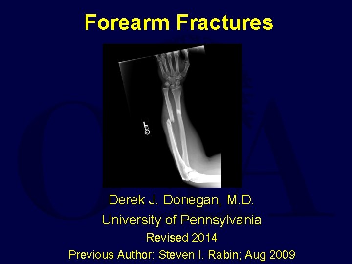
Forearm Fractures Derek J. Donegan, M. D. University of Pennsylvania Revised 2014 Previous Author: Steven I. Rabin; Aug 2009
Problem • Fractures of adult forearm are inherently unstable • According to the AO documentation center, forearm fractures accounted for 10 -14% of all fractures between 1980 and 1996 • Mistreatment can lead to malunions and nonunions – Cosmetically unappealing – Functionally impeding
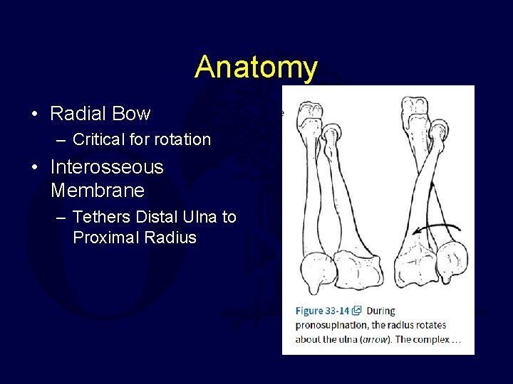
Anatomy • Radial Bow – Critical for rotation • Interosseous Membrane – Tethers Distal Ulna to Proximal Radius
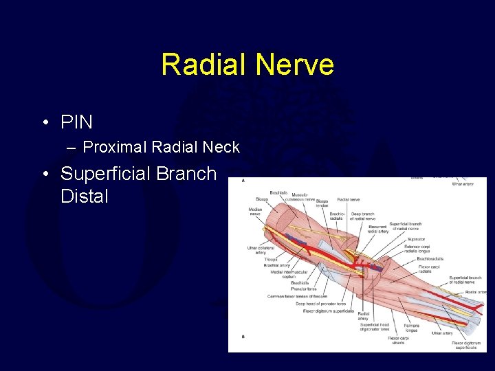
Radial Nerve • PIN – Proximal Radial Neck • Superficial Branch Distal
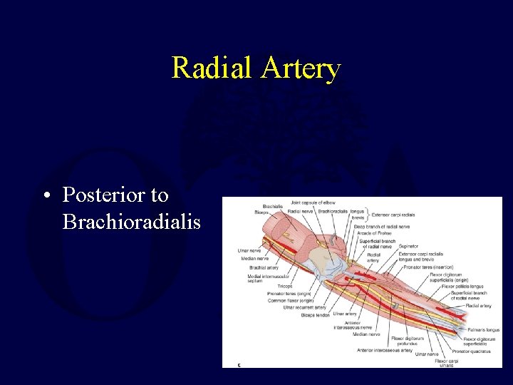
Radial Artery • Posterior to Brachioradialis
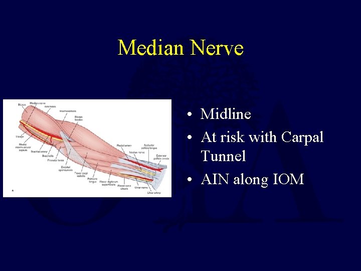
Median Nerve • Midline • At risk with Carpal Tunnel • AIN along IOM
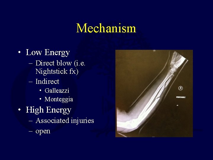
Mechanism • Low Energy – Direct blow (i. e. Nightstick fx) – Indirect • Galleazzi • Monteggia • High Energy – Associated injuries – open
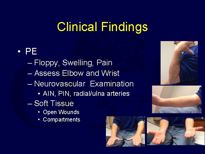
Clinical Findings • PE – Floppy, Swelling, Pain – Assess Elbow and Wrist – Neurovascular Examination • AIN, PIN, radial/ulna arteries – Soft Tissue • Open Wounds • Compartments
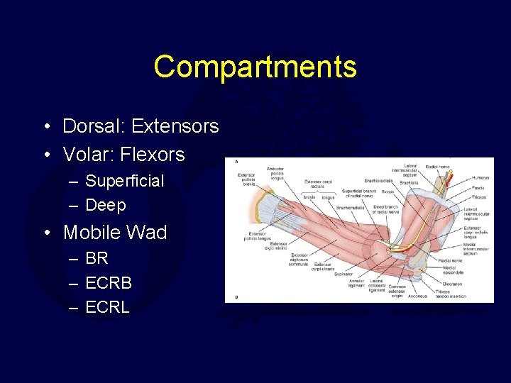
Compartments • Dorsal: Extensors • Volar: Flexors – Superficial – Deep • Mobile Wad – BR – ECRB – ECRL
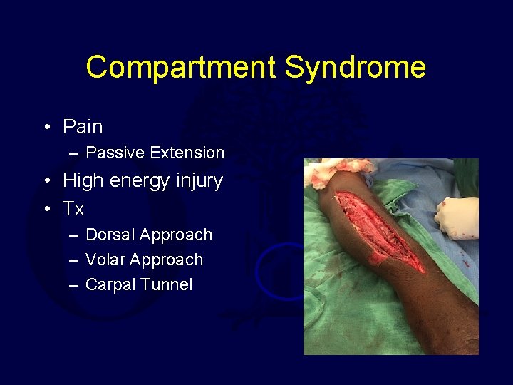
Compartment Syndrome • Pain – Passive Extension • High energy injury • Tx – Dorsal Approach – Volar Approach – Carpal Tunnel
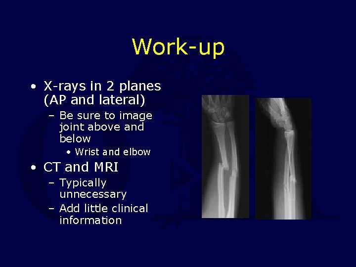
Work-up • X-rays in 2 planes (AP and lateral) – Be sure to image joint above and below • Wrist and elbow • CT and MRI – Typically unnecessary – Add little clinical information
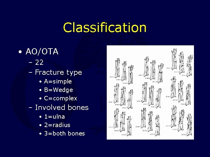
Classification • AO/OTA – 22 – Fracture type • A=simple • B=Wedge • C=complex – Involved bones • 1=ulna • 2=radius • 3=both bones
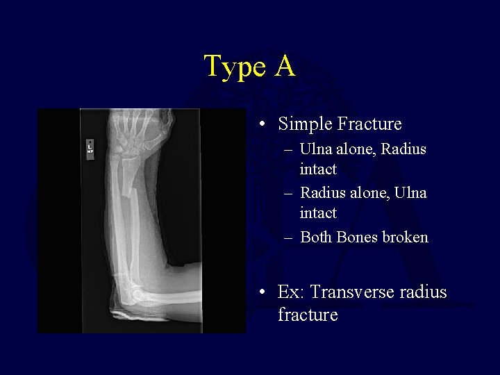
Type A • Simple Fracture – Ulna alone, Radius intact – Radius alone, Ulna intact – Both Bones broken • Ex: Transverse radius fracture
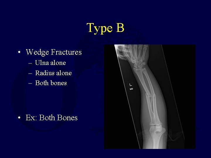
Type B • Wedge Fractures – Ulna alone – Radius alone – Both bones • Ex: Both Bones
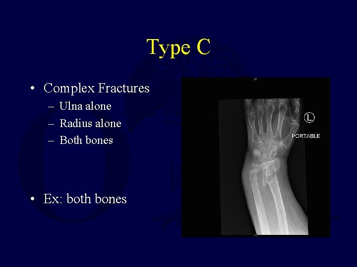
Type C • Complex Fractures – Ulna alone – Radius alone – Both bones • Ex: both bones
Non-Operative Treatment • Non-operative – Poor – Nonunion – Malunion • Non-operative – Functional Brace / Cast – Ulna • • Stable Closed Distal 1/3 < 10 Degrees – Radius • Nondisplaced • Radial bow maintained
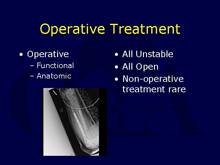
Operative Treatment • Operative – Functional – Anatomic • All Unstable • All Open • Non-operative treatment rare
Treatment • Early surgical intervention (within the first 6 -8 hours) is optimal to avoid radioulnar synostosis • Goals – Anatomic reduction – Rigid fixation – Stable construct – Restoration of radial bow
Timing of Surgery • Early Surgery is Desirable but not Essential – Easier reduction especially if shortening – Avoids pre-op immobilization • Delayed Surgery – If poor soft tissues – If other injuries or medical problems prevent
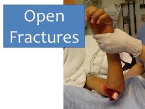
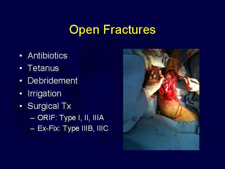
Open Fractures • • • Antibiotics Tetanus Debridement Irrigation Surgical Tx – ORIF: Type I, IIIA – Ex-Fix: Type IIIB, IIIC
Treatment • Fixation options include – IM nailing – External fixation – plate fixation
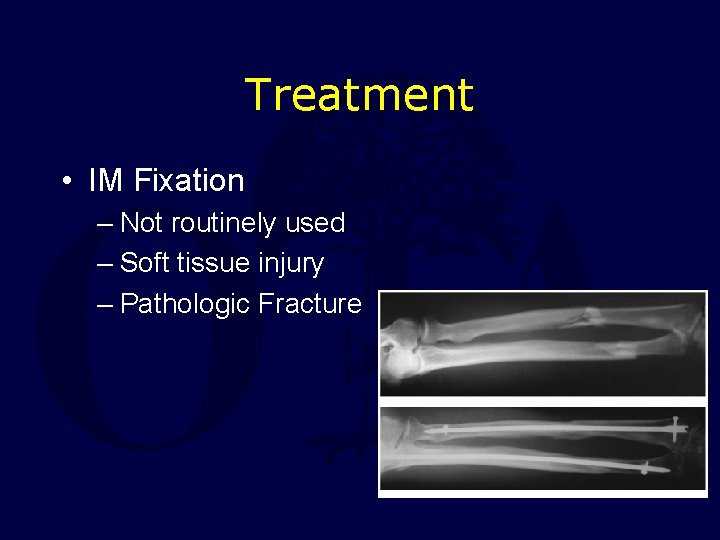
Treatment • IM Fixation – Not routinely used – Soft tissue injury – Pathologic Fracture
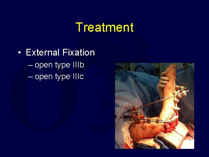
Treatment • External Fixation – open type IIIb – open type IIIc
Treatment • Plate Fixation – provides stable strong anatomic fixation – eliminates need for external casting – allows early functional motion with union rates over 95%. • Obtain anatomic reduction • Restore ulna & radial length – Prevents subluxation of either proximal or distal radioulnar joints • Restore rotational alignment • Restore radial bow – Essential for rotational function of forearm
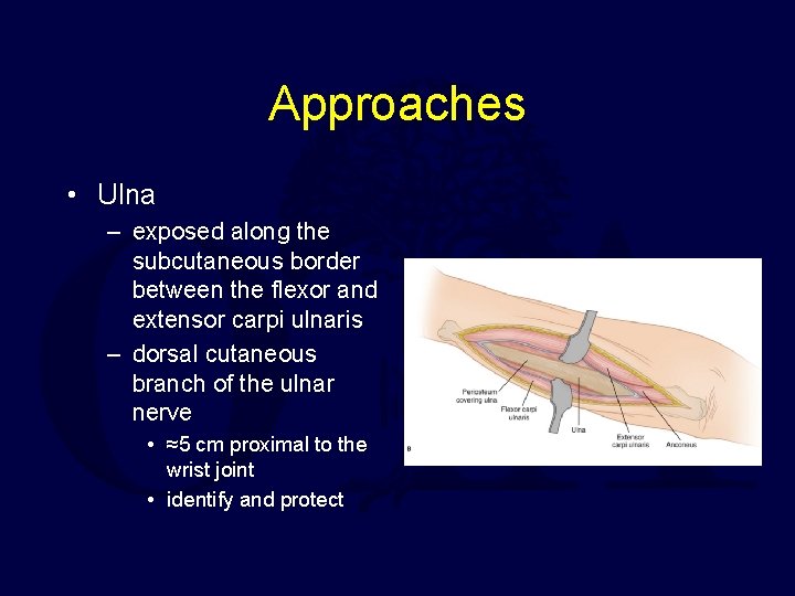
Approaches • Ulna – exposed along the subcutaneous border between the flexor and extensor carpi ulnaris – dorsal cutaneous branch of the ulnar nerve • ≈5 cm proximal to the wrist joint • identify and protect
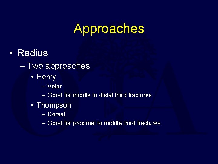
Approaches • Radius – Two approaches • Henry – Volar – Good for middle to distal third fractures • Thompson – Dorsal – Good for proximal to middle third fractures
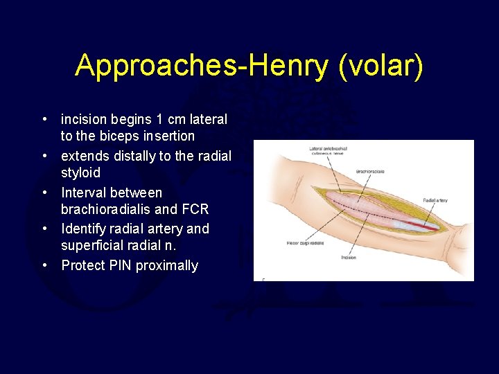
Approaches-Henry (volar) • incision begins 1 cm lateral to the biceps insertion • extends distally to the radial styloid • Interval between brachioradialis and FCR • Identify radial artery and superficial radial n. • Protect PIN proximally
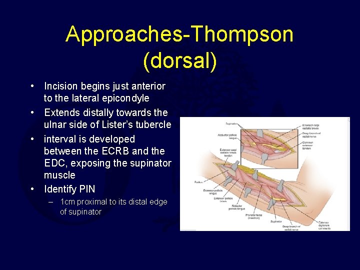
Approaches-Thompson (dorsal) • Incision begins just anterior to the lateral epicondyle • Extends distally towards the ulnar side of Lister’s tubercle • interval is developed between the ECRB and the EDC, exposing the supinator muscle • Identify PIN – 1 cm proximal to its distal edge of supinator
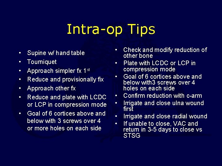
Intra-op Tips • • • Supine w/ hand table Tourniquet Approach simpler fx 1 st Reduce and provisionally fix Approach other fx Reduce and plate with LCDC or LCP in compression mode • Goal of 6 cortices above and below with 3 screws over 4 or more holes on each side • Check and modify reduction of other bone • Plate with LCDC or LCP in compression mode • Goal of 6 cortices above and below with 3 screws over 4 holes on each side • Confirm reduction with c-arm • Irrigate and close ulna wound first • Irrigate and close radial wound • If unable to close, VAC and return in 3 -5 days to close vs STSG
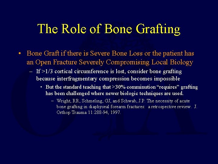
The Role of Bone Grafting • Bone Graft if there is Severe Bone Loss or the patient has an Open Fracture Severely Compromising Local Biology – If >1/3 cortical circumference is lost, consider bone grafting because interfragmentary compression becomes impossible • But the standard teaching that >30% comminution “requires” grafting has been challenged where newer biologic techniques are used. – Wright, RR, Schmeling, GJ, and Schwab, J. P. The necessity of acute bone grafting in diaphyseal forearm fractures: a retrospective review. J. Orthop Trauma 11: 288 -94, 1997.
Technical Tips for Plate Fixation of Forearm Fractures • Use Indirect Reduction Techniques Preserving Soft Tissue Attachments – Periosteal stripping must be minimized – Narrow retractors placed to avoid penetration of interosseous membrane • Close or Skin Graft Open Wounds within 35 days
Post-op • • Sterile dressing and sugartong splint Closely monitor compartments Low threshold to split dressing POD#1 – Initiate digital ROM • Delay Wrist/Elbow ROM 3 -5 days – Prevents hematoma formation
Follow-up • Forearm rotation is initiated as the patient's comfort allows – Usually 1 st or 2 nd week post-op • RTC @ 2 weeks, 6 weeks, 12 weeks, and 4 -6 months postoperatively – AP/lat X-rays each visit • Activity modification to ADL’s only until fracture healed – 8 -12 weeks • progressively return to a normal lifestyle.
Complications • • Refracture after plate removal Symptomatic hardware Nonunion Malunion Infection Neurologic injury Compartment syndrome Radioulnar synostosis
Pain & Hardware Removal • Two Years • Bone Density Does Not Normalize for 21 months – Rossen, JW et al, JBJS 1991: 73 B: 65 -7. • 4 to 20% Refracture Risk – Usually through original fracture or screw hole – Large plate (4. 5 mm DCP) – Nonunion – Infection & Nerve Injury – Pain may persist after plate removal • Post-removal – – – 67% Residual Symptoms 9% Worse Weather Exercise Skin or Tendon Irritation – Mih, AD et al, CORR 1994: 299: 256 -8
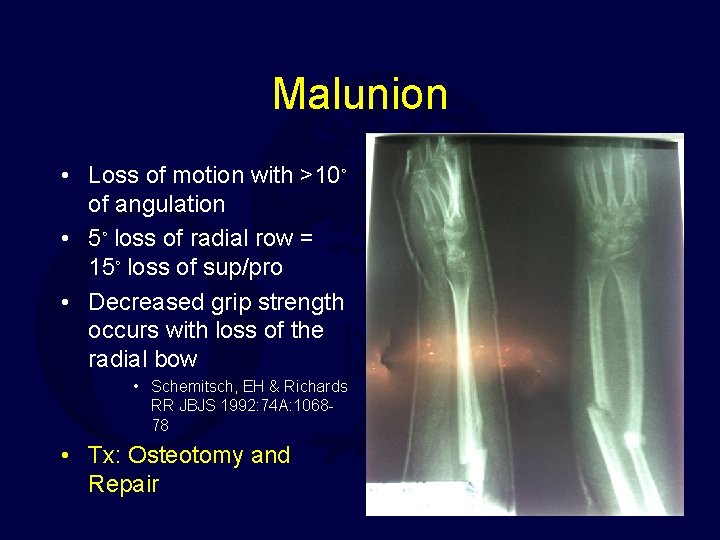
Malunion • Loss of motion with >10◦ of angulation • 5◦ loss of radial row = 15◦ loss of sup/pro • Decreased grip strength occurs with loss of the radial bow • Schemitsch, EH & Richards RR JBJS 1992: 74 A: 106878 • Tx: Osteotomy and Repair
Nonunion • Poor biomechanics • Tx • Poor Technique – Stable construct • Too few screws • Improper compression – Soft tissue management • Initial Fracture – Open Injury – Comminuted fracture – Revision Fixation – Bone Grafting – Segmental bone loss • Iliac crest <3. 5 cm • Consider vascularized fibular graft >3. 5 cm
Neurologic Injury • Closed Fracture – Usually Iatrogenic – PIN: Proximal approach – AIN: Vigorous Radial Reduction – Radial Sensory Branch: Anterior dorsal exposure • Open Fracture – AIN Most Common
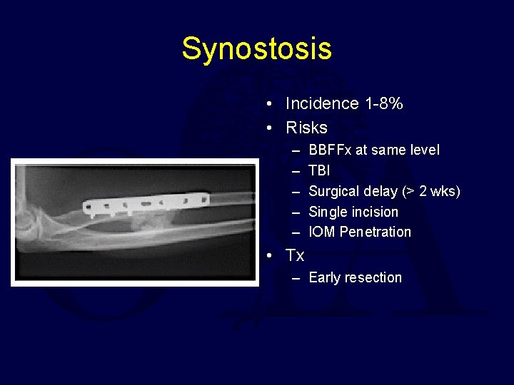
Synostosis • Incidence 1 -8% • Risks – – – BBFFx at same level TBI Surgical delay (> 2 wks) Single incision IOM Penetration • Tx – Early resection
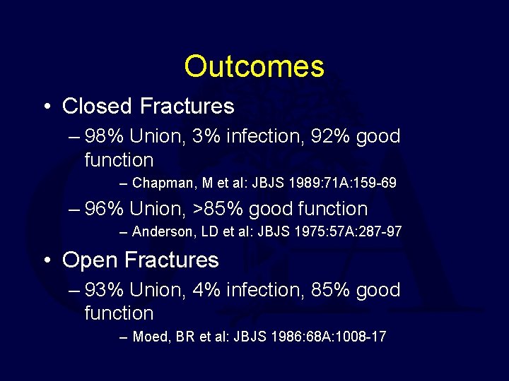
Outcomes • Closed Fractures – 98% Union, 3% infection, 92% good function – Chapman, M et al: JBJS 1989: 71 A: 159 -69 – 96% Union, >85% good function – Anderson, LD et al: JBJS 1975: 57 A: 287 -97 • Open Fractures – 93% Union, 4% infection, 85% good function – Moed, BR et al: JBJS 1986: 68 A: 1008 -17
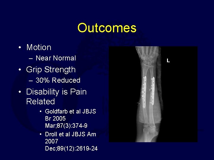
Outcomes • Motion – Near Normal • Grip Strength – 30% Reduced • Disability is Pain Related • Goldfarb et al JBJS Br 2005 Mar; 87(3): 374 -9 • Droll et al JBJS Am 2007 Dec; 89(12): 2619 -24
Special Cases • Fractures Associated with Joint Disruption – Galleazzi Fracture – Monteggia Fracture – Combined Patterns • Fractures Associated with other Injury – Floating Elbow (Ipsilateral Humerus Fracture) – Open Fractures
Fractures Associated with Joint Disruption Galeazzi & Monteggia • Best Treatment – ORIF w. Plate Fixation of Diaphyseal Fracture – Joint Usually Reduces Indirectly and is stable – If Unstable: require open reduction of joint – If irreducible – it is usually because the diaphyseal fracture has been mal-reduced
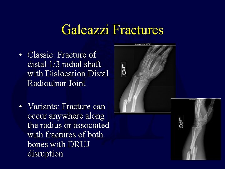
Galeazzi Fractures • Classic: Fracture of distal 1/3 radial shaft with Dislocation Distal Radioulnar Joint • Variants: Fracture can occur anywhere along the radius or associated with fractures of both bones with DRUJ disruption
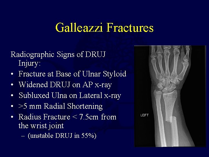
Galleazzi Fractures Radiographic Signs of DRUJ Injury: • Fracture at Base of Ulnar Styloid • Widened DRUJ on AP x-ray • Subluxed Ulna on Lateral x-ray • >5 mm Radial Shortening • Radius Fracture < 7. 5 cm from the wrist joint – (unstable DRUJ in 55%)
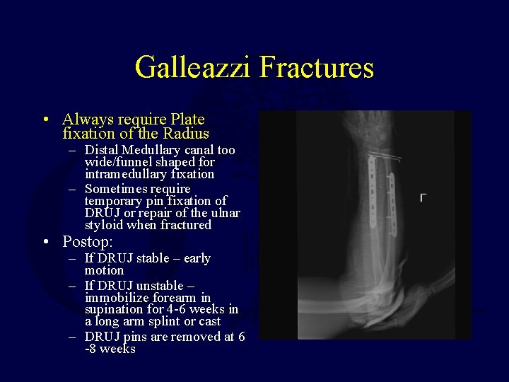
Galleazzi Fractures • Always require Plate fixation of the Radius – Distal Medullary canal too wide/funnel shaped for intramedullary fixation – Sometimes require temporary pin fixation of DRUJ or repair of the ulnar styloid when fractured • Postop: – If DRUJ stable – early motion – If DRUJ unstable – immobilize forearm in supination for 4 -6 weeks in a long arm splint or cast – DRUJ pins are removed at 6 -8 weeks
Galeazzi fractures • May be associated damage to triangular fibrocartilage, which may require early or late repair with open or arthroscopic techniques – Can Occur with Low Velocity Gunshots • Lenihan, MR et al J. O. T. 1992: 6: 32 -35.
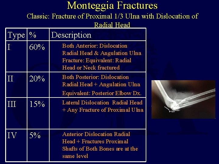
Monteggia Fractures Classic: Fracture of Proximal 1/3 Ulna with Dislocation of Radial Head Type % Description Both Anterior: Dislocation I 60% Radial Head & Angulation Ulna Fracture: Equivalent: Radial Head or Neck fractured II 20% Both Posterior: Dislocation Radial Head + Angulation Ulna Equivalent: Posterior Elbow Dx. III 15% Lateral Dislocation Radial Head + Any Fracture of Proximal Ulna IV 5% Anterior Dislocation Radial Head + Fractures Proximal Shafts of Both Bones are at the same level
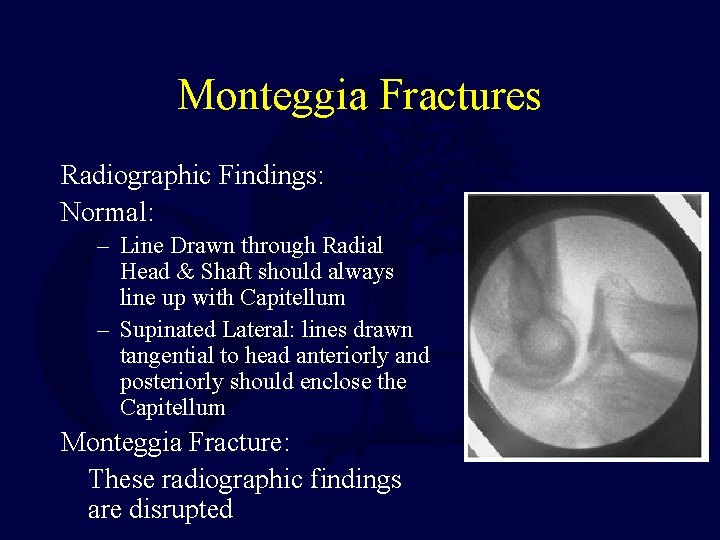
Monteggia Fractures Radiographic Findings: Normal: – Line Drawn through Radial Head & Shaft should always line up with Capitellum – Supinated Lateral: lines drawn tangential to head anteriorly and posteriorly should enclose the Capitellum Monteggia Fracture: These radiographic findings are disrupted
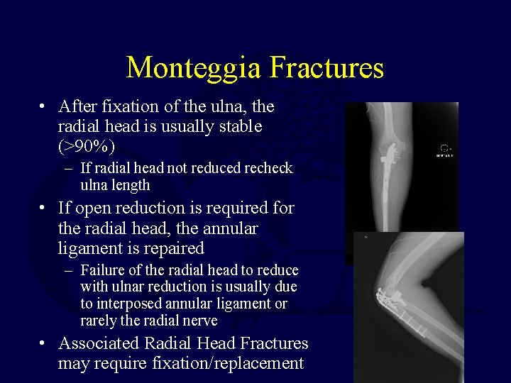
Monteggia Fractures • After fixation of the ulna, the radial head is usually stable (>90%) – If radial head not reduced recheck ulna length • If open reduction is required for the radial head, the annular ligament is repaired – Failure of the radial head to reduce with ulnar reduction is usually due to interposed annular ligament or rarely the radial nerve • Associated Radial Head Fractures may require fixation/replacement
Monteggia Fractures • Postoperative treatment depends on rigidity of ulnar fixation and stability of the radial head – Casting with more than 90 degrees of elbow flexion is rarely needed to maintain the radial head reduction (6 weeks)
Literature • • • • Calkins MS, Burkhalter W, Reyes F. Traumatic segmental bone defects in the upper extremity. Treatment with exposed grafts of corticocancellous bone. J Bone Joint Surg Am. 1987 Jan; 69(1): 19 -27 PMID: 3543018 (Link to Abstract) Chapman MW, Gordon JE, Zissimos AG. Compression-plate fixation of acute fractures of the diaphyses of the radius and ulna. J Bone Joint Surg Am. 1989 Feb; 71(2): 159 -69. PMID: 2918001 (Link to Abstract) Falder S, Sinclair JS, Rogers CA, Townsend PL. Long-term behaviour of the free vascularised fibula following reconstruction of large bony defects. Br J Plast Surg. 2003 Sep; 56(6): 571 -84. PMID: 12946376 (Link to Abstract) Ring D, Allende C, Jafarnia K, Allende BT, Jupiter JB. Ununited diaphyseal forearm fractures with segmental defects: plate fixation and autogenous cancellous bone-grafting. J Bone Joint Surg Am. 2004 Nov; 86 -A(11): 2440 -5. PMID: 15523016 (Link to Abstract) Schemitsch EH, Richards RR. The effect of malunion on functional outcome after plate fixation of fractures of both bones of the forearm in adults. J Bone Joint Surg Am. 1992 Aug; 74(7): 1068 -78. PMID: 1522093 (Link to Abstract) Street DM. Intramedullary forearm nailing. Clin Orthop Relat Res. 1986 Nov; (212): 219 -30. PMID: 3769288 (Link to Abstract) Wei SY, Born CT, Abene A, Ong A, Hayda R, De. Long WG Jr. Diaphyseal forearm fractures treated with and without bone graft. J Trauma. 1999 Jun; 46(6): 1045 -8. PMID: 10372622 (Link to Abstract) Wright RR, Schmeling GJ, Schwab JP. The necessity of acute bone grafting indiaphyseal forearm fractures: a retrospective review. J Orthop Trauma. 1997 May; 11(4): 288 -94. PMID: 9258828 (Link to Abstract) Level of Evidence 5 and Other Journal Articles (includes Case Reports, Expert Opinions, Personal Observations, and Biomechanic Studies) Levin LS. Early versus delayed closure of open fractures. Injury. 2007 Aug; 38(8): 896 -9. PMID: 17585912 (Link to Abstract) Noda K, Goto A, Murase T, Sugamoto K, Yoshikawa H, Moritomo H. Interosseous membrane of the forearm: an anatomical study of ligament attachment locations. J Hand Surg Am. 2009 Mar; 34(3): 415 -22. Epub 2009 Feb 11 PMID: 19211201 (Link to Abstract) Pfaeffle HJ, Stabile KJ, Li ZM, Tomaino MM. Reconstruction of the interosseous ligament restores normal forearm compressive load transfer in cadavers. J Hand Surg Am. 2005 Mar; 30(2): 319 -25. PMID: 15781355 (Link to Abstract)
Literature • • • • Bauer G, Arand M, Mutschler W. Post-traumatic radioulnar synostosis after forearm fracture osteosynthesis. Arch Orthop Trauma Surg. 1991; 110(3): 142 -5. PMID: 2059537 (Link to Abstract) Beingessner DM, Patterson SD, King GJ. Early excision of heterotopic bone in the forearm. J Hand Surg Am. 2000 May; 25(3): 483 -8. PMID: 10811753 (Link to Abstract) Deluca PA, Lindsey RW, Ruwe PA. Refracture of bones of the forearm after the removal of compression plates. J Bone Joint Surg Am. 1988 Oct; 70(9): 1372 -6. PMID: 3182889 (Link to Abstract) Egol KA, Kubiak EN, Fulkersojn E, et. al: Biomechanics of locked plates and screws. J Orthop Trauma, 2004; 18: 488 -493 PMID: 15475843 (Link to Abstract) Koval KJ (ed): Orthopaedic Knowledge Update 7. Rosemont, IL, American Academy of Orthopaedic Surgeons, 2002, pp 307 -316 Mc. Auliffe JA, Wolfson AH. Early excision of heterotopic ossification about the elbow followed by radiation therapy. J Bone Joint Surg Am. 1997 May; 79(5): 749 -55 PMID: 9160948 (Link to Abstract) Moed BR, Kellam JF, Foster RJ, et al: Immediate internal fixation of open fractures of the diaphysis of the forearm. J Bone Joint Surg Am 1986; 68: 1008 -1017 PMID: 3745238 (Link to Abstract) Pollock FH, Pankovich AM, Prieto JJ, Lorenz M. The isolated fracture of the ulnar shaft. Treatment without immobilization. J Bone Joint Surg Am. 1983 Mar; 65(3): 339 -42. PMID: 6826596 (Link to Abstract) Rumball K, Finnegan M. Refractures after forearm plate removal. J Orthop Trauma. 1990; 4(2): 124 -9 PMID: 2358925 (Link to Abstract) Sommer C, Babst R, Muller M, et. al: Locking compression plate loosening and plate breakage: A report of 4 cases. J Orthop Trauma, 2004; 18: 571 -577. PMID: 15475856 (Link to Abstract) Vince KG, Miller JE. Cross-union complicating fracture of the forearm. Part I: Adults. J Bone Joint Surg Am. 1987 Jun; 69(5): 640 -53. PMID: 3110165 (Link to Abstract) Wood MB. Upper extremity reconstruction by vascularized bone transfers: results and complications. J Hand Surg Am. 1987 May; 12(3): 4227. PMID: 3584891 (Link to Abstract) Wright RR, Schmeling GJ, Schwab JP: The necessity of acute bone grafting in diaphyseal forearm fractures: A retrospective review. J Orthop Trauma 1997; 11: 288 -294 PMID: 9258828 (Link to Abstract)
Literature • • • Bado JL. The Monteggia lesion. Clin Orthop Relat Res 1967; 50: 71 -86. PMID: 6029027 (Link to Abstract) Fowles JV, Sliman N, Kassab MT. The Monteggia lesion in children: Fracture of the ulna and dislocation of the radial head. J Bone Joint Surg Am 1983; 65: 1276 -1282 PMID: 6654941 (Link to Abstract) Tan JW, Mu MZ, Liao GJ, Li JM. Pathology of the annular ligament in pediatric Monteggia fractures. Injury. 2008 Apr; 39(4): 451 -5. Epub 2007 Nov 19. PMID: 18005963 (Link to Abstract) Korompilias AV, Lykissas MG, Kostas-Agnantis IP, Beris AE, Soucacos PN. Distal radioulnar joint instability (Galeazzi type injury) after internal fixation in relation to the radius fracture pattern. J Hand Surg Am. 2011 May; 36(5): 847 -52. Epub 2011 Mar 23. PMID: 21435802 (Link to Abstract) Rettig ME, Raskin KB. Galeazzi fracture-dislocation: a new treatment-oriented classification. J Hand Surg Am. 2001 Mar; 26(2): 228 -35. PMID: 11279568 (Link to Abstract) Biyani A, Bhan S: Dual extensor tendon entrapment in Galeazzi fracture-dislocation: A case report. J Trauma 1989; 29: 1295 -1297. PMID: 2769817 (Link to Abstract) Budgen A, Lim P, Templeton P, Irwin LR. Irreducible Galeazzi injury. Arch Orthop Trauma Surg. 1998; 118(3): 176 -8. PMID: 9932197 (Link to Abstract) Giannoulis FS, Sotereanos DG. Galeazzi fractures and dislocations. Hand Clin. 2007 May; 23(2): 153 -63, v. PMID: 17548007 (Link to Abstract) Giannoulis FS, Sotereanos DG. Galeazzi fractures and dislocations. Hand Clin. 2007 May; 23(2): 153 -63, v. Review. PMID: 17548007 (Link to Abstract) Paley D, Mc. Murtry RY, Murray JF. Dorsal dislocation of the ulnar styloid and extensor carpi ulnaris tendon into the distal radioulnar joint: The empty sulcus sign. J Hand Surg Am 1987; 12: 1029 -1032. PMID: 3693829 (Link to Abstract)
Conclusion • • Forearm fxs are inherently unstable fxs Vast majority require operative fixation Goal is anatomic reduction with stable fixation Restore ulna length Restore radial bow Respect the soft tissue Don’t miss injury to joint above or below If you would like to volunteer as an author for the Resident Slide Project or recommend updates to any of the following slides, please send an email to ota@ota. org E-mail OTA about Questions/Comments Return to Upper Extremity Index