
The Forearm Wrist Hand and Fingers Chapter 24



- Slides: 35
The Forearm, Wrist, Hand, and Fingers Chapter 24
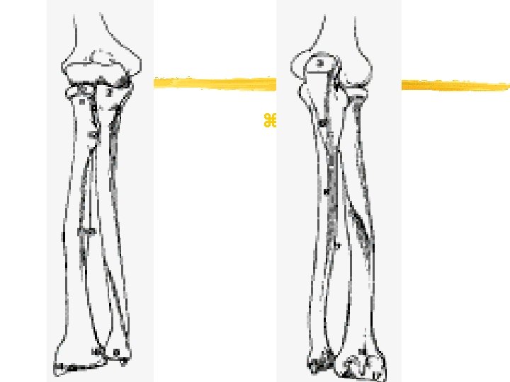
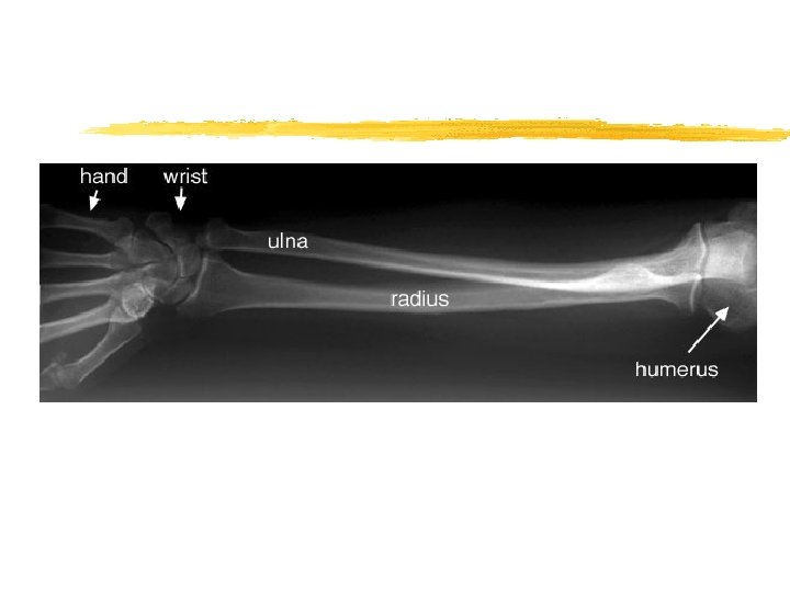
Forearm Anatomy z Radius and Ulna: Elbow y. Joints: radioulnar joint (superior, middle, and distal) z Bone: proximal radial head, olecranon process, radial shaft, ulnar shaft, distal radius, radial styloid process, ulnar head, ulnar styloid z Musculature: flexors& pronators (lie anteriorly. ulnar side), extensors & supinators (lie posteriorly, medial side) z Nerve/Blood Supply: median and radial nerve and brachial, radial, and ulnar artery
z
z
Forearm Assessment z. History z. Observation y. Visually inspect, including wrsit and elbow y. If no deformity present, observe while they supinate and pronate z. Palpation z. Special Tests
Management of Forearm Injuries z Contusion y Etiology: direct blow x. Why more common to ulna? y Signs and Symptoms y Management z Forearm Splints y Etiology: repeated severe static contraction y Signs and Symptoms: dull ache between extensors, interosseous membrane y Management: early season vs late in season? y Note: Acute / Chronic exertional compartment syndrome: deep compartment most common and associated with avulsions, distal radius fracture, or crushing injuries; management same as in lower leg
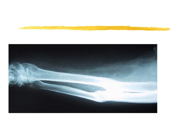
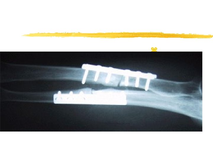
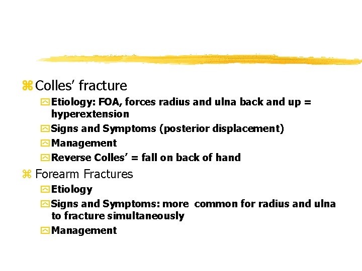
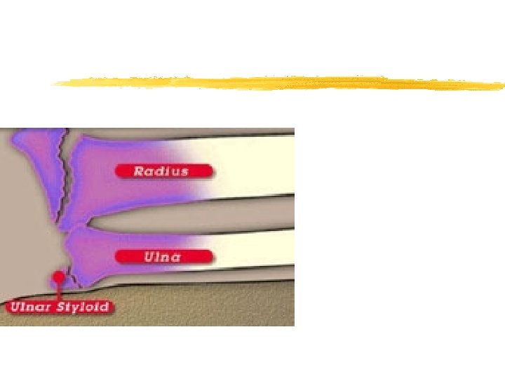
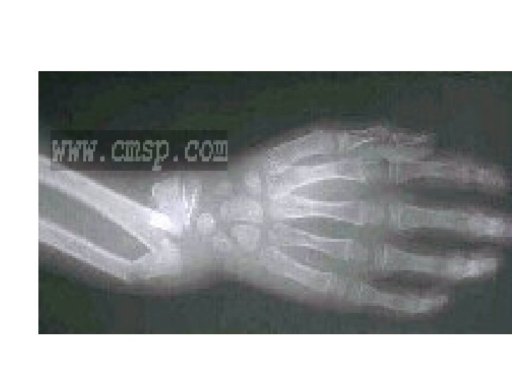
z Colles’ fracture y Etiology: FOA, forces radius and ulna back and up = hyperextension y Signs and Symptoms (posterior displacement) y Management y Reverse Colles’ = fall on back of hand z Forearm Fractures y Etiology y Signs and Symptoms: more common for radius and ulna to fracture simultaneously y Management
Wrist, Hand, and Finger Anatomy z Bones: carpals and metacarpals z Joints: radiocarpal, metacarpal, and phalangeal joints z Ligaments: “many at each joint in the hand” y. TFCC (triangular fibrocartilage complex); b/t head of ulna and triquetrial bone z Musculature: “many intrinsic and extrinsic muscles” z Blood and Nerve Supply: ulnar, median, radial nerve and radial and ulnar superficial and deep palmar arch arteries.
Assessment of Wrist, Hand, and Finger Injuries z History z Observation z Palpation z Special Tests: Finklestein’s test, Tinel’s Sign, Phalen’s test, valgus and varus stress test, z Circulatory and Neurological Evaluation y. Allen test z Functional Evaluation
Special Tests z Finklesteins’ Test De Quervains (tenosynovitis) Thumb tucked inside fist with ulnar deviation z Tinel’s Sign y Tap over transverse carpal ligament y Pain numbness and tingling indicates median nerve disruption and presence of carpal tunnel z Phalen’s Test y Carpal tunnel y Bilateral wrist flexion and press them together; pain is positive sign z Valgus/varus at wrist, MCP, and IP joints z Circulatory / neurological evaluations y Allen's test: test function of radial and ulnar arteries y Athlete makes fist 4 -5 times; while holding final fist, evaluator pinches off both arteries; hand should be blanched y Release arties individually
Recognition and Management of Wrist, Hand, and Finger Injuries z Wrist Sprain y. Etiology y. Signs and Symptoms y. Management z Triangular Fibrocartilage Complex Injury y. Etiology: forced hyperextension or compression of radioulnar joint and proximal row of carpals y. Signs and Symptoms y. Management
z Tenosynovitis y. Etiology: repeated wrist acceleration and deceleration y. Signs and Symptoms: pain w/ passive stretching y. Management: may need splinting and strengthening z Tendinitis y. Etiology: repetitive pulling motions and pressure on palm of hand y. Signs and Symptoms: pain with AROM and passive stretching y. Management z Nerve Compression, Entrapment, Palsy y. Etiology: median (carpal tunnel) and ulnar (pisiform and hamate) y. Signs and Symptoms: deformities(bishop’s, claw and drop wrist) y. Management: if chronic, may require surgical
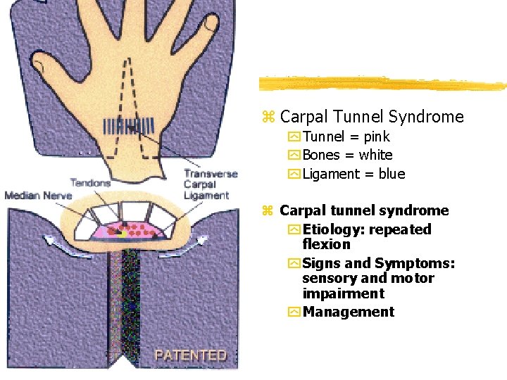
z Carpal Tunnel Syndrome y Tunnel = pink y Bones = white y Ligament = blue z Carpal tunnel syndrome y Etiology: repeated flexion y Signs and Symptoms: sensory and motor impairment y Management
Recognition and Management of Wrist, Hand, and Finger Injuries z Dislocation of the Lunate Bone y Etiology: forced hyperextension of wrist y Signs and Symptoms: difficulty with wrist and finger flexion; may have impaired nerves y Management: referral for reduction z Hamate Fracture y Etiology: contact while holding something(racket) y Signs and Symptoms y Management z Wrist Ganglion(synovial cyst) y Etiology: herniation of joint capsule or tendon y Signs and Symptoms y Management
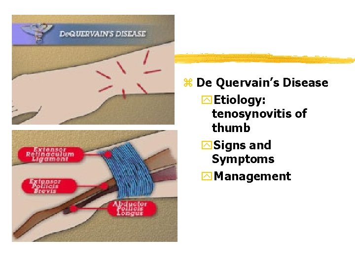
z De Quervain’s Disease y. Etiology: tenosynovitis of thumb y. Signs and Symptoms y. Management
Scaphoid Fracture z. Etiology: compression of scaphoid b/t radius and ulna y. Concerns: portion of scaphoid has decreased vascular supply; improper healing can occur and result in aseptic necrosis of the scaphoid bone z. Signs and Symptoms y. Anatomical snuffbox pain z. Management
Finger anatomy z Bones z Ligaments y PIP and DIP have the same design y Collateral ligaments, palmar fibrocartilage, and loose posterior capsule or synovial membrane (protected by extensor expansion)
Finger anatomy z. Musculature y. PIP: Flex. Digitorium Superficialis y. DIP: Flex. Digitorium Profundus y. PIP & DIP: Exten. Digitorium Longus (becomes extensor expansion after MCP) y. Intrinsics: x. Dorsal and palmar interosseei: x. Lumbricals: volar surface; MCP flex. , IP exten. x. Thenar (4 that act on thumb) & hypothenar (4 that act on 5 th)
Recognition and Management of Wrist, Hand, and Finger Injuries z Contusion to hand fingers y. Etiology y. Signs and Symptoms: fingernail? y. Management z Bowler’s Thumb y. Etiology: fibrosis of the ulnar digital nerve form pressure y. Signs and Symptoms: pain, numbness, tingling y. Management: pad area, decrease activity; surgery PRN z Jersey finger y. Etiology: FDP rupture, grabbing jersey y. Signs and Symptoms: DIP cannot flex y. Management: SURGERY
z Trigger finger or thumb y. Etiology: stenosing tendon by repeated movements y. Signs and Symptoms: resistance to reextension after thumb and finger flexed y. Management: possible injections; splinting z Dupuytren’s Contracture y. Etiology: idiopathic development of nodules in palmer aponeurosis y. Signs and Symptoms: flexion deformity; cannot extend y. Management: surgical removal
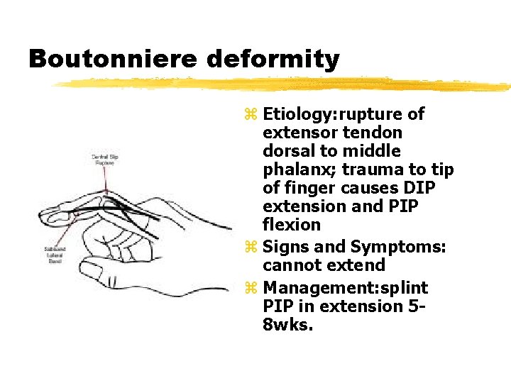
Boutonniere deformity z Etiology: rupture of extensor tendon dorsal to middle phalanx; trauma to tip of finger causes DIP extension and PIP flexion z Signs and Symptoms: cannot extend z Management: splint PIP in extension 58 wks.
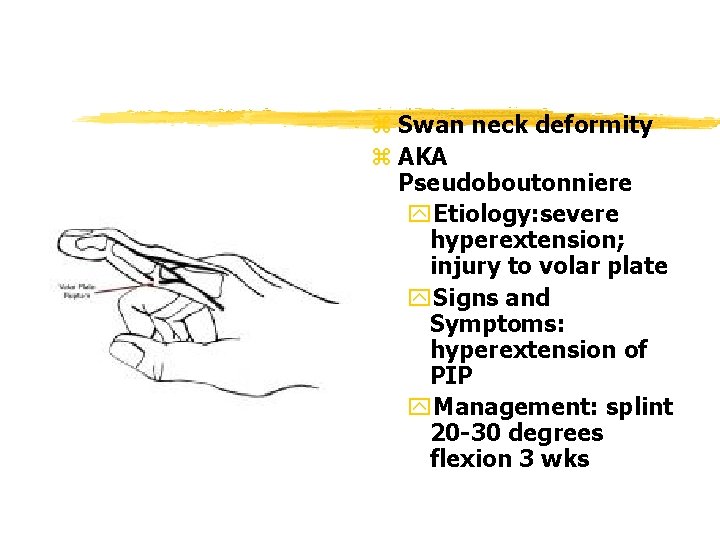
z Swan neck deformity z AKA Pseudoboutonniere y. Etiology: severe hyperextension; injury to volar plate y. Signs and Symptoms: hyperextension of PIP y. Management: splint 20 -30 degrees flexion 3 wks
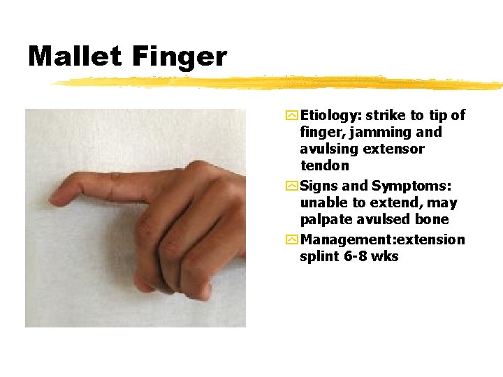
Mallet Finger y Etiology: strike to tip of finger, jamming and avulsing extensor tendon y Signs and Symptoms: unable to extend, may palpate avulsed bone y Management: extension splint 6 -8 wks
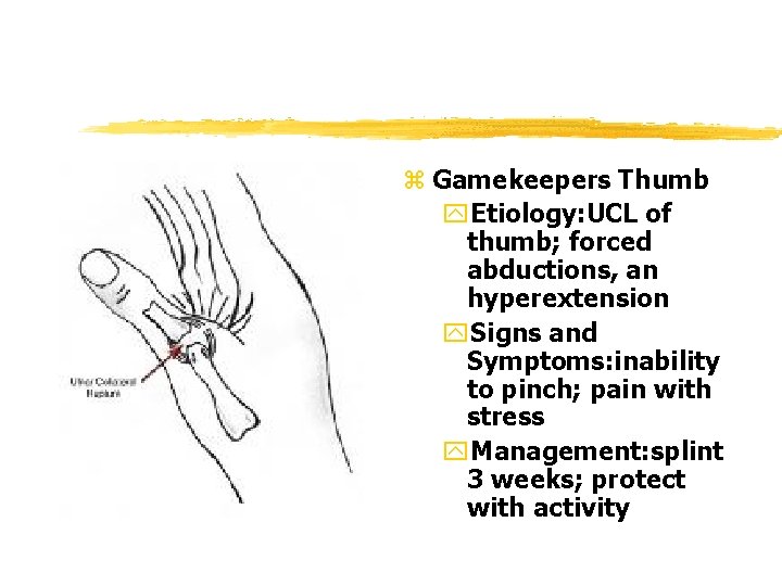
z Gamekeepers Thumb y. Etiology: UCL of thumb; forced abductions, an hyperextension y. Signs and Symptoms: inability to pinch; pain with stress y. Management: splint 3 weeks; protect with activity
Recognition and Management of Wrist, Hand, and Finger Injuries z Sprains, Dislocations, and Fractures y Etiology y Signs and Symptoms y Management z Sprains PIP and DIP joint y Etiology y Signs and Symptoms y Management z PIP Doral Dislocation y Etiology: twist while semiflexed y Signs and Symptoms y Management: splint in ext z PIP Dorsal dislocation y Etiology: hyperext. y Signs and symptoms: deformity; inability to move y Management: reduce and splint 20 -30 degrees flex
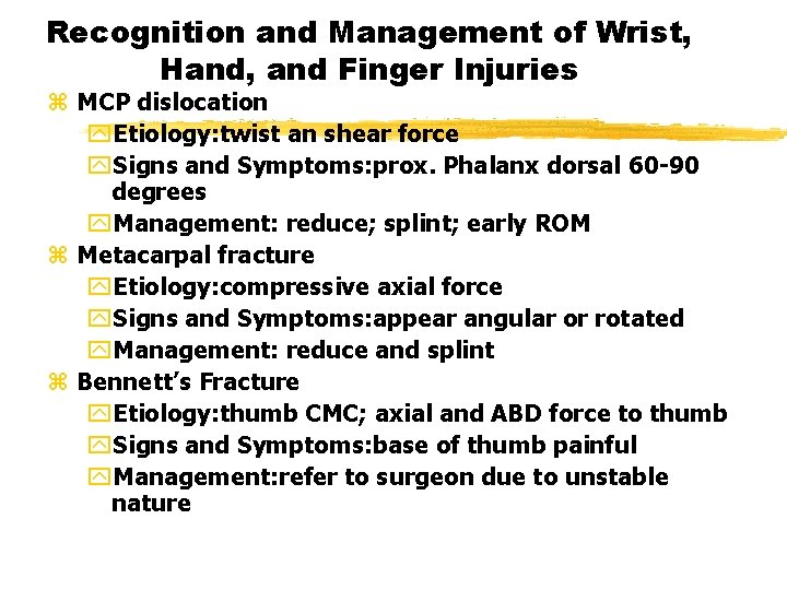
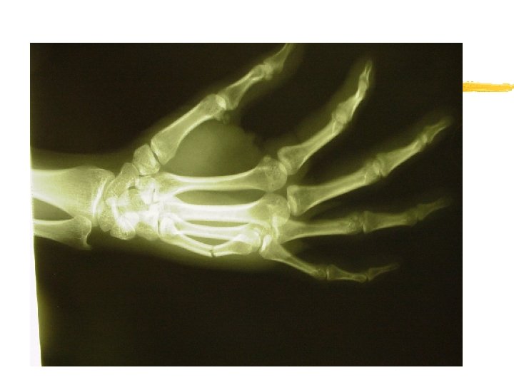
Recognition and Management of Wrist, Hand, and Finger Injuries z MCP dislocation y. Etiology: twist an shear force y. Signs and Symptoms: prox. Phalanx dorsal 60 -90 degrees y. Management: reduce; splint; early ROM z Metacarpal fracture y. Etiology: compressive axial force y. Signs and Symptoms: appear angular or rotated y. Management: reduce and splint z Bennett’s Fracture y. Etiology: thumb CMC; axial and ABD force to thumb y. Signs and Symptoms: base of thumb painful y. Management: refer to surgeon due to unstable nature
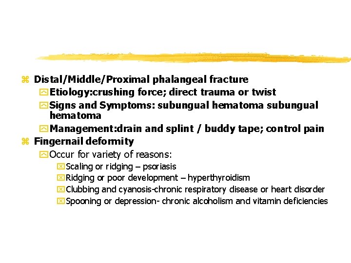
z Distal/Middle/Proximal phalangeal fracture y Etiology: crushing force; direct trauma or twist y Signs and Symptoms: subungual hematoma y Management: drain and splint / buddy tape; control pain z Fingernail deformity y Occur for variety of reasons: x. Scaling or ridging – psoriasis x. Ridging or poor development – hyperthyroidism x. Clubbing and cyanosis-chronic respiratory disease or heart disorder x. Spooning or depression- chronic alcoholism and vitamin deficiencies
Rehabilitation Principles for the Forearm, Wrist, Hand, and Fingers z General Body Conditioning z Joint Mobilization: traction and mobilization help restore ROM z Flexibility: full ROM is measure of good rehab z Strength: equal z Neuromuscular Control: great dexterity required z Return to Activity: Goals: full dexterity, full ROM, full strength