Endocrine Pathophysiology Dr Gary Mumaugh Essentials of Endocrinology


")
 and Triiodothyronine (T-3) �Causes of hypothyroidism")


 o Cushing’s Syndrome: cortisol hypersecretion o Hyperaldosteronism: hypersecretion")


 o Corticosteroid deficiency- destruction of cortex or")


- Slides: 64
Endocrine Pathophysiology Dr. Gary Mumaugh
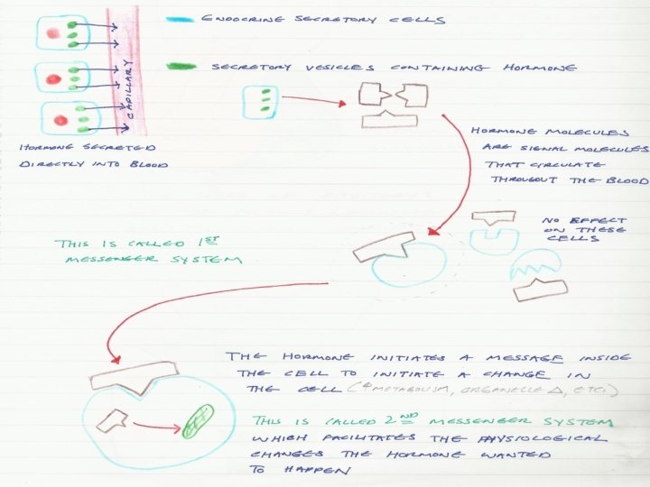
Essentials of Endocrinology • Main function: releases hormones to control cellular activities of target cells • Autocrine cells: secrete substances that control their own function • Paracrine cells: secrete hormones that diffuse to adjacent cells and regulate their action
Essentials of Endocrinology • Neuroendocrine: nervous system exerts regulatory and control functions on endocrine glands • Tropic hormones: secretions that influence the secretions of another endocrine gland (ex. releasing hormones from hypothalamus to anterior pituitary)
Endocrine Specificity • Most hormones are delivered to their target tissues via the bloodstream, where they bind to a receptor. • Intracellular receptors: steroid and thyroid hormones pass through cell membrane and bind to receptors in the cytoplasm o Cause increase in the production of enzymes that enhance a metabolic pathway’s activity, altering the target tissue’s function
Endocrine Specificity • Membrane receptors: binding site for protein, peptide, and amino acid hormones, which cannot enter the cell o Hormones active receptor systems - G proteins or protein kinases o Alter cytoplasmic concentrations of molecules or ions on which cell processes depend • Hormone binding to either type of receptors causes alteration of the target cell’s level of activity.
Plasma Hormone Level • Plasma hormone level determined by rate of entering and leaving the blood o Rate of entering blood stream determined primarily by secretion rate. • 2 components: o Hormone synthesis from dietary or endogenous precursors o Rate of release from the endocrine cell
Plasma Hormone Level • Cleared from blood stream either by inactivation or excretion o Inactivation at target tissue into nonfunctional forms or in liver into chemically converted inactive forms o Excretion by kidneys • Increased binding rates at target cell induce an increased response.
Endocrine Dysfunction • 2 types of dysfunction: – Hypofunction – Hyperfunction • These two concepts are the basis for much of endocrine pathophysiology and should be the first factors considered in clinical situations.
Elevated or Depressed Hormone Levels • Failure of feedback systems • Dysfunction of an endocrine gland • Secretory cells are unable to produce, obtain, or convert hormone precursors • The endocrine gland synthesizes or releases excessive amounts of hormone • Increased hormone degradation or inactivation
Endocrine Hypofunction • Defined as inadequate target tissue response • Causes: hyposecretion or hormone resistance • Hyposecretion may be due to: o Agenesis: lack of gland development o Genetic defect that prevents hormone synthesis o Dietary deficiency
Endocrine Hypofunction • Atrophy of the endocrine gland. • Replacement of normal endocrine tissue with tumor tissue. • Surgery to remove part of a over-secreting gland. • Damage to a functioning gland that is then unable to maintain secretions. • Often accompanied by high levels of control hormones
Endocrine Hypofunction • Decreased or insufficient function of gland • Hormone resistance: insensitivity of a target tissue to its hormone • May be due to: o Hereditary defect that affects the tissue’s ability to synthesize hormone receptors o Autoimmune mechanism in which an antibody binds to the hormone receptors o If faced with a chronically elevated hormone level, the target tissue might reduce the number of hormone receptors
Endocrine Hyperfunction • Exaggerated target tissue responses • Usual cause: hypersecretion - circulating hormone is present in inappropriately high levels o Causes of hypersecretion: § Exposure to high levels of tropic hormones § Defective feedback control § Tumors in the gland- occurs if neoplastic cells retain the ability to secrete functional hormone • Signs and symptoms of endocrine diseases are often puzzling because of altered functions in
Alterations of Thyroid Function • Hyperthyroidism o Thyrotoxicosis o Graves disease o Hyperthyroidism resulting from nodular thyroid disease o Manifestations related to hypermetabolic state o Thyrotoxic crisis
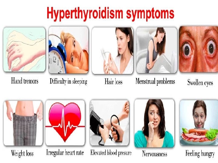
Hyperthyroidism • Thyroid gland produces thyroxine hormone • An autoimmune disorder • Significantly accelerates metabolism o Sudden weight loss, a rapid or irregular heartbeat, sweating, nervousness or irritability o Fatigue, muscle weakness, difficulty sleeping o Tremor, sweating o Changes in menstrual patterns o Increased sensitivity to heat • 8 times more common in women
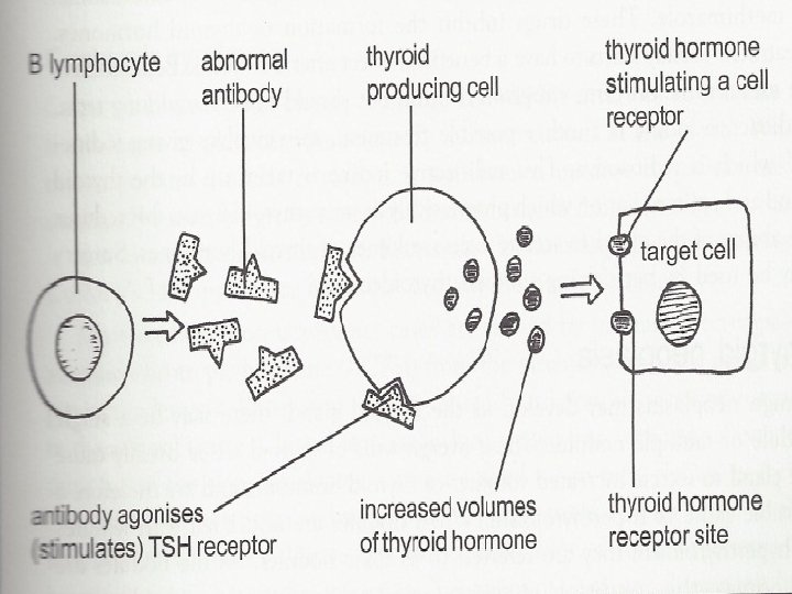
• Etiology of Grave’s Disease o For autoimmune reasons, a group of B lymphocytes secrete Ig. G which fits into and stimulates the TSH receptors present on cell membranes which increases the production of thyroid hormone.
• The characteristic exopthalmus is caused by inflammation of the tissue lining the orbit and extraocular muscles. This causes edema and swelling and fibrosis. • The increased metabolic rate increases appetite and weight gain. The increased rate increases O 2 consumption and patient is short of breath. • Increased sympathetic stimulation is present.
• Causes o Graves' disease, an autoimmune disorder, is the most common cause of hyperthyroidism § Antibodies produced by your immune system stimulate your thyroid to produce too much thyroxine o Hyperfunctioning thyroid nodules o Thyroiditis • Diagnosis o o Radioactive iodine uptake test Thyroid scan Increased T 3 & T 4 Increased ANA titers
Thyrotoxicosis (Graves Disease)
Hypothyroidism �Low levels of thyroid hormones �Thyroxine (T-4) and Triiodothyronine (T-3) �Causes of hypothyroidism �Autoimmune disease - Hashimoto thyroiditis �Treatment for hyperthyroidism �Radiation therapy �Thyroid surgery �Medications (lithium) �Less common causes �Congenital disease �Pituitary disorder �Iodine deficiency �Pregnancy
• Etiology of Hypothroidism • Iodine is essential component to synthesize T 3 & T 4 • As the thyroid hormone levels fall in the blood, the pituitary produces more TSH, which generates enlargement of thyroid goiter • In some areas low in iodine it is called endemic goiter
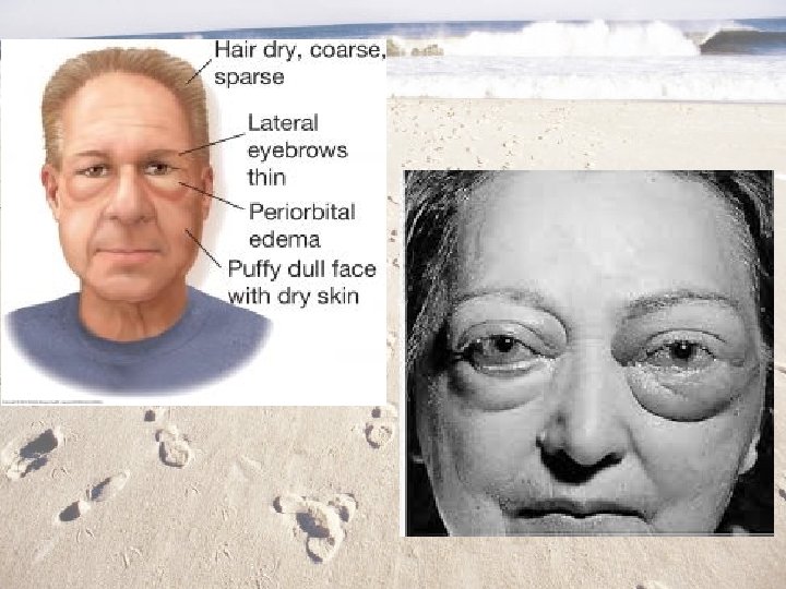
• Pathophysiology of Hypothroidism • Develops slowly with an insidious onset • The lowered metabolic rate causes weight gain, lethargy, tiredness, difficulty concentrating, and cold. • Can affect the adult brain leading to memory loss, slowed mentation, depression and paranoia. • Severe cases is called myxedema madness • Decreased metabolic rate reduces heart rate and stroke volume and over time can cause cardiomegaly. • Decreased metabolic rate causes decreased GI function and decreased sexual function.
• Hypothroidism in pregnancy is serious – Thyroid hormones are essential for development and maturation of the infant and child’s brain. – Called cretinism in children • Stunted growth, large head, learning difficulties, dwarfism, pug nose, short neck.
Cretinism: hypothyroid in newborns • Effects if untreated: physical and mental retardation, and stocky, thick body with infantile proportions
�Risk factors �Mainly in women over 50 �Close relative, with an autoimmune disease �Prior treatment with radioactive iodine or antithyroid medications �Received radiation to your neck or upper chest �Have had thyroid surgery (partial thyroidectomy)
�S & S �Tiredness, weakness, slow reaction time, hypotension, cold intolerance, weight gain even when dieting �Sluggishness, constipation, muscle weakness �Joint pain, stiffness and swelling �Brittle fingernails and hair �Depression
Manifestations of Thyroid Alterations
Goiter: thyroid gland enlargement • Diffuse colloid goiter: generalized enlargement of the thyroid due to increased thyroid stimulating hormone secretions. • Multinodular goiter: nodules form from follicular atrophy and fibrosis • Nontoxic goiter: enlarged gland but hyposecretes hormones • Hashimoto’s Thyroiditis: inflammatory cells overtake functional tissue
33
• Hypothyroidism o Myxdema: chronic hypothyroid in adults causes glycoproteins to be deposited in the dermis • Facial puffiness is characteristic sign • May lead to myxdema coma- can be fatal
Pancreatic Islet Cells • Function: regulate blood glucose levels through the production of insulin and glucagon • Diabetes Mellitus: most common endocrine disorder • Two forms, with differing pathogenesis: o Juvenile onset DM, type I DM, insulindependent DM o Maturity onset DM, type II DM, non-insulindependent DM
Type 1 Diabetes Mellitus • Genetic susceptibility • Failure of beta cells by autoimmune destruction, requires insulin therapy • Immunologically mediated destruction of beta cells • Manifestations: – – – Hyperglycemia Polydipsia Polyuria Polyphagia Weight loss Fatigue
Type 2 Diabetes Mellitus • Beta cells lose their capacity to produce insulin slowly while target tissue also show reduced sensitivity • Can be controlled with diet and exercise for a while, insulin therapy required less often • Maturity-onset diabetes of youth (MODY) • Gestational diabetes mellitus (GDM) • Common form of diabetes mellitus type 2
Type 2 Diabetes Mellitus • Initial insulin resistance • Later loss of beta cells • Diagnosis (fasting glucose, postprandial glucose) • Manifestations (non-specific): fatigue, pruritus, recurrent infections, visual changes, or symptoms of neuropathy; often overweight, dyslipidemic, hyperinsulinemic, and hypertensive
• Insulin physiology – Produced by the beta cells in the islets and lowers blood glucose. – When glucose levels rise, this is detected by the beta cells and secretory granules of insulin emerge from the cell membrane. – The insulin then travels in the hepatic portal vein to the liver and then on to all the body tissues in the systemic circulation. – Insulin is eventually removed from the blood by being broken down by the liver and kidneys.
– Insulin lowers blood glucose levels by converting glucose into insoluble glycogen for storage in the liver and muscles. – Insulin is needed to transfer glucose in tissue fluids through a gate into the cytosol of the cell. – Without this insulin action, glucose cannot enter the cell and cannot be used by the mitochondria in energy production. – The irony is that the tissue fluids have to much glucose and the intracellular mitochondria does not have enough.
• Insulin and proteins – Insulin stimulates protein metabolism and increases the movement of amino acid into cells. – Insulin also prevents the catabolism of proteins. • Insulin and fats – Insulin promotes the synthesis of fatty acids and glycerol in the blood causing hyperlipidemia.
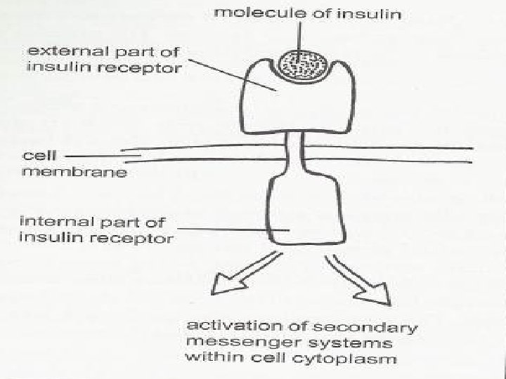
• Insulin receptors o Insulin can only exert a physiological effect when it is combined with a specific receptor. These are transmembrane proteins which means that part of the receptor is inside the cell and part is outside the cell.
Chronic Complications of Diabetes Mellitus • Macrovascular disease o Affects large artery walls with fatty deposits that leads to fibrous collagen and plaque formation. o Coronary artery disease o Stroke o Peripheral arterial disease § Leads to ischemia and possible gangrene. • Diabetic neuropathies • Infection
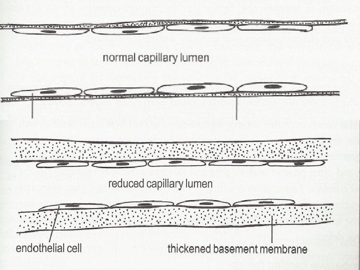
Chronic Complications of Diabetes Mellitus • Microvascular disease – There is a progressive thickening of the basement membranes which narrow the lumen and lowers elasticity. – This leads to localized ischemia and hypoxia, which causes more vascular compromise. – Retinopathy – Diabetic nephropathy
Hypoglycemia • Low blood sugar o Normal levels = 80 -110 mg/d. L o Mild = <80 Significant symptoms <60 • Causes o Most are diabetics with to much insulin usage o Fasting o Alcoholics and liver disease § Drinking alcohol with sugar causes temporarilly o Overeating o Pancreatic tumor o Addison’s disease
• S & S – occurs when blood sugar < 60 mg o o Sweatiness, dizziness, nervousness, shaking Anxiety, faintness, weakness Palpitations, hunger Confusion, inappropriate behavior • Diagnosis o Patient’s history o Glucose tolerance test o Fasting glucose and insulin levels • Treatment o Glucose now!!
Adrenal Cortex • Adrenocortical Hypersecretion (Hyperadrenalism) o Cushing’s Syndrome: cortisol hypersecretion o Hyperaldosteronism: hypersecretion by aldosterone-secreting cells. o Adrenal virilism: androgen hypersecretion in females, induces various masculine traits o Sexual precocity: androgen hypersecretion in young males leads to rapid and premature sexual development
Cushing Disease
Abdominal striae of Cushings
Virilization
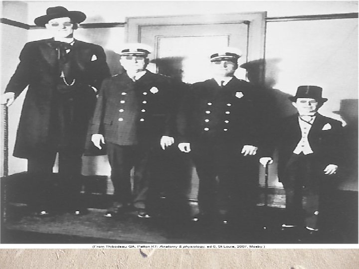
Sexual Precocity 9 year old twins 4 year old
• Adrenocortical Hyposecretion (Hypoadrenalism, Addison’s disease) o Corticosteroid deficiency- destruction of cortex or suppression of ACTH by therapeutic doses of glucocorticoids o Characteristic feature: excess pigmentation and high vulnerability to stress
Hyperparathyroidism �Usually caused by a parathyroid adenoma �More common in women �Often causes bone pain from high calcium �Hypercalcemia is also seen in metastatic bone disease (from breast, lung, prostate) and sometimes in pregnancy �S & S �Bones – bone pain from high calcium �Stones – kidney stones common �Groans – pain and slow muscle contractions �Moans – psychiatric and mental changes
Hypoparathyroidism • Often seen after surgery of thyroid and parathyroid o If the parathyroid glands have been removed, then the diagnosis will be permanent • Results in low serum calcium & high serum phosphate • S&S o Low calcium causes muscle cramps, tetany, & paresthesias o Convulsions and arrhythmias o Acute onset, especially after thyroid surgery, could lead to respiratory spasm and suffocation – needing tracheostomy
Hypopituitarism �Pituitary gland fails to produce one or more of its hormones, or doesn't produce enough of them �Causes �Pituitary ademomas �Strokes �Metastatic carcinomas �Primary brain tumors �Autoimmune disorders �Brain trauma �Encephalitis �Idiopathic
Signs & Symptoms �Depending on which hormones are deficient �Fatigue , Headaches , Low tolerance for stress �Muscle weakness , Nausea �Constipation , Weight loss or gain �A decline in appetite , Abdominal discomfort �Sensitivity to cold or difficulty staying warm �Visual disturbances �Loss of underarm and pubic hair �Joint stiffness �Hoarseness �Facial puffiness �Thirst and excess urination �Low blood pressure �Lightheadedness when standing
• Men may experience o Loss of interest in sexual activity o Erectile dysfunction o Decrease in facial or body hair • Women mat experience o Irregular or no menstrual periods o Infertility o Inability to produce milk for breast-feeding • Children may experience o Stunted growth o Short stature o Slowed sexual development
Hyperpituitarism �Excessive production of growth hormone, which continues to be produced well into adulthood �In adults, since the growth plates are closed, excessive levels cause abnormal growth of hands, feet, and internal organs – called acromegaly �In children, excess growth hormone causes increased height known as gigantism �Diagnosis �Elevated GH in blood test �Pituitary tumor on CT or MRI