Pathophysiology of the most common symptoms and signs


 – pain is feeling in places which")


 Pulmonary vascular congestion disturbances of gas exchange in the lung hypoxemia hypoxia of")
 Edema of small airway wall obstruction wheeze cardial asthma e) Developmet of pleural")


: - result of aortic stenosis")
 Mechanical alternans (pusus alternans) b) Electrical")
 ABI = Ankle systolic BP/ Arm systolic BP – test comparing")


- Slides: 39
Pathophysiology of the most common symptoms and signs of cardiovascular diseases Prof. Jan Hanacek, MD, Ph. D
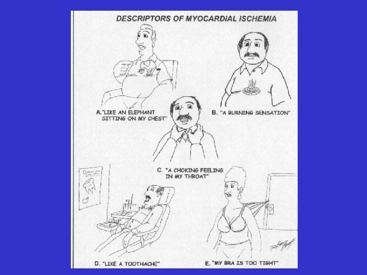
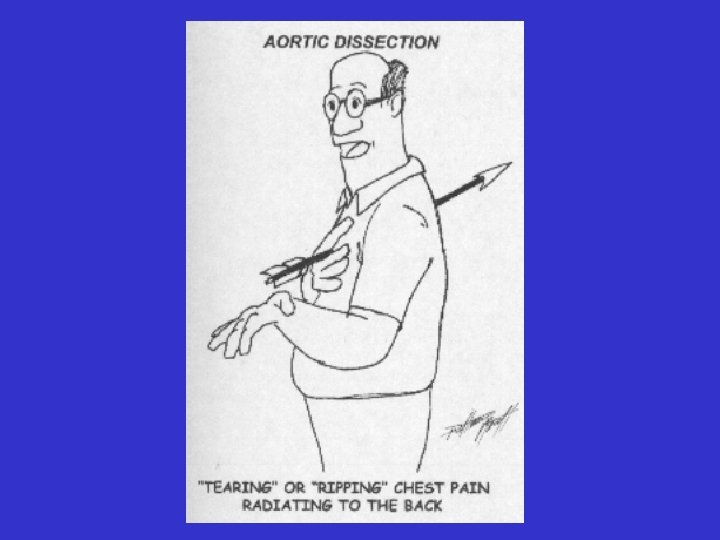
I. Main symptoms and signs of heart diseases chest pain/discomfort, dyspnoea, palpitations, cyanosis, dizziness and syncope, edema, cough, hemoptysis, fatigue and tiredness, puls changes, urination during day, urination during night (nocturia) Chest pain and/or discomfort • Angina pectoris – consequence of myocardial oxygen lack Character: dull and deep (sometimes sharp and superficial) Pathomechanisms: – change of oxidative to unoxidative metabolism – overproduction of toxic metabolites and their accumulation in myocardium and coronary vessel wall (K+, adenosin, lactic acid, bradykinin, PGE 2 , histamin, serotonin)
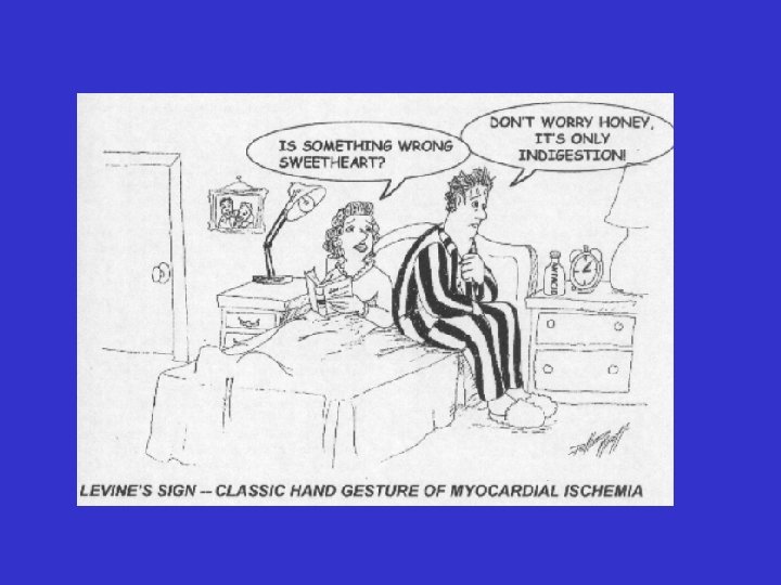
– chemical stimulation of free nerve endings of afferent limb of sympathetic nerve fibres activation of the ascending spinothalamic neurons as part of nociceptive system into the CNS development of pain Visceral pain – pain arising from visceral organs induced by myocardial ischemia Pain localisation – behind the sternum (Levine's sign) – in epigastrium
• Referred visceral pain (transferred pain) – pain is feeling in places which are remoted from the place of its origin, e. g. pain referred to neck, left arm, back, right arm. . . Pathomechanism: – when an algogenic process affecting the viscus recurs frequently or becomes more intense and prolonged the painful sensation is progressively felt in more superficial structures (its localisation becomes more exact) – convergence of sensitive informations comming from viscera and from skin – both converge into the same spinothalamic neurons in segment of spinal cord transport into region of CNS responsible for pain processing
Sensory impulses from the viscera create an irritable focus in the segment at which they enter the spinal cord Afferent impulses from the skin entering the same segment as sensory inputs (convergence), are thereby facilitated, giving rise to true somatic pain • Senzitization of neurons in dorsal horn – different kinds of neurons in the dorsal horn are more sensitive to comming afferent impulses (even not painful) – central sensitization
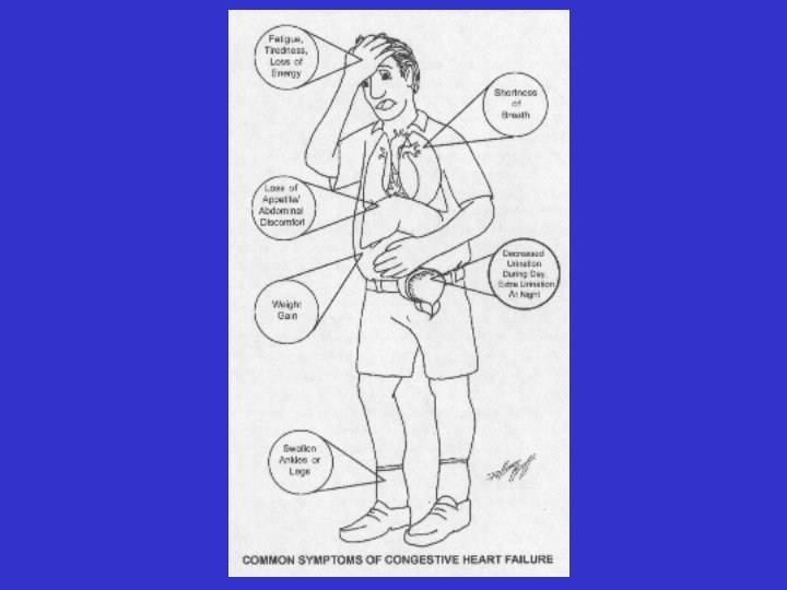
Abdominal pain/dyscomfort in right heart failure – expansion of fluid in the liver capsula distension pain (feeling of pressure) in upper right abdominal quadrant – feeling dyscomfort in abdomen – it is a result of disturbed digestion due to GIT congestion
• Dyspnoea – difficulty in breathing, due to different pathomechanisms activated by disturbances associated with pulmonary vascular congestion (LV failure) Pathomechanisms: a) – overdistension of capilaries and venules in the lung – incresed amount of fluid in pericapilary space – stimulation of J-receptors in the lung (rapid shallow breathing) – stimulation of RAR in the mucous membrane of small airways (cough) aferentation to CNS activatin of system involved in development of dyspnea
b) Pulmonary vascular congestion disturbances of gas exchange in the lung hypoxemia hypoxia of respiratory muscles decreased strength of muscles decresed pulmonary ventilation overburden of respiratory muscles afferentation to the central nervous system development of dyspnea c) Pulmonary vascular congestion lung weight lung resistance to expansion overburden of respiratory muscles aferentation to CNS development of dyspnoe
d) Edema of small airway wall obstruction wheeze cardial asthma e) Developmet of pleural effusions – in congestive heart failure Pleural fluid is secreted by the parietal layer of the pleura and reabsorbed by the visceral layer of the pleura Pleural effusions appear on chest X-rays as white space at the base of the lung.
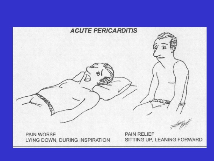
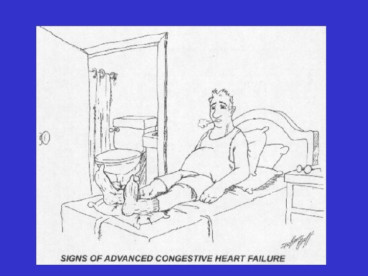
• Orthopnea - difficulty in breathing in the recumbent position releived by assuming an upright or sitting position Pathomechanisms: – congestion of lung circulation in recumbent position – due to blood return to right heart – decresede activity SNS during sleep heart performance congestion of lung circulation Paroxysmal nocturnal dyspnea – sudden attack of dyspnea at rest during the night Pathomechanism: – result of LV failure due to: - serious types of dysrhythmias - activity of SNS and activity of PSNS during night
Dyspnoe in patients with right heart failure – in serious lung diseases – when massive pulmonary artery embolisation – when there is restriction in diaphragm movement due to ascites and/or liver enlargement – development of hypoxia and metabolic acidosis due to right heart failure
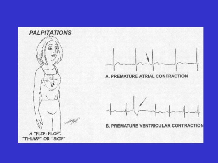
• Palpitation – patient s awareness of the heartbeat that occures with sudden changes in rate, rhythm, and stroke volume accompanied with unpleasant sensation with the heart beats Pathomechanisms: – changes in the heart rate (tachycardia, bradykardia) – irregularity of the heart beats (PAB, PVB, heart blocks) – increased force of ventricular contractions ( stroke volume) Main forms of palpitation - intermittent, irregular, lasting seconds or fractions of seconds – -„skipped“ beats, „flip-flop“ sensation (due to premature beats) -„the heart stop“, „stopped beating“ – due to compensatory pause following PVB - slow regular palpitation – due to sinus bradykardia, – due to junctional rhythm – due to 3 rd degree of AV-block
– abrupt onset and termination of palpitation – due to paroxysmal supraventricular tachycardia – fast, irregular palpitations – due to atrial fibrilation – palpitations induced by specific diseses – valvular regurgitations, thyreotoxicosis, anemia, hepatal failure – palpitation associated with the use of tabacco, coffee, tea, alcohol – „holiday-heart syndrome“ Another symptoms and signs associated with – supraventricular tachykardia: • dizziness, dyspnea, sweating, chest discomfort, polyuria (due to ihibition of ADH secretion and stimulaation of ANF secretion) (during prolonged attack of palpitation) – ventricular tachycardia: • nausea, sweating, chest discomfort, dizziness and syncope
• Edema – can be caused by both cardiac and non - cardiac conditions – accumulation of fluids and swelling of tissues in the lung (lung edema), and lower part of the body (dependent edema) ankles, feet, legs, abdominal cavity) Pathomechanisms: – left or right heart failure – fluid accumulation in the interstitial spaces usually in mentioned areas as a result of bad drenage or gravity, and preceded by the respiratory symptoms and signs (when LV failure), and weight gain
Pulmonary edema – accumulation of a fluid in the lung Causes and pathomechanisms: left heart failure, mitral stenosis accumulation of blood in front of LV increased hydrostatic pressure in pulmonary vessels development of pulmonary congestion development of interstitial pulmonary edema development of alveolar pulmonary edema It is facilitated by: – limited lymphatic drenage of the lung, – permeability of the alveolo – capillary membrane, – oncotic pressure of the blood
Symptoms and signs of pulmonary edema – reduced pulmonary perfusion and decreased transfer factor impaired maximal O 2 uptake hypoxemia hypoxia of different tissue in the body disturbancies of metabolism, cyanosis – distension of congested vessels prevention of enlargement of alveoli and decrease of lung compliance dyspnoea – distention of congested vessels bronchi are narrowed rezistance to breathing maximal breathing capacity progression of dyspnoea on exercise – rapid shallow breathing – cough, haemoptysis
E Edemas in right heart failure – accumulation of blood in systemic venous circulation venous congestion – edemas of feet, legs – congestion of GIT system, ascites – anasarca (generalized edemas)
Cyanosis – blue or blue-gray discolloration of a mucosa and/or skin due to an abnormal amount of deoxyganated Hb, met. Hb and sulph. Hb in capillary vascular bed Mechanisms: – 5 g and more of deoxygenated Hb per 1 dl of blood in small superficial vessels, especially capillaries – 1. 5 g/dl of met. Hb or 0. 5 g/dl of sulf. Hb – slate blue discoloration of the skin – argyria Cyanosis can be caused by different mechanisms: - decreased oxygenation of blood in the lungs - increased consumption of O 2 by tissue - decresed speed of blood flow - drug overdose-nitrates, nitrites - due to deposition of melanin stimulated by silver iodide
• Development of cyanosis is less probable in people suffering from anemia and more probable in people with polycythemia • Syncope – transient loss of cosciousness associated with weakness and inability to maintain an upright position Pathomechanism: – result of inadequate cerebral blood flow and reduced perfusion of the brain – another symptoms and signs associated with syncope: loss of vision, aphasia, muscular weakness, confusion, generalized convulsive movements
• Fatigue and weakness – skeletal muscles indurance and strength is decreased Pathomechanism: – low cardiac output – redistribution of blood flow peripheral vasoconstriction centralisation of blood flow oxygen and substrates supply to the working muscles
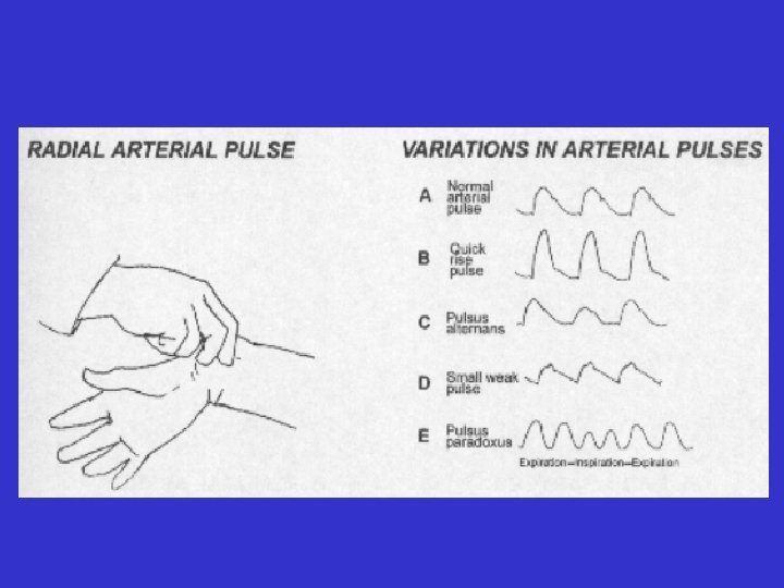
Changes of arterial pulse and blood pressure • Changes of the rate, regularity, amplitude, and quality of arterial pulse should be taken into account – radial pulse deficit: the consequence of irregular heart activity some diastolic pauses are too short to normal filling of the LV some LV contractions are ineffective (no systolic blood ejection) – weak thready pulse: the consequence of a low stroke volume or incrased/decreased peripheral arterial resistance – forceful bounding pulse: consequence of high stroke volume and reduced peripheral arterial rersistance
– small pulse with a slow upstroke (pulsus tardus): - result of aortic stenosis – bounding, rapidly rising and collapsing pulse (reffered to a waterhammer pulse or Corrigan s pulse: result of aortic regurgitation – pulsus alternans: alterning strong and weak pulses at regular intervals: - it frequently reflects LVF – pulsus bigeminus: alterning strong and weak pulses at irregular intervals: - consequence of extrasystolic bigeminia – pulsus paradoxus: smaller pulse amplitude during inspirium: - it is the consequence of exaggerated fall in systolic BP of greater than 10 mm. Hg during inspiration – it is present in heart tamponade and constrictive pericarditis
Pulsus alternans Cosequence of ventricular function changes a) Mechanical alternans (pusus alternans) b) Electrical alternans (alternation of tall and short QRS kompexes) a) Mechanical alternans – beat-to-beat oscilation in the strength of ventricular myocardium contraction at a constant rate (sinus rhythm) Manifestation: - in patients with heart failure - in patients with aortic or subaortic stenosis Mechanisms responsible: 1) based on Frank-Starling mechanism 2) based on alternation of myocardial contractility: - due to alternation of i. Ca 2+ concentration caused by alternation of Ca 2+ release from sarcoplasmatic reticulum (e. g. in hypotermia, ischemia)
Ankle-brachial index (ABI) ABI = Ankle systolic BP/ Arm systolic BP – test comparing the BP in feet to BP in arm – it is used for diagnosis of peripheral arterial diseases Normal ABI: 1. 0 – 1. 3 Supranormal: > 1. 30 (OK!) A normal resting ankle-brachial index is 1. 0 to 1. 3. This means that your BP at your ankle is the same or greater than the pressure at your arm, and suggests that you do not have significant narrowing or blockage of blood flow. Abnormal An abnormal resting ankle-brachial index is 0. 9 or lower. If the ABI is 0. 91 to 0. 99, it is considered borderline abnormal.
Venous pressure and pulsation Jugular venous pressure and pulsation reflect the function of the right side of the heart: – pressure in the internal jugular vein (IJV) is taken as the central venous pressure(CVP) – CVP is increased when pulsation in IJA is present higher than 3 cm over the sternal angle: it is the consequence of right side heart failure – paradoxic increase in CVP during inspiration (Kussmaul s sign): consequence of venous return impediment to the right heart – it is present in severe right heart failure – positive hepatojugular reflux: result of right HF
Pulsation in internal jugular vein - Norm – up to 7 cm over - Pathologic – more than 10 cm Record of Int jug art pressure a - right atrium contraction c - transmission from right ventricular pressure during its isometric contraction x - TK in atrium during its relaxation and shift of fibrous anulus downword v – end of atrium filling y - atrium empties
Precordial movements – sign of ventricular hypertrophy and incresed myocardial contractility Pathomechanism: – LV hypertrophy the apical impulse is more sustained, more forceful, and larger point of maximal impulse done by LV is displaced laterally to the left and down-ward – RV hypertrophy produces substernal heave or a systolic lift of the sternum
Abnormal heart sounds – third heart sound – develops during fast filling of ventricle in early phase of diastole – forth heart sound – it is present at the end of ventricular distole due to pushing the blood from atrium by its contraction
Abnormal heart sounds