Endocrine hormone System Hendra Wijaya Hormones are chemical
 System Hendra Wijaya")


 • Regulates")

~bones √gigantism/dwarfism")


: • epinephrine & norepinephrine~ increase basal metabolic")
 • sperm formation")
~ sperm formation; male")


- Slides: 49
Endocrine (hormone) System Hendra Wijaya
Hormones are chemical messengers secreted by certain tissues into the blood or institial fluid, serving to regulate the activity of other tissues.
Chemical Classification of Hormones • Amine hormones are derived from tyrosine or tryptophan – Include NE, Epi, thyroxine, melatonin • Polypeptide/protein hormones are chains of amino acids – Include ADH, GH, insulin, oxytocin, glucagon, ACTH, PTH • Glycoproteins – Long polypeptide bound to a carbohydrate group – Include LH, FSH, TSH • Steroids are lipids derived from cholesterol – Include testosterone, estrogen, progesterone & cortisol 11 -7
Chemical Classificaton of Hormones • Steroid Hormones: – Lipid soluble – Diffuse through cell membranes – Endocrine organs • • Adrenal cortex Ovaries Testes placenta
Chemical Classification of Hormones • Nonsteroid Hormones: – Not lipid soluble – Received by receptors external to the cell membrane – Endocrine organs • • • Thyroid gland Parathyroid gland Adrenal medulla Pituitary gland pancreas
Regulatory Systems • • Target cells~ body cells that respond to hormones Endocrine system/glands~ hormone secreting system/glands (ductless); exocrine glands secrete chemicals (sweat, mucus, enzymes) through ducts Neurosecretory cells~ actual cells that secrete hormones Feedback mechanisms ~ negative and positive
Endocrine System
Major Endocrine Glands 1. Pineal Gland 2. Hypothalamus 3. Pituitary Gland 1. Anterior 2. Posterior 4. Thyroid Gland 5. Parathyroid Glands 6. Adrenal Glands 6. Cortex 7. Medulla 7. Thymus Gland 8. Pancreas 9. Gonads 6. Ovaries 7. Testes
There is a hierarchical chain of command in hormonal signaling. Coordination center of the endocrine system. Via direct neuronal connection
Hypothalamus-pituitary axis
Feedback control
Feedback control of thyroid function
Mode of Action: Chemical Signaling • 1 - Plasma membrane reception • signal-transduction pathways (neurotransmitters, growth factors, most hormones) • 2 - Cell nucleus reception • steroid hormones, thyroid hormones, some local regulators
Hypothalamus • Part of brain – Regulates ANS, emotions, feeding/satiety, thirst, body temperature, etc. – Hormones related to these functions • “Releasing hormones” • Axonal transport to posterior lobe
Hypotalamus • Hypothalamus and pituitary gland secrete hormones and regulate other endocrine organs. They are the main regulatory organs of the endocrine system.
Hypothalamus • Located below the thalamus and above the pituitary gland (=epiphysis) • Regulates the pituitary gland secretions through two different mechanisms
Hypothalamus - neurohypophysis • 1 - Neurons, receiving information from receptors, fire APs which travel down to the post pituitary gland stimulate the release of stored neurohormones – Oxytocin (OT) and anti-diuretic hormone (ADH)
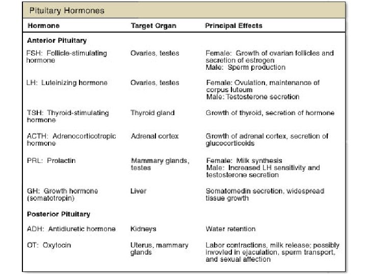
Pituitary Gland • • • Releasing and inhibiting hormones Anterior pituitary: Growth (GH)~bones √gigantism/dwarfism √acromegaly Prolactin (PRL)~mammary glands; milk production Follicle-stimulating (FSH) & Luteinizing (LH)~ovaries/testes Thyroid-stimulating (TSH)~ thyroid Adrenocorticotropic (ACTH)~ adrenal cortex Melanocyte-stimulating (MSH) Endorphins~natural ‘opiates’; brain pain receptors
Hormones of the Anterior Pituitary
Hypothalamus & Pituitary Gland
Posterior Region of the Pituitary Gland • The posterior pituitary: • Oxytocin~ uterine and mammary gland cell contraction • Antidiuretic (ADH)~ retention of water by kidneys
The Pineal, Thyroid, & Parathyroid • Melatonin~ pineal gland; biological rhythms • Thyroid hormones: Calcitonin~ lowers blood calcium Thyroxine~ metabolic processes • Parathyroid (PTH)~ raises blood calcium
The thyroid gland - Chp 21 p 623 -625 • Located in the neck, just below the larynx • Secrete 2 types of hormone: - thyroid hormones stimulate cell metabolism, triiodothyronine (T 3) and thyroxine (T 4) – iodine is needed to synthesize these hormones - calcitonin decrease blood calcium Figure 6. 8 a
Thyroid hormones • T 3 and T 4 secreted by the follicular cells • Stored as colloid • Parafollicular cells (C cells) secrete calcitonin (Chp 19)
Thyroid Hormones T 3 and T 4 • Target organs: all cells • Role: Increase cell metabolism, oxygen consumption • Permissive role for some other hormones (growth hormone)
Thyroid hormone regulation Figure 6. 7
Goiter • Both hypo and hyperthyroidism can have goiter as a symptom • Goiter is a swelling of the neck due to hypertrophy of the thyroid gland • How can one explain that?
Goiter in hypothyroidism • • • Most often due to a lack of dietary iodine The thyroid hormone is unable to synthesize a functional thyroid hormone (T 3 and T 4) The person express symptoms of hypothyroidism The nonfunctional T 3/T 4 cannot promote a negative feedback on TRH and TSH the hypotalamus and pituitary gland increase their secretions the thyroid gland is stimulated to secrete more T 3 and T 4 … In children, the lack of functional T 3/T 4 result in cretinism, a form a mental retardation
Goiter in hyperthyroidism • The cells secreting TRH or TSH on the hypothalamus and pituitary gland (respectively) have become abnormal and no longer are sensitive to the negative feedback they continue to secrete TRH or TSH continuous stimulation of the thyroid gland with excess thyroid hormones being formed • symptoms of hyperthyroidism
Parathyroid glands • Four nodules located in the back of the thyroid gland • Secreted parathyroid hormone or parathormone or PTH • Action of PTH opposes action of calcitonin • Both hormones play a role in calcium metabolism
Roles of calcium • Most calcium ions are stored in the bones • Calcium is an important cofactor for enzymatic activity, plays a role in blood coagulation and action potentials. • Calcitonin and PTH participate in calcium regulation • Vitamin D helps PTH activity
Calcium regulation: • Calcitonin promotes blood calcium decrease, by: - 1. calcium deposition on bone - 2. calcium dumping by the kidney • PTH promotes blood calcium increase by: - 1. bone resorption - 2. calcium reabsorption by kidney - 3. increase calcium absorption by intestine
Calcium Metabolism: Figure 23 -20: Calcium balance in the body
Figure 19. 20
The Pancreas • Islets of Langerhans • Alpha cells: • glucagon~ raises blood glucose levels • Beta cells: • insulin~ lowers blood glucose levels • Type I diabetes mellitus (insulin-dependent; autoimmune disorder) • Type II diabetes mellitus (non-insulin-dependent; reduced responsiveness in insulin targets)
The pancreas • Located in the left upper abdominal cavity • Exocrine and endocrine glands • The endocrine function is due to the cells of the islets of the Langerhans -- α cells glucagon -- β insulin -- δ somatostatin
Glucose regulation • Glucose level controlled by insulin and glucagon • Insulin promotes a decrease in blood glucose • Glucagon promotes an increase in blood glucose
Glucose regulation
Fate of glucose Figure 3. 21
Diabetes mellitus • Type I: autoimmune disease beta cells of the islets of Langerhans are destroyed by antibodies • Type II: The cells become insulin-resistant glucose does not enter the cells as readily • http: //faculty. weber. edu/nokazaki/Human_Physiol ogy/Class%20 notes/diabetes. htm
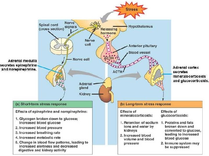
The Adrenal Glands • Adrenal medulla (catecholamines): • epinephrine & norepinephrine~ increase basal metabolic rate (blood glucose and pressure) • Adrenal cortex (corticosteroids): • glucocorticoids (cortisol)~ raise blood glucose • mineralocorticoids (aldosterone)~ reabsorption of Na+ and K+
The Gonads • Steroid hormones: precursor is cholesterol – Androgens (testosterone) • sperm formation • male secondary sex characteristics; gonadotropin – Estrogens (estradiol) • uterine lining growth • female secondary sex characteristics • gonadotropin – Progestins (progesterone) • uterine lining growth
The Gonads • Steroid hormones: precursor is cholesterol • androgens (testosterone)~ sperm formation; male secondary sex characteristics; gonadotropin • estrogens (estradiol)~uterine lining growth; female secondary sex characteristics; gonadotropin • progestins (progesterone)~uterine lining growth
Revision: Major endocrine conditions Endocrine Gland Hormones Endocrine Disorders Anterior Pituitary Growth Hormone Gigantism, acromegaly Dwarfism Thyroid Thyroxine (T 4) Triodothronine (T 3) Thyrotoxicosis Goitre Exopthalmos Hypothroidism Cretinism Myxoedema Goitre Parathyroid Parathormone Osteoporosis Kidney stones Tetany Adrenal Cortex Glucacorticoids Cushings syndrome Addisons disease Adrenal Medulla Epinephrine Norepinephrine Increased metabolism Hypertension Pancreatic Islets Insulin Hyper Hypo Diabetes mellitus
Pituitary Dwarfism
Hyper secretion of Growth hormone: Gigantism