Endocrine system Cells tissues and organs that synthesize


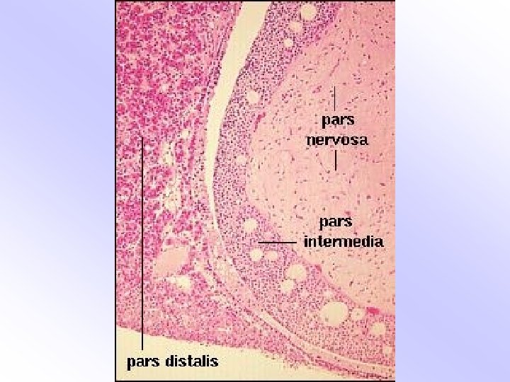
 Adenohypophysis Pars distalis, Pars tuberalis, Pars intermedia Neurohypophysis Median")


 ü Lighter staining cytoplasm ü Bigger in size,")

 cells ü Round, Pale, slightly acidophilic cytoplasm ü")


 Outer most layer – zona glomerulosa (1/5 th")


- Slides: 51
Endocrine system
• Cells, tissues and organs that synthesize and secrete hormones • Bloodstream • No excretory ducts
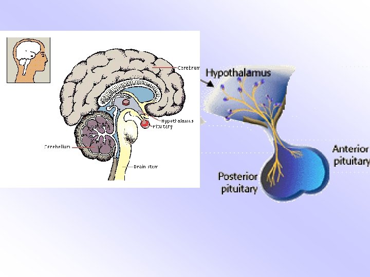
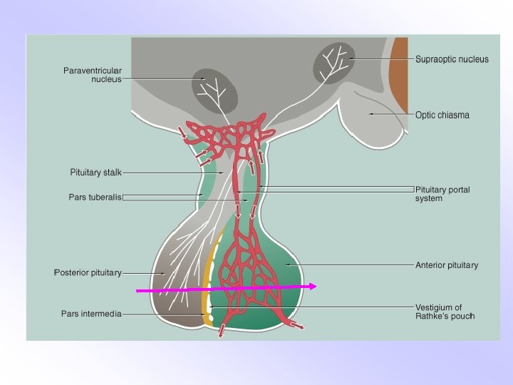
The pituitary gland (Hypophysis cerebri) Adenohypophysis Pars distalis, Pars tuberalis, Pars intermedia Neurohypophysis Median eminence, Infundibulum, Pars nervosa
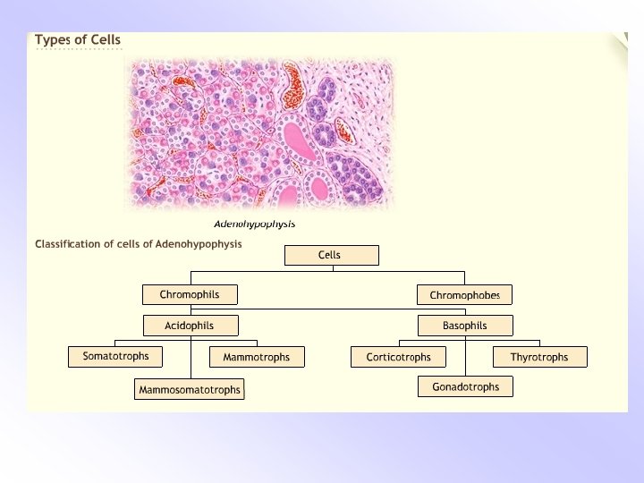
Pars distalis • Chromophobe cells • Chromophil cells
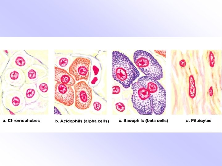
Chromophobes: ü Non-granular, homogenous cytoplasm ü Cell boundaries indistinct
Chromophils Acidophils: ü Granular Cytoplasm ü Cell outlines distinct ü Lie in the vicinity of sinusoidal capillaries Basophils: ü Granular cytoplasm ü Vary in shape
Pars intermedia: ü Colloid filled cysts ü Follicles with basophilic cells
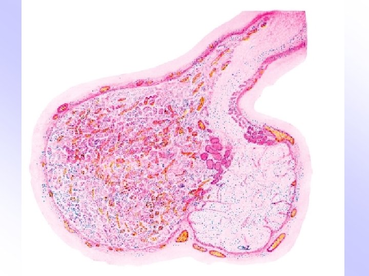
Hypophysis cerebri
Pars nervosa: ü Unmyelinated axons ü Supraoptic and paraventricular nuclei ü Hypothalamo hypophyseal tract ü Pituicytes are neuroglial cells ü Herring bodies (terminal ends of unmyelinated axons)
The hormones • Pars distalis – Acidophils: GH, Prolactin – Basophils: ACTH, TSH, LH, FSH – Chromophobes: Give rise to chromophils/resting cells • Pars intermedia – MSH • Neurohypophysis – Vasopressin, Oxytocin
Pars nervosa Pars intermedia Pars distalis
Acidophils Basophils Chromophobes
Pituicytes
Hypophysis cerebri
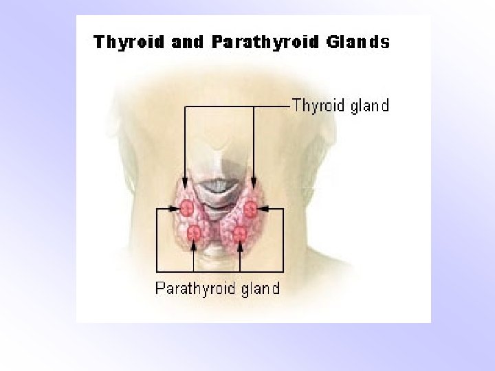
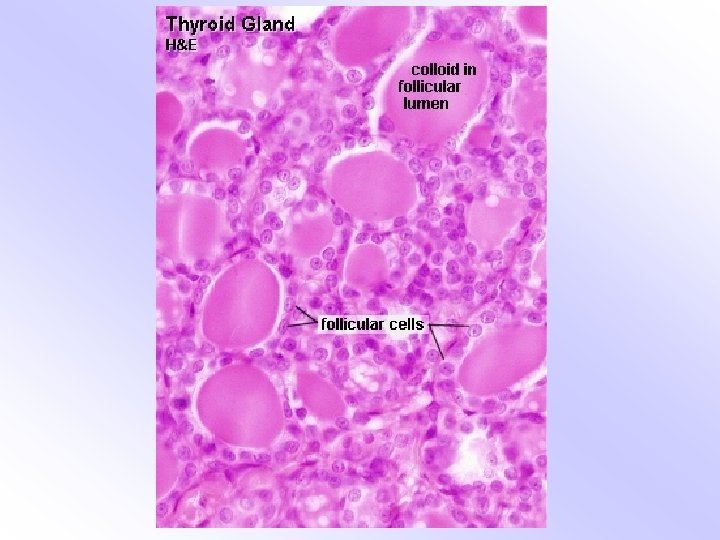
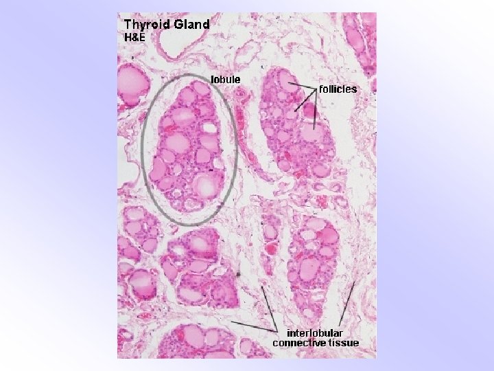
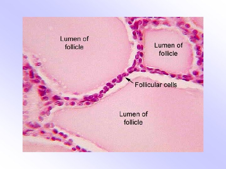
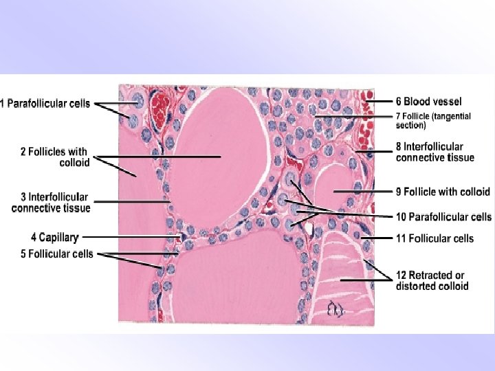
Thyroid ü Covered by capsule ü Connective tissue septa , lobules ü Colloid filled follicles, lined by simple cuboidal cells ü Thyroglobulin (colloid) ü Hormones: T 3 and T 4
Parafollicular cells (clear or c cells) ü Lighter staining cytoplasm ü Bigger in size, oval in shape ü Present between follicular cells ü Hormone: Calcitonin
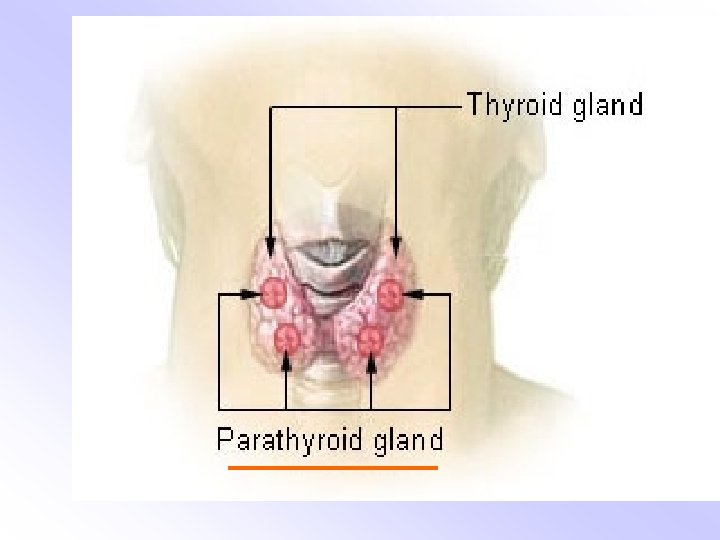
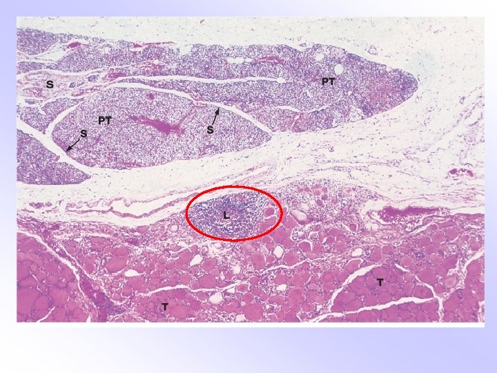
Parathyroid ü Associated with the thyroid ü Generally four in number ü Cells arranged in clumps
Two types of cells Chief (principal) cells ü Round, Pale, slightly acidophilic cytoplasm ü Most numerous ü Small dark-staining nuclei Oxyphilic cells ü Larger in size, less numerous ü Strongly acidophilic
Hormones Chief cells: Parathormone Oxyphil cells: not known
Thyroid and parathyroid
Thyroid and parathyroid
Micro Structural Anatomy of pancreas
• Covered with a very thin layer of loose connective tissue capsule. • Thin septa arising from capsule and divide the gland into many lobules. • Inter lobular connective tissue contains large ducts, blood vessels and nerve fibers. • Interlobular- surrounds the acini & Islets
Exocrine Pancreas • It is the serous gland. • Secretory units are acinar or tubuloacinar in shape. • Formed by simple epithelium of pyramidal serous cells.
Duct system of exocrine part Main Pancreatic Duct Interlobular Duct Intralobular Duct Intercalated Duct Centro Acinar
Endocrine pancreas • Diffuse organ & secretes hormone that regulates the glucose level • Islets of Langerhans scattered throughout organ. • 1 – 3 million • Polygonal cells are arranged in short, irregular cords
• Zenkar formal fixation & Mallory Azan method • A- alpha-20%, Densely packed cytoplasm • B- beta-70%, dense polyhedral core and pale matrix. • D- delta- 5 -10%, Secretory granules large than A & B cells. • Minor cells- PP & D 1 cells
SUPRARENAL GLAND
SUPRARENAL GLAND Ø A pair of endocrine glands Ø Situation: -on the posterior abdominal wall over the upper pole of the kidneys behind the peritoneum Ø Shape: -right suprarenal gland – triangular left suprarenal gland -semilunar
Histology ØCovered by connective tissue capsule ØSepta from capsule extend into the gland substance ØParts: -superficial-cortex (9/10) deeper-medulla (1/10)
Cortex Ø Layers of cortex a) Outer most layer – zona glomerulosa (1/5 th of the cortex) b) Middle layer – zona fasciculata (3/5 th of the cortex) c) Innermost layer – zona reticularis (1/5 th Of the cortex)
Zona glomerulosa Ø Cells are arranged as inverted u shaped formations Ø Cells are polyhedral or columnar. Basophilic cytoplasm and deeply stained nuclei. Ø Hormones – mineralocarticoidsaldosterone, deoxycorticost rone.
Zona fasciculata Ø Cells are arranged in straight columns in two cell thickness. Ø Sinusoids intervenes between the columns. Ø Cells are large polyhedral basophilic cytoplasm and vasicular nuclei. Ø Hormones – glucocorticoidscortizone, cortisol dehydroepiandrosterone(DHA).
• Zona reticularisØ Cords will branch and anastomose with each other to form a kind of reticulum. Ø Cells are large polyhedral eosinophilic cytoplasm. Ø Hormones – glucocorticoids sex hormones-eastrogens androgens.
Medulla Ø Distinct from the cortex both functionally and embryologically Ø Made up of groups of cells seperated by wide sinusoids. Ø Cells are columnar or polyhedral with basophilic cytoplasm (chromaffin cells). Ø Functionally these are modified post ganglionic sympathetic neurons. Ø Hormones – noradrenalin and adrenalin.
summary Zona glomerulosa cortex Zona fasciculata Zona reticularis medulla Chromaffin cells