Chapter 45 Hormones and the Endocrine System Power

 Endocrine signaling Response (b) Paracrine signaling Response")


 Different receptors")


 is an autoimmune disorder in which")


 is secreted by the anterior pituitary gland has")


 and norepinephrine")


- Slides: 38
Chapter 45 Hormones and the Endocrine System Power. Point® Lecture Presentations for Biology Eighth Edition Neil Campbell and Jane Reece Lectures by Chris Romero, updated by Erin Barley with contributions from Joan Sharp Copyright © 2008 Pearson Education, Inc. , publishing as Pearson Benjamin Cummings
Fig. 45 -2 Blood vessel Response (a) Endocrine signaling Response (b) Paracrine signaling Response (c) Autocrine signaling Synapse Neuron Response (d) Synaptic signaling Neurosecretory cell Blood vessel (e) Neuroendocrine signaling Response
Fig. 45 -3 Water-soluble Lipid-soluble 0. 8 nm Polypeptide: Insulin Steroid: Cortisol Amine: Epinephrine Amine: Thyroxine
• Signaling by any of these hormones involves three key events: – Reception – Signal transduction – Response Copyright © 2008 Pearson Education, Inc. , publishing as Pearson Benjamin Cummings
Fig. 45 -5 -2 Binding of a hormone to its receptor initiates a signal transducti on pathway leading to responses in the cytoplasm, enzyme activation, or a change in gene expression Watersoluble hormone Transport protein Signal receptor TARGET CELL Cytoplasmic response (a) OR Gene regulation NUCLEUS Signal receptor Cytoplasmic response (b) The response to a lipid-soluble hormone is usually a change in gene Fat-soluble hormone expression Steroids, thyroid hormones, and the hormonal form of vitamin D Protein-receptor complexes then act as transcription factors in the nucleus, regulating transcription of specific genes Gene regulation
• The hormone epinephrine has multiple effects in mediating the body’s response to short-term stress • Epinephrine binds to receptors on the plasma membrane of liver cells • This triggers the release of messenger molecules that activate enzymes and result in the release of glucose into the bloodstream Copyright © 2008 Pearson Education, Inc. , publishing as Pearson Benjamin Cummings
Fig. 45 -6 -1 Epinephrine Adenylyl cyclase G protein-coupled receptor GTP ATP c. AMP Second messenger
Fig. 45 -6 -2 Epinephrine Adenylyl cyclase G protein-coupled receptor GTP ATP c. AMP Inhibition of glycogen synthesis Promotion of glycogen breakdown Protein kinase A Second messenger
Multiple Effects of Hormones • The same hormone may have different effects on target cells that have – Different receptors for the hormone – Different signal transduction pathways – Different proteins for carrying out the response • A hormone can also have different effects in different species Copyright © 2008 Pearson Education, Inc. , publishing as Pearson Benjamin Cummings
Fig. 45 -8 -2 Same receptors but different intracellular proteins (not shown) Different receptors Epinephrine receptor Glycogen deposits Glycogen breaks down and glucose is released. (a) Liver cell Vessel dilates. (b) Skeletal muscle blood vessel Vessel constricts. (c) Intestinal blood vessel
Signaling by Local Regulators • In paracrine signaling, nonhormonal chemical signals called local regulators elicit responses in nearby target cells • Types of local regulators: – Cytokines and growth factors – Nitric oxide (NO) – Prostaglandins help regulate aggregation of platelets, an early step in formation of blood clots Copyright © 2008 Pearson Education, Inc. , publishing as Pearson Benjamin Cummings
Fig. 45 -10 Major endocrine glands: Hypothalamus Pineal gland Pituitary gland Thyroid gland Parathyroid glands Organs containing endocrine cells: Thymus Heart Adrenal glands Testes Liver Stomach Pancreas Kidney Small intestine Ovaries
Hormonal control 1. Positive Feedback
Fig. 45 -11 – Negative feedback A negative feedback loop inhibits a response by reducing the initial stimulus Negative feedback regulates many hormonal pathways involved in homeostasis Pathway Example Stimulus Low p. H in duodenum S cells of duodenum secrete secretin ( ) Endocrine cell Blood vessel Target cells Response Pancreas Bicarbonate release
Insulin and Glucagon: Control of Blood Glucose • Insulin and glucagon are antagonistic hormones that help maintain glucose homeostasis • The pancreas has clusters of endocrine cells called islets of Langerhans with alpha cells that produce glucagon and beta cells that produce insulin
Fig. 45 -12 -5 Insulin reduces blood glucose levels by – Promoting the cellular uptake of glucose – Slowing glycogen breakdown in the liver – Promoting fat storage Glucagon increases blood glucose levels by - Stimulating conversion of glycogen to glucose in the liver – Stimulating breakdown of fat and protein into glucose Body cells take up more glucose. Insulin Beta cells of pancreas release insulin into the blood. Liver takes up glucose and stores it as glycogen. STIMULUS: Blood glucose level rises. Blood glucose level declines. Homeostasis: Blood glucose level (about 90 mg/100 m. L) STIMULUS: Blood glucose level falls. Blood glucose level rises. Alpha cells of pancreas release glucagon. Liver breaks down glycogen and releases glucose. Glucagon
Diabetes Mellitus • Type I diabetes mellitus (insulindependent) is an autoimmune disorder in which the immune system destroys pancreatic beta cells • Type II diabetes mellitus (noninsulin-dependent) involves insulin deficiency or reduced response of target cells due to change in insulin receptors • Best-known endocrine disorder • It is caused by a deficiency of insulin or a decreased response to insulin in target tissues • It is marked by elevated blood glucose levels
Coordination of Endocrine and Nervous Systems in Vertebrates • Signals from the nervous system initiate and regulate endocrine signals • The hypothalamus receives information from the nervous system and initiates responses through the endocrine system • Attached to the hypothalamus is the pituitary gland composed of the posterior pituitary and anterior pituitary
Fig. 45 -14 Cerebrum Pineal gland Thalamus Cerebellum Pituitary gland Hypothalamus Spinal cord The posterior pituitary stores and secretes hormones that are made in the hypothalamus The anterior pituitary makes and releases hormones under regulation of the hypothalamus Hypothalamus Posterior pituitary Anterior pituitary
Fig. 45 -15 Hypothalamus Neurosecretory cells of the hypothalamus Axon Posterior pituitary Anterior pituitary HORMONE ADH Oxytocin TARGET Kidney tubules Mammary glands, uterine muscles
Fig. 45 -16 Pathway Example Stimulus Suckling + Sensory neuron Positive feedback Hypothalamus/ posterior pituitary Neurosecretory cell Blood vessel Target cells Response Posterior pituitary secretes oxytocin ( ) Smooth muscle in breasts Milk release
Fig. 45 -17 Tropic effects only: FSH A tropic hormone regulates the function of LH endocrine cells or glands TSH ACTH Neurosecretory cells of the hypothalamus Nontropic effects only: Prolactin MSH Nontropic and tropic effects: GH Hypothalamic releasing and inhibiting hormones Portal vessels Endocrine cells of the anterior pituitary Posterior pituitary Pituitary hormones HORMONE FSH and LH TSH ACTH Prolactin MSH GH TARGET Testes or ovaries Thyroid Adrenal cortex Mammary glands Melanocytes Liver, bones, other tissues Promotes milk excretion Skin pigmentation
Fig. 45 -18 -3 Example Stimulus Cold Sensory neuron – Hypothalamus secretes thyrotropin-releasing hormone (TRH ) Neurosecretory cell Blood vessel – Anterior pituitary secretes thyroid-stimulating hormone (TSH or thyrotropin ) Negative feedback A hormone can stimulate the release of a series of other hormones, the last of which activates a nonendocrine target cell; this is called a hormone cascade pathway The release of thyroid hormone results from a hormone cascade pathway involving the hypothalamus, anterior pituitary, and thyroid gland Hormone cascade pathways are usually regulated by negative feedback Pathway Thyroid gland secretes thyroid hormone (T 3 and T 4 ) Target cells Response Body tissues Increased cellular metabolism
Growth Hormone • Growth hormone (GH) is secreted by the anterior pituitary gland has tropic and nontropic actions • It promotes growth directly and has diverse metabolic effects • It stimulates production of growth factors • An excess of GH can cause gigantism, while a lack of GH can cause dwarfism
Thyroid Hormone: Control of Metabolism and Development • The thyroid gland consists of two lobes on the ventral surface of the trachea • It produces two iodine-containing hormones: triiodothyronine (T 3) and thyroxine (T 4)
• Thyroid hormones stimulate metabolism and influence development and maturation • Hyperthyroidism, excessive secretion of thyroid hormones, causes high body temperature, weight loss, irritability, and high blood pressure • Graves’ disease is a form of hyperthyroidism in humans • Hypothyroidism, low secretion of thyroid hormones, causes weight gain, lethargy, and intolerance to cold
• Proper thyroid function requires dietary iodine for hormone production
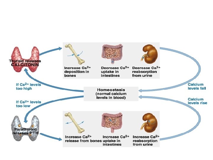
Parathyroid Hormone and Vitamin D: Control of Blood Calcium • Two antagonistic hormones regulate the homeostasis of calcium (Ca 2+) in the blood of mammals – Parathyroid hormone (PTH) is released by the parathyroid glands – Calcitonin is released by the thyroid gland
Adrenal Hormones: Response to Stress • The adrenal glands are adjacent to the kidneys • Each adrenal gland actually consists of two glands: the adrenal medulla (inner portion) and adrenal cortex (outer portion)
Catecholamines from the Adrenal Medulla • The adrenal medulla secretes epinephrine (adrenaline) and norepinephrine (noradrenaline) • These hormones are members of a class of compounds called catecholamines • They are secreted in response to stressactivated impulses from the nervous system • They mediate various fightor-flight responses • Epinephrine and norepinephrine – Trigger the release of glucose and fatty acids into the blood – Increase oxygen delivery to body cells – Direct blood toward heart, brain, and skeletal muscles, and away from skin, digestive system, and kidneys • The release of epinephrine and norepinephrine occurs in response to nerve signals from the hypothalamus
Fig. 45 -21 Stress Spinal cord Nerve signals Releasing hormone Nerve cell Hypothalamus Anterior pituitary Blood vessel ACTH Adrenal medulla Adrenal cortex Adrenal gland Kidney (a) Short-term stress response Effects of epinephrine and norepinephrine: 1. Glycogen broken down to glucose; increased blood glucose 2. Increased blood pressure 3. Increased breathing rate 4. Increased metabolic rate 5. Change in blood flow patterns, leading to increased alertness and decreased digestive, excretory, and reproductive system activity (b) Long-term stress response Effects of mineralocorticoids: Effects of glucocorticoids: 1. Retention of sodium 1. Proteins and fats broken down ions and water by and converted to glucose, leading kidneys to increased blood glucose 2. Increased blood volume and blood pressure 2. Possible suppression of immune system
Gonadal Sex Hormones • The gonads, testes and ovaries, produce most of the sex hormones: androgens, estrogens, and progestins • All three sex hormones are found in both males and females, but in different amounts
• The testes primarily synthesize androgens, mainly testosterone, which stimulate development and maintenance of the male reproductive system • Testosterone causes an increase in muscle and bone mass and is often taken as a supplement to cause muscle growth, which carries health risks
• Estrogens, most importantly estradiol, are responsible for maintenance of the female reproductive system and the development of female secondary sex characteristics • In mammals, progestins, which include progesterone, are primarily involved in preparing and maintaining the uterus • Synthesis of the sex hormones is controlled by FSH and LH from the anterior pituitary
Melatonin and Biorhythms • The pineal gland, located in the brain, secretes melatonin • Light/dark cycles control release of melatonin • Primary functions of melatonin appear to relate to biological rhythms associated with reproduction
Table 45 -1
Table 45 -1 a