Dysregulation of innate and adaptive immunity in patients


- Slides: 38

Dysregulation of innate and adaptive immunity in patients with atopic dermatitis: Impact of IL-31/IL-31 R & staphylococcal α-toxin Sadaf Kasraie, Ph. D. Division of Immunodermatology and Allergy Research, Department of Dermatology and Allergy

Activation IL-31/IL-31 R CD 4+ T- Cells Induces pro-inflammatory cytokines in some human cell lines so far 1, 2 (24 KDa) Receptor 1: IL-31 RA (GPL) (120 KDa) Receptor 2: OSMR (180 KDa) Epithelial cells Keratinocytes Jak/STAT, PI 3 kinase/Akt and MAP kinase signalling pathway Sadaf Kasraie Division of Immunodermatology and Allergy Research Activated monocytes and macrophages Eosinophils 1 Dambacher J. et al. 2007. Gut 56: 257 -65. Modified from Le Saux S. et al. J Biol Chem. 2010. 285: 3470 -7 2 Yagi Y. et al. 2007. Int J Mol Med 19: 941 -6.

Role of IL-31 overexpression may lead to : • Pruritus • Alopecia • Skin lesions • Airway hypersensitivity • Atopic dermatitis (atopic eczema) • Allergy Sadaf Kasraie Division of Immunodermatology and Allergy Research

Review • Transgenic overexpression of IL-31 in lymphocytes induces severe pruritus in mice (Dillon et al. , Nat Immunol 2004). • IL-31 is overexpressed in the skin of patients with atopic dermatitis (Sonkoly et al. , JACI 2006). • Serum IL-31 levels are significantly higher in patients with AD and correlated with disease activity in AD (Raap et al. , JACI 2008). • Staphylococcal superantigens rapidly induce IL-31 expression in atopic individuals in vivo and in leukocytes in vitro (Sonkoly et al. , JACI 2006). Sadaf Kasraie Division of Immunodermatology and Allergy Research

Approach To investigate • the expression, • the regulation of IL-31 R, • functional effects of IL-31 in human primary cells (monocytes, macrophages, keratinocytes and eosinophils): Regulation of STAT and MAPK signalling pathways as well as cytokine secretion Sadaf Kasraie Division of Immunodermatology and Allergy Research

Expression of IL-31 RA on Immune Cells & Keratinocytes Non Stimulated* Monocytes - ++1, 2 Macrophages - ++1, 2 DC: mo. DC - - DC: IDEC - - Lymphocytes - - Keratinocytes +3 ++ Eosinophils +4 + Kasraie S. et al. 2010. Allergy. 65: 712– 721. 2 Kasraie S. et al. 2011. Allergy. 66: 845 -52. 3 Kasraie S. et al. 2013. Allergy. 68: 739 -47. 4 Raap U, Gehring M, Kasraie S. et al. Submitted. 1 Sadaf Kasraie Division of Immunodermatology and Allergy Research * SEB, α-toxin or TLR-2 agonists

Macrophages Kasraie and Werfel. 2013. Mediators Inflamm 2013: 942375 Modified from Werfel T. 2009. J Invest Dermatol 129: 1878 -91 Sadaf Kasraie Division of Immunodermatology and Allergy Research

Staphylococcal exotoxins significantly up-regulate IL-31 RA expression in human monocytes and macrophages at the m. RNA level A) Monocytes Sadaf Kasraie Division of Immunodermatology and Allergy Research B) Macrophages

Staphylococcal exotoxins up-regulate IL-31 RA Staphylococcal expression in human monocytes and at the expression in human monocytes atmacrophages the protein level A) Monocytes Sadaf Kasraie Division of Immunodermatology and Allergy Research B) Macrophages

Signalling pathways of IL-31 in human macrophages p. STAT-1 β-actin p. STAT-5 β-actin p. ERK 1/2 β-actin Sadaf Kasraie Division of Immunodermatology and Allergy Research

Summary of IL-31 effects upon staphylococcal exotoxins stimulation in monocytes & macrophages Cells Cytokine MΦ & PBMCs IL-6 MΦ & PBMCs IL-1β MΦ IL-18 MΦ IL-10 MΦ TNF-α MΦ MIP-1α MΦ MCP-1α MΦ IL-12 p 40 MΦ IL-12 p 70 MΦ IL-23 p 19/p 40 Sadaf Kasraie Division of Immunodermatology and Allergy Research SEB α-toxin

IL-31 induces pro-inflammatory cytokines in human macrophages following up-regulation of the IL-31 R with staphylococcal exotoxins Sadaf Kasraie Division of Immunodermatology and Allergy Research

IL-31 down-regulates IL-12 in human macrophages following up-regulation of the IL-31 R with staphylococcal exotoxins Sadaf Kasraie Division of Immunodermatology and Allergy Research

Keratinocytes Kasraie and Werfel. 2013. Mediators Inflamm 2013: 942375 Modified from Werfel T. 2009. J Invest Dermatol 129: 1878 -91 Sadaf Kasraie Division of Immunodermatology and Allergy Research

TLR-2 ligand Pam 3 Cys up-regulates IL-31 RA expression in human primary keratinocytes at the m. RNA level * *Pam 3 Cys-SK 4: N-Palmitoyl-S-[2, 3 -bis (palmitoyloxy)-(2 RS)-propyl]-[R]-cysteinyl-[S]-seryl-[S]-lysyl-[S]-lysine Sadaf Kasraie Division of Immunodermatology and Allergy Research

Pam 3 Cys or IFN-γ up-regulates IL-31 R expression on human primary keratinocytes at the protein level NS= Not Stimulated Sadaf Kasraie Division of Immunodermatology and Allergy Research

Signalling pathway of IL-31 in human keratinocytes p. STAT-3 Sadaf Kasraie Division of Immunodermatology and Allergy Research

Summary of IL-31 effects upon Pam 3 Cys or IFN-γ stimulation in keratinocytes HPKs Chemokine/Cytokine Pam 3 cys IFN-γ Foreskin & Hair CCL 2 + + Foreskin VEGF Foreskin CCL 22 + + - Foreskin CCL 20 - - Foreskin EGF Foreskin MMP-9 Foreskin & Hair IL-1β - - Foreskin & Hair IL-6 - - Foreskin & Hair IL-8 - - Sadaf Kasraie Division of Immunodermatology and Allergy Research

IL-31 induces CCL 2 secretion in human primary keratinocytes following up-regulation of the IL-31 R with Pam 3 Cys or IFN-γ Sadaf Kasraie Division of Immunodermatology and Allergy Research

IL-31 Staphylococcal exotoxins TLR-2 ligands or IFN-γ Activated monocytes and macrophages STAT-1/5, ERK 1/2 pro-inflammatory cytokines (IL-1β, IL-6, IL-18) Sadaf Kasraie Division of Immunodermatology and Allergy Research Eosinophils Keratinocytes STAT-3 CCL 2, VEGF STAT-3, ERK 1/2 CCL 26, release of superoxide species

Atopic dermatitis (AD) vs healthy controls (HC) Patient 1 with AD Sadaf Kasraie Division of Immunodermatology and Allergy Research Patient 2 with AD

IFN-IL-31 γ leads to significantly higher of IL-31 RA induces greater levels of expression IL-1β secretion in on monocytes patients AD compared to monocytes from patients with AD compared to HC healthy controls (HC) Sadaf Kasraie Division of Immunodermatology and Allergy Research

Pam 3 Cys IL-31 RA in keratinocytes IL-31 up-regulates induced lower levels expression of CCL 2 secretion in both from AD and keratinocytes from patients with ADHC compared to HC Sadaf Kasraie Division of Immunodermatology and Allergy Research

Impaired TLR-2 expression in keratinocytes from inflamed AD skin compared with healthy controls Sadaf Kasraie Division of Immunodermatology and Allergy Research

Conclusion IL-31 Homey et al. 2006. J Allergy Clin Immunol. Review. • • The Th 2 cytokine IL-31 induces itch. IL-31 has inflammatory properties. The IL-31 R is expressed on immune cells and keratinocytes. The IL-31 R is regulated by staphylococcal-derived molecules. Sadaf Kasraie Division of Immunodermatology and Allergy Research

Conclusion & Outlook • Evidence for a functional role of IL-31 on human eosinophils. • Novel implications for treatment options in eosinophil associated diseases including AD. • Colonization with S. aureus and presence of IL-31 in the skin may represent important trigger factors in the complex amplification cycle of atopic skin inflammation. • Antiseptic strategies as well as targeting IL-31 and its receptor in atopic individuals may represent useful tools to support therapeutic management of patients suffering from AD. Sadaf Kasraie Division of Immunodermatology and Allergy Research

Acknowledgment Ulrike Raap Margarete Niebuhr Kathrin Baumert Gabriele Begemann Maria Gschwandtner Manuela Gehring Jana Zeitvogel Christina Hartmann Hari Balaji Susanne Mommert Susanne Hradetzky Thomas Werfel Armin Braun Wolfgang Baeumer All members of the Dept. of Dermatology & Allergy Family! Sadaf Kasraie Division of Immunodermatology and Allergy Research

Acknowledgment Ulrike Raap Margarete Niebuhr Kathrin Baumert Gabriele Begemann Maria Gschwandtner Manuela Gehring Jana Zeitvogel Christina Hartmann Hari Balaji Susanne Mommert Susanne Hradetzky Thomas Werfel Armin Braun Wolfgang Baeumer All members of the Dept. of Dermatology & Allergy Family! Sadaf Kasraie Division of Immunodermatology and Allergy Research

RE Schmidt Susanne Kruse Marlies Daniel Sadaf Kasraie Division of Immunodermatology and Allergy Research HBRS et al. 2010. MHH info.

Overview Atopic Dermatitis (atopic eczema) is a chronically relapsing inflammatory skin disease Prevelance: 10 to 20% in children and 1% to 3% in adults Causes: Although it is such a common disease, the immunopathogenesis is not completely understood Sadaf Kasraie Division of Immunodermatology and Allergy Research

Trigger factors in Atopic Dermatitis Food allergens Hormones & related Factors Autoantigens Irritable substances Skin colonizing microorganisms Genetic background Inhalant allergens Environment Psychological stress Werfel et Sadaf Kasraie Division of Immunodermatology and Allergy Research Werfel et al. Leitline Atopisches Ekzem www. awmf. de 2002 (Revision 2007) al. HTA Bericht Neurodermitis www. egms. de 2006

IL-31 induces IL-12 suppression via ERK 1/2 phosphorylation in human macrophages following up-regulation of the IL-31 R with staphylococcal exotoxins Sadaf Kasraie Division of Immunodermatology and Allergy Research

Eosinophils Modified from Werfel T. 2009. J Invest Dermatol 129: 1878 -91 Sadaf Kasraie Division of Immunodermatology and Allergy Research

Human eosinophils express the IL-31 RA A) At the m. RNA level Sadaf Kasraie Division of Immunodermatology and Allergy Research B) At the protein level

Signalling pathways of IL-31 in human eosinophils p. STAT-3 Sadaf Kasraie Division of Immunodermatology and Allergy Research p. ERK 1/2

Human eosinophils are activated by IL-31 with release of CCL 26 and reactive oxygen species Sadaf Kasraie Division of Immunodermatology and Allergy Research

Functional effect of IL-31 in eosinophils 2 Raap U, Gehring M, Kasraie S. et al. Submitted. Sadaf Kasraie Division of Immunodermatology and Allergy Research

Th 2 Th 1 MHC II Th 1 Exotoxins APC atopic dermatitis CCL 2 Exotoxins TLR 2 ligand IL-31 RA Th 2 IL-4 IL-13 IL-31 Keratinocytes Sadaf Kasraie Division of Immunodermatology and Allergy Research
 Difference between acquired immunity and innate immunity
 Innate immunity examples
 Innate immunity examples
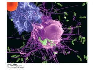
 Neutrophil extracellular traps
 Innate immunity first line of defense
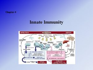
 Innate immunity
 Innate immunity first line of defense
 Innate immunity first line of defense
 Innate immunity first line of defense
 Phagocytr
 Innate immunity
 Waldeyer's ring
 Adaptive noise immunity
 Adaptive immunity
 Dmdd diagnosis
 Bpd quotes
 Disruptive mood dysregulation
 Bdt emotional dysregulation
 Mood dysregulation disorder
 The difference between humoral and cell mediated immunity
 Chapter 13 lymphatic system and immunity
 Immunity assignment slideshare
 Chapter 16 lymphatic system and immunity
 Chapter 22 lymphatic system and immunity
 Euglena etymology
 Active artificial immunity
 Innate vs learned behavior
 Proximate cause and ultimate cause
 Innate behavior biology
 Innate and acquirable qualities with examples
 Taxis and kinesis
 Effector mechanism of humoral immunity
 Specific vs nonspecific immunity
 Do mandated reporters have immunity under canra
 Keva immunity booster
 En 61000-6-3
 Wepapers
 Acquired immunity definition
 Odibate


