Diseases of TMJ Dr Maji Jose TMJ diseases


 or fixation (fusion) of the")


")
 • Rare benign condition where nodular cartilagenous or osteocartilagenous")
 • Tempero-mandibular syndrome • Second most common cause of facial pain(after tooth")
 • It is defined as")


 Age-20 -40 years. Symptoms •")


- Slides: 35
Diseases of TMJ Dr Maji Jose
TMJ diseases Congenital anomalies Developmental defects Traumatic disturbances Inflammatory conditions Benign and malignant tumors Dysfunction of articular discs and ligaments Disorders associated with mandible or temporal bone 80% of the population suffers from TMJ diseases • •
Developmental disturbances • Condylar Hypoplasia / Aplasia – Underdevelopment of condyle – Unilateral/Bilateral – Congenital/primary • Failure of development • Part of a syndrome( Treacher Collins syndrome) – acquired /secondary • Due to Trauma/ infection or therapeutic irradiation
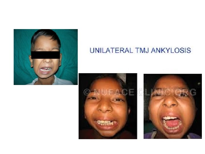
C/F • Bilateral – Micrognathia – Crowding of teeth – Anterior open bite • Unilateral – obvious facial asymmetry – Growth of mandible on affected side is retarded – Deviation of mandible to affected side while opening
• Condylar hyperplasia • Bifid condyle
Traumatic disturbances
1. TMJ Ankylosis • Ankylosis is the stiffening (immobility) or fixation (fusion) of the joint which leads to chronic, painless limitation of the movements of the joint. • Adhesion of joint components by fibrous or bony union leads to loss of function • Can be two types – Intra-articular (true) ankylosis – extra-articular (false) ankylosis. • False ankylosis may be caused by enlargement of the coronoid process, depressed fracture of the zygomatic arch, scarring from surgery, irradiation, infection, etc.
• • • The causes of TMJ Ankylosis Trauma to the joint during an accident, fall etc Trauma during birth, forceps delivery etc Congenital or birth defect Disease or infection in the joint, ear infection etc Enlargement of the coronoid process Depressed fracture of the zygomatic arch, condylar neck fracture etc Surgery to or around the joint Rheumatoid arthritis Ankylotic conditions such as ankylosing spondylitis Destruction of the joint cartilage inflammatory process in TMJ region
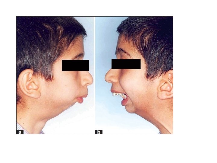
The problems associated with TMJ Ankylosis Functional – Restricted jaw movements – `Inadequate masticatory function – `Restricted mouth opening – `Inhibited facial and physical growth – `Impaired speech • Aesthetic (Cosmetic), – Reduced growth of mandible resulting in micrognathia & “Bird Face” – Facial asymmetry if only one side is affected – Deviation of jaw to ankylosed side – Malaligned teeth because of lack of space for the eruption of the normal component of teeth, Increased over jet • Other emotional, social and psychological disturbances
Radiographic features • Abnormal or irregular shape of condyle • Radiopacity indicationg dense bone filling the joint space. • Prominent antigonial angle.
Treatment for TMJ ankylosis • Surgical correction
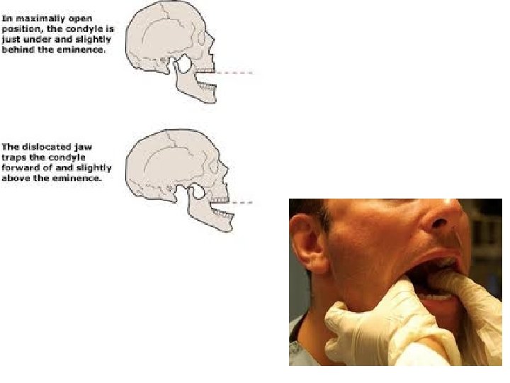
2. Dislocation • Dislocation of condyle from glenoid fossa • Anterior dislocation – More common condyle moves anteriorly over articular eminence to such aposition that it can not be returned voluntarily to normal position – Further divided to Luxation & subluxation • Cranial dislocation – Dislocation of condyle posteriorly to cranial fossa due to trauma
2. Luxation & subluxation • Luxation – refers to complete dislocation of TMJ with head of the condyle moves anteriorly over the articular eminence into such a position that it cannot be returned to voluntarily to its normal position. • Luxation can be caused due to traumatic injury or is a result of yawning or opening mouth too wide for dental procedures etc.
• Luxation is characterized by sudden locking & immobilization of the jaw when the mouth is open, accompanied by prolonged spasmodic contraction of masticatory muscles with protrusion of mandible. • Patient will not be able to close the mouth & all activities such as talking, eating are impossible.
Subluxation – refers to partial or incomplete dislocation of TMJ, where the condyle may lie well anterior to the articular eminence. • Such anterior positioning is normal for many people. • This is a type of hypermobility of joint Treatment • Reduction of dislocated condyle by guiding the condyle by inferior & posterior pressure.
Inflammatory disturbances of TMJ • Osteoarthritis- degenerative joint diseases • Rheumatoid arthritis • Septic(infectious) arthritis
Synovial chondromatosis/ (Loose joint bodies) • Rare benign condition where nodular cartilagenous or osteocartilagenous entities proliferating in synovium
Tempero-mandibular disorder(TMD) • Tempero-mandibular syndrome • Second most common cause of facial pain(after tooth ache) • TMD secondary to Myofacial pain dysfunction – Pain without noticeable destructive change in TMJ • TMD secondary to true articular disease
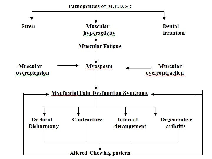
MYOFASCIAL PAIN DYSFUNCTION SYNDROME • (TMJ pain dysfunction syndrome) • It is defined as psychophysiologic disorder that involves the masticatory muscles and is characterized by dull, aching and radiating pain that is exacerbated by mandibular function, tenderness on muscle palpation and limited movement of joint.
Causes of MPDS • • Bilateral loss of posterior teeth Excessive alveolar bone resorption in patients with complete dentures Parafunctional habits like bruxism or clenching Malocclusion Improperly occluding restorations Psychosomatic causes- personality disorders, stress, anxiety
LASKIN – “psycho-physiologic theory”
Clinical features Gender- Females are more commonly(4 times more) Age-20 -40 years. Symptoms • pain of unilateral origin, preauricular region radiating to head • The pain is severe in the morning and worsens as the day progresses. • Masseter is most frequently involved and the patient refers to the pain as a jaw ache. • Temporalis muscle is next commonly involved muscle and produces pain on the side of the head. • Involvement of the lateral pterygoid muscle causes an ear ache and/or pain behind the eye. • Medial pterygoid muscle involvement causes discomfort while swallowing and the feeling of a painful gland behind the angle of mandible.
Cardinal signs of MPDS Positive Signs • Pain • Muscle tenderness • Limitation of mouth opening, unilaterally of bilaterally in approximately an equal ratio, sometimes with deviation of the jaw to the affected side. • Clicking or popping sound or crepitations on opening and closing the jaws.
Negative signs • Patients with M. P. D. S usually have absence of clinical, radiographic and biochemical evidence of organic change in the joint. • Lack of pain while palpating when TMJ is palpated through the external auditory meatus
Differential diagnosis a. Otitis media b. Trigeminal neuralgia c. Myositis ossificans d. Parotitis e. Eagle's syndrome f. Pericoronitis
Diagnosis of M. P. D. S a. Clinical examination b. Radiographs to rule out other diseases c. Sonography and colour doppler tests
Management of M. P. D. S 1. Pharmacological Management a. N. SAID, Steroids, Local anaesthesia, Muscle relaxants, Antidepressants etc 2. Psychiatric Therapy a. Biofeedback Therapy, b. Relaxation therapy 3. Physical Medicine a. Heat, b. Cryotherapy, c. Counterirritants, d. Therapeutic exercises, e. Joint mobilization f. Ultrasound etc.
Important questions • Essay 7 Marks • Classify fibro-osseous lesions of jaw bones. Discuss in detail fibrous dysplasia *** (2+5=7 marks) • Classify fibro-osseous lesions of jaw bones. Discuss in detail Paget’s disease ** (2+5=7 marks)
Short essays 5 Marks • Cleidocranial dysplasia/ Dysostosis** • Radiographic &histopathological features of Paget’s disease *** • Radiographic&histopathological features of Fibrous dysplasia *** • Cherubism*** • Myofacial pain dysfunction syndrome *** • Osteogenesisimperfect* • TMJ Ankylosis *
Short notes • • • • Blue sclera *** Treacher Collins syndrome ** Pierre Robin syndrome ** Marfan’ssyndrome ** Cotton wool appearance *** Ground glass appearance *** Chinese letter pattern *** Albright’s syndrome *** Café-Au- Lait spots *** Down’s syndrome * Achondroplasia* Luxation and sublaxation ** Dislocation ** Brittle bone * Marble bone disease * Mosaic pattern *** 2 Marks
Objective type questions 1 Mark • Blue sclera is a characteristic finding in osteogenesisimperfecta* • Pierre Robin syndrome is characterized by micrognathia, cleft palate and glossoptosis* • Cotton wool appearance characteristic radiographic finding in Paget’s disease*** • Ground glass appearance characteristic radiographic finding in fibrous dysplasia *** • Chinese letter pattern characteristic histologic finding in fibrous dysplasia *** • Café-Au- Lait spots characteristic cutaneous manifestation in Polyostotic fibrous dysplasia and neurofibromatosis ** • “Floating tooth “ phenomenon is a radiological feature of Cherubism** • Marble bone disease is also called as Osteopetrosis ** • Brittle bone is seen in osteogenesisimperfecta ** • Mandibulofacialdyostosis is other name of Treacher Collins syndrome ** • Jigsaw puzzle and mosaic pattern is a feature seen in Pagets disease *** • Trisomy 21 syndrome is also known as Down’s syndrome ** • Bird or fish like face is a feature seen is. Mandibulofacialdyostosis**