Temporomandibular joint Types of Joints Fibrous Two bones

Temporomandibular joint

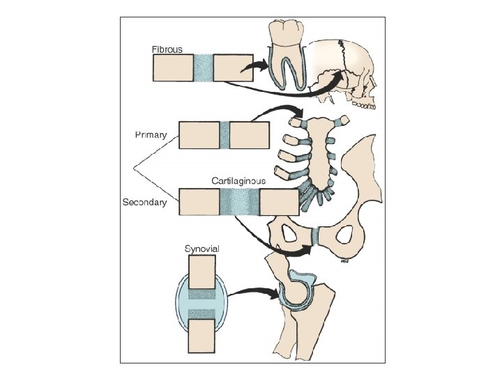
Types of Joints • Fibrous – Two bones connected with fibrous tissue – Limited movement Examples suture (little or no movement) gomphosis (tooth - PDL - bone) syndesmosis (fibula & tibia, radius and ulna; interosseous ligament)

Types of Joints • Cartilagenous – Little or no movement – Primary • Bone-Cartilage (costochondral junction) – Secondary • Bone-Cartilage-FT-Cartilage-Bone (pubic symphysis)

Types of Joints • Synovial – Two bones; each articular surface covered with hyaline cartilage in most cases – The bones are united with a capsule (joint cavity) In the capsule there is presence of synovial fluid – The capsule is lined by a synovial membrane In many synovial joints there maybe an articular disk – Synovial joints are characterized by the presence of ligaments – Movement affected by the muscles – Same innervation of muscle and joint

Synovial joints are classified according to the • Number of axes of bone movement: uniaxial, biaxial, multiaxial • Shapes of articulating surfaces: planar, ginglymoid (=hinged), pivot, condyloid
")
The temporomandibular joint is a synovial, slidingginglymoid joint (humans)

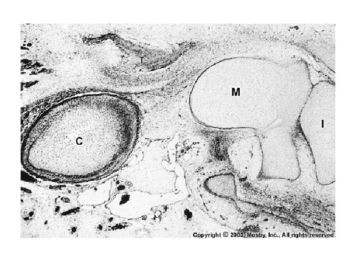
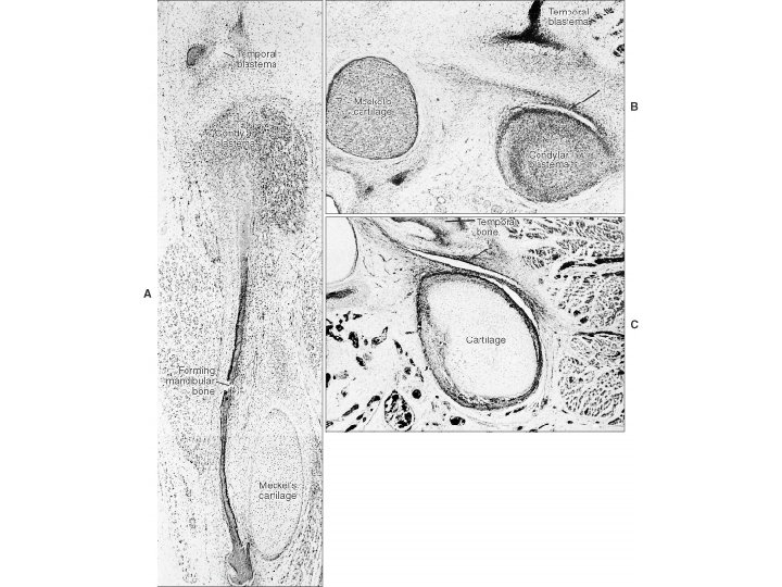
Embryology of the TMJ • Primary TMJ: Meckel's cartilage --> malleus & incal cartilage. It lasts for 4 months • Secondary TMJ: Starts developing around the third month of gestation Two blastemas (temporal and condylar); condylar grows toward the temporal (temporal appears and ossifies first)

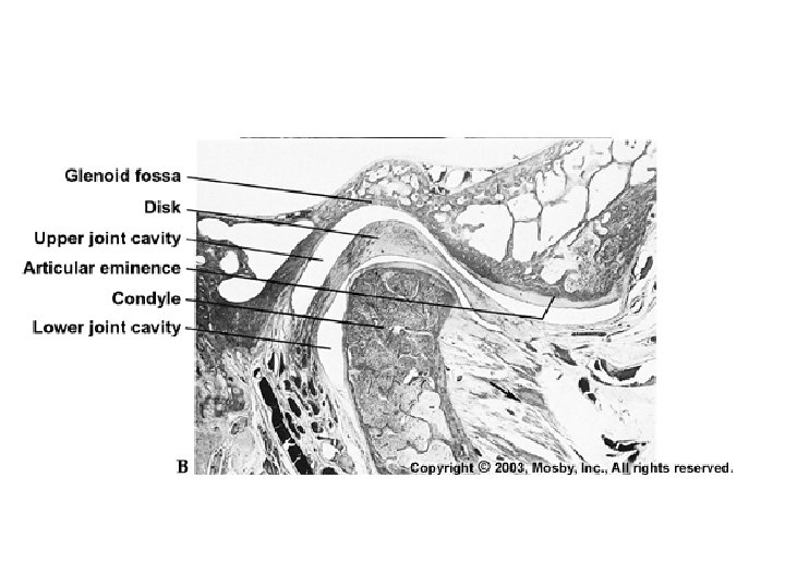
Secondary TMJ • Formation of two cavities: inferior and upper • Appearance of disk • Bones: glenoid fossa (temporal bone) and condyle (mandible) • ARTICULAR SURFACES COVERED BY FIBROUS TISSUE – TMJ is an exception from other synovial joints • acromio- and sternoclavicular joints
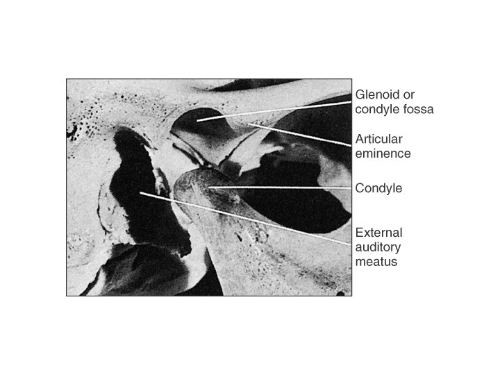

Glenoid Fossa • • Medially is the sphenoid process Laterally is the zygomatic process Anteriorly is the articular eminence Posteriorly are the squamotympanic and petrotympanic fissures (temporal bone)
 –")
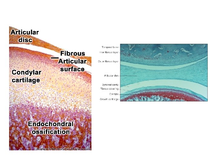
Histologic topography • Fibrous layer: collagen type I, avascular (self -contained and replicating) – • • Proliferating zone that formes condylar cartilage Condylar cartilage is fibrocartilage that does not play role in articulation, no formal function – • Lamina splendens Growth site Capsule: dense collagenous tissue (includes the articular eminence)

Condylar cartilage • Absence of ordered cartilagenous cell columns • Multidirectional proliferation
;")
Histologic topography • Synovial membrane: lines capsule (does not cover disk except posterior region); contains folds (increase in # in pathologic conditions) and villi Two layers: a cellular intima (synovial cells in fiber-free matrix) and a vascular subintima Synovial cells A (macrophage-like) Phagocytosis B (fibroblast-like) add hyaluronate and other proteins in the fluid
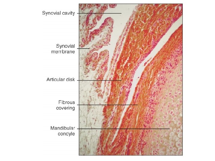

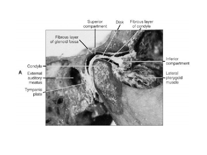
Histologic topography • Synovial fluid: plasma with mucin and proteins, cells Liquid environment: lubrication, ? nutrition • Disk: separates the cavity into two compartments, type I collagen – anterior and posterior portions – anteriorly it divides into two lamellae one towards the capsule, the other towards the condyle – vascular in the periphery, avascular in the center

Histologic topography • Ligaments: nonelastic collagenous structures. One ligament worth mentioning is the lateral or temporomandibular ligament. This restricts displacement of the mandible in three different planes – Lateral and medial dislocation – Inferior displacement (oblique component) – Posterior displacement (horizontal component) • 2 other ligaments (no functional role)

1 Glenoid fossa 2 Superior synovial cavity 3 Articular disc 4 Inferior synovial cavity 5 Condylar head

1 Articular disc 2 Synovial cavity 3 Fibrous zone 4 Proliferative zone & Fibrocartilage 5 Cartilage

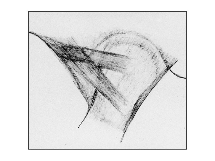
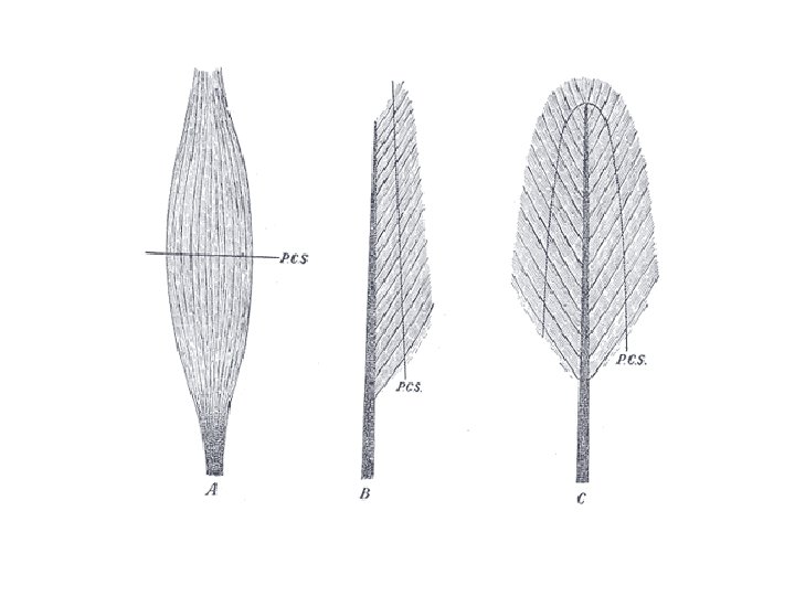
Muscles • Strap muscles – Fasciculi parallel • Fusiform muscles – Bundles converge in the areas of origin and insertion • Fan-shaped muscles • Unipennate and bipennate muscles

Motor unit • Innervation of muscles achieved through motor end plate

Two other neuronal structures for muscle contraction 1. Muscle spindle Encapsulated proprioreceptor Detects changes in length Intrafusal fibers 2. Golgi tendon organ Junction between muscle and tendons Smaller than spindles

Muscle Spindle Intrafusal fibers assume two forms: a. Nuclear bag fiber b. Nuclear chain fiber Nuclear bag fiber innervated by primary afferent that spirals around the bag Nuclear chain fiber innervated y primary afferent in the central portion and a secondary terminal on either side Primary afferent: degree and rate of stretch Secondary terminal: only degree of stretch

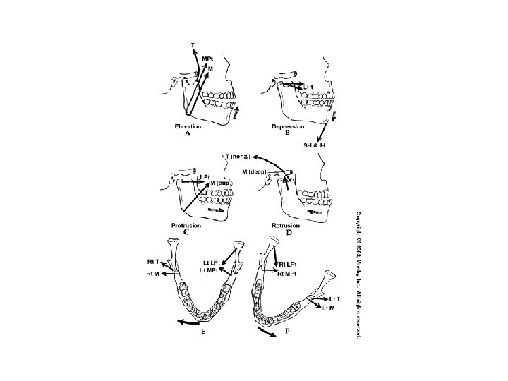
Muscles of mastication bi Retractor Elevator Disk control Closing Opening multi

Function of muscles • Mas. , Med. Pter. , ant. Temp, upper head of Lat. Pter closing • Inferior head of Lat. Pter. , anterior belly digastric, mylohyoid opening • Inf head of Lat. Pter and the elevator group protrusion • Posterior Temp and elevator group retrusion • Elevator, post Temp retrusion working side • Elevator, Lat. Pter protrusion non-working side

Innervation Ruffini Posture Dynamic and static balance Pacini Movement Dynamic mechanoreception accelerator Golgi Static mechanoreception Protection (ligament) Free Pain Protection joint

Blood supply • External carotid – Superficial temporal – Anterior tympanic – Ascending pharyngeal – Deep auricular
- Slides: 35