Diagnostic Methods in Virology Mgr Ludk Eyer Ph


- Slides: 49
Diagnostic Methods in Virology Mgr. Luděk Eyer, Ph. D. Veterinary Research Institute, Brno Department of Virology Emerging Viral Diseases
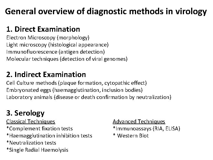
General overview of diagnostic methods in virology 1. Direct Examination Electron Microscopy (morphology) Light microscopy (histological appearance) Immunofluorescence (antigen detection) Molecular techniques (detection of viral genomes) 2. Indirect Examination Cell Culture methods (plaque formation, cytopathic effect) Embryonated eggs (haemagglutination, inclusion bodies) Laboratory animals (disease or death confirmation by neutralization) 3. Serology Classical Techniques *Complement fixation tests *Haemagglutination inhibition tests *Neutralization tests *Single Radial Haemolysis Advanced Techniques *Immunoassays (RIA, ELISA) * Western Blot
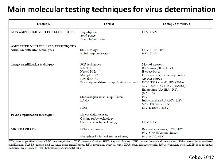
Main molecular testing techniques for virus determination Cobo, 2012
Why? • Virus identification • Virus subtyping • Virus genotypization • Identification of drug-resistant mutants more effective therapy
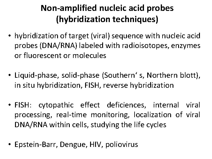
Non-amplified nucleic acid probes (hybridization techniques) • hybridization of target (viral) sequence with nucleic acid probes (DNA/RNA) labeled with radioisotopes, enzymes or fluorescent or molecules • Liquid-phase, solid-phase (Southern‘ s, Northern blott), in situ hybridization, FISH, reverse hybridization • FISH: cytopathic effect deficiences, internal viral processing, real-time monitoring, localization of viral DNA/RNA within cells, studying the life cycles • Epstein-Barr, Dengue, HIV, poliovirus
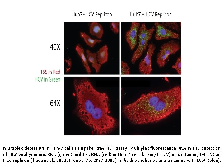
Multiplex detection in Huh-7 cells using the RNA FISH assay. Multiplex fluorescence RNA in situ detection of HCV viral genomic RNA (green) and 18 S RNA (red) in Huh-7 cells lacking (-HCV) or containing (+HCV) an HCV replicon (Ikeda et al. , 2002, J. Virol. , 76: 2997 -3006). In both panels, nuclei are stained with DAPI (blue).
Amplified nucleic acid techniques Signal amplification techniques • b. DNA assays • hybrid capture assays Target amplification techniques • PCR techniques • transcription-based amplification methods • strand displacement amplification Probe amplification techniques • cleavase-invander technology • ligase chain reaction • cycling probe technology
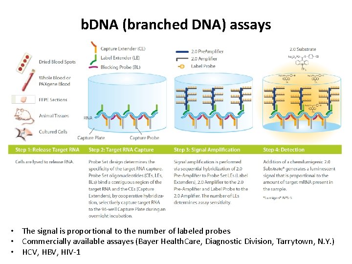
b. DNA (branched DNA) assays • The signal is proportional to the number of labeled probes • Commercially available assayes (Bayer Health. Care, Diagnostic Division, Tarrytown, N. Y. ) • HCV, HBV, HIV-1
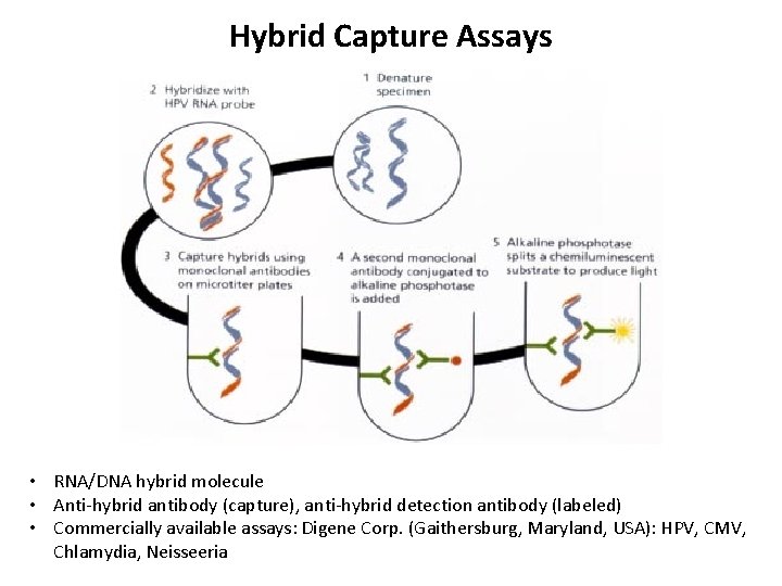
Hybrid Capture Assays • RNA/DNA hybrid molecule • Anti-hybrid antibody (capture), anti-hybrid detection antibody (labeled) • Commercially available assays: Digene Corp. (Gaithersburg, Maryland, USA): HPV, CMV, Chlamydia, Neisseeria
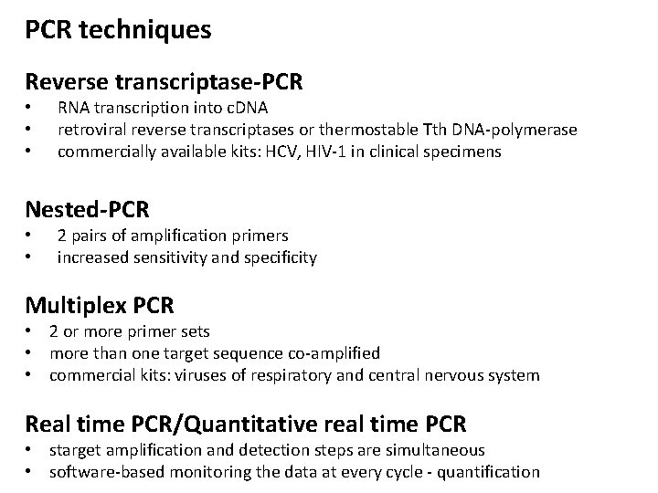
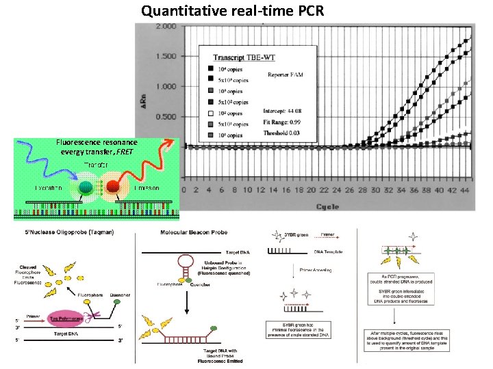
PCR techniques Reverse transcriptase-PCR • • • RNA transcription into c. DNA retroviral reverse transcriptases or thermostable Tth DNA-polymerase commercially available kits: HCV, HIV-1 in clinical specimens Nested-PCR • • 2 pairs of amplification primers increased sensitivity and specificity Multiplex PCR • 2 or more primer sets • more than one target sequence co-amplified • commercial kits: viruses of respiratory and central nervous system Real time PCR/Quantitative real time PCR • starget amplification and detection steps are simultaneous • software-based monitoring the data at every cycle - quantification
Quantitative real-time PCR Fluorescence resonance evergy transfer, FRET
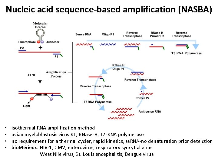
Nucleic acid sequence-based amplification (NASBA) • • isothermal RNA amplification method avian myeloblastosis virus RT, RNase-H, T 7 -RNA polymerase no requirement for a thermal cycler, rapid kinetics, ss. RNA-no denaturation prior deteiction bio. Mérieux: HIV-1, CMV, enterovirus, respiratory syncytial virus West Nile virus, St. Louis encephalitis, Dengue virus
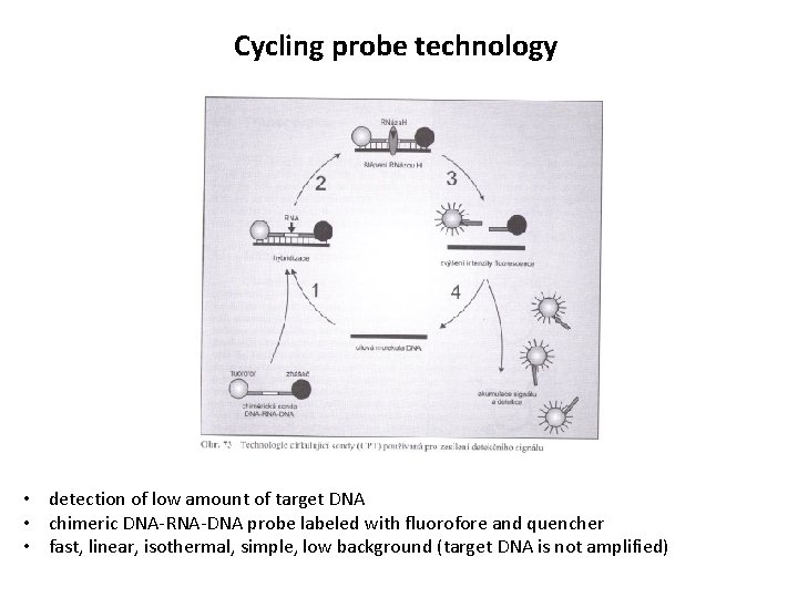
Cycling probe technology • detection of low amount of target DNA • chimeric DNA-RNA-DNA probe labeled with fluorofore and quencher • fast, linear, isothermal, simple, low background (target DNA is not amplified)
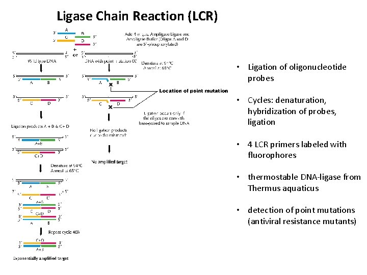
Ligase Chain Reaction (LCR) • Ligation of oligonucleotide probes • Cycles: denaturation, hybridization of probes, ligation • 4 LCR primers labeled with fluorophores • thermostable DNA-ligase from Thermus aquaticus • detection of point mutations (antiviral resistance mutants)
Isothermal amplification methods • • simple operation, rapid reaction, easy detection not require thermal cycler performed in a heating block or water bath single uniform temperature • Loop-Mediated Isothermal Amplification (LAMP) • Helicase-Dependent Amplification (HDA) • LAMP: Dengue, SARS, influenza A/B, CMV, HSV, VZV. . . • HDA: HIV-1 in human plasma, HSV 1 and 2 from genital lesions
Amplification methods: advantages and drawbacks • high sensitivity • rapid methods • cost-effective diagnostic technques • can not be used for identification of new viruses • not convenient for viruses showing huge genetic variability • reverse transcription step of forming c. DNA is not efficient enough (efficacy ~ 20%)
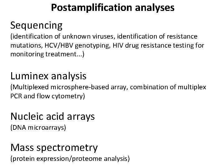
Postamplification analyses Sequencing (identification of unknown viruses, identification of resistance mutations, HCV/HBV genotyping, HIV drug resistance testing for monitoring treatment. . . ) Luminex analysis (Multiplexed microsphere-based array, combination of multiplex PCR and flow cytometry) Nucleic acid arrays (DNA microarrays) Mass spectrometry (protein expression/proteome analysis)
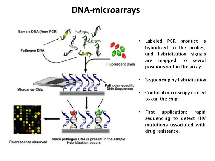
DNA-microarrays • Labeled PCR product is hybridized to the probes, and hybridization signals are mapped to sevral positions within the array. • Sequencing by hybridization • Confocal microscopy is used to can the chip. • First application: rapid sequencing to detect HIV mutations associated with drug resistance.
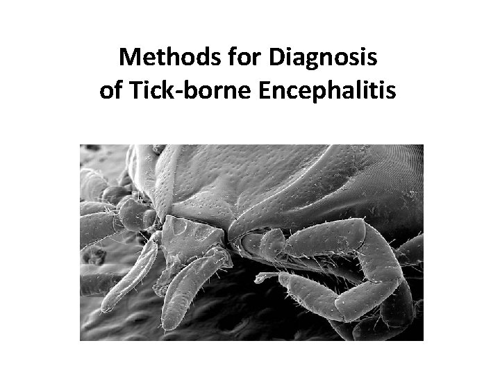
Methods for Diagnosis of Tick-borne Encephalitis
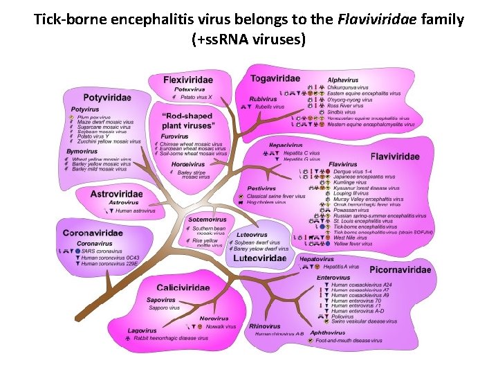
Tick-borne encephalitis virus belongs to the Flaviviridae family (+ss. RNA viruses)
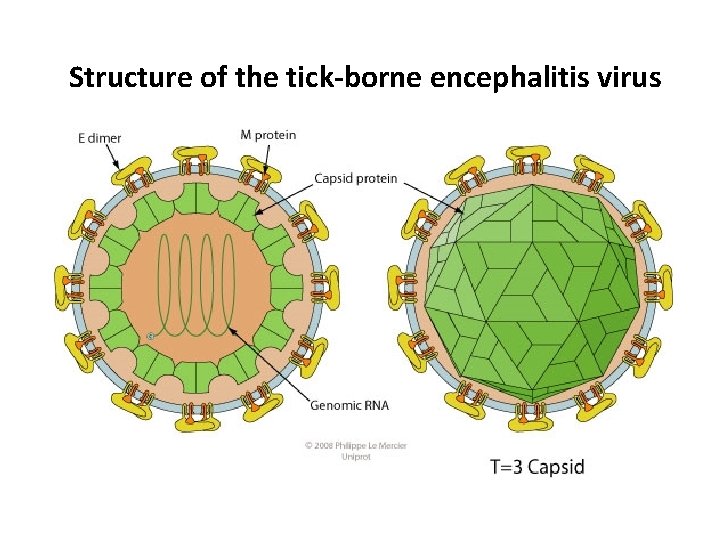
Structure of the tick-borne encephalitis virus
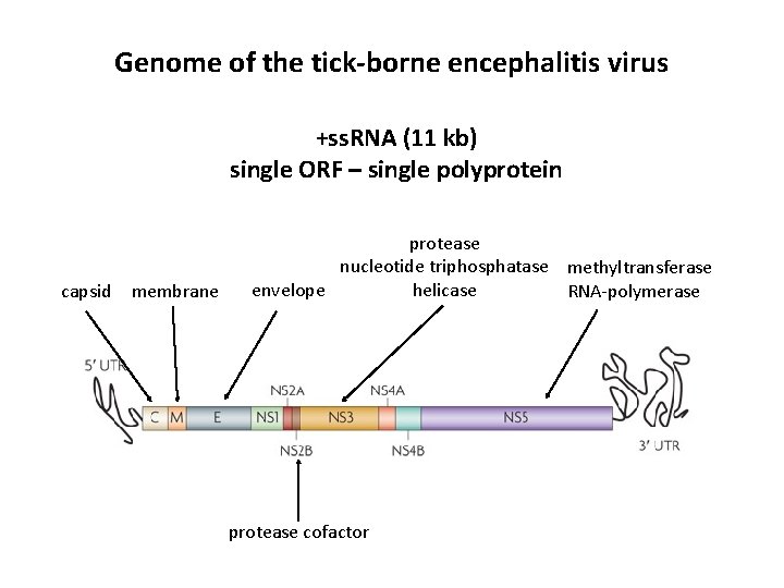
Genome of the tick-borne encephalitis virus +ss. RNA (11 kb) single ORF – single polyprotein capsid membrane protease nucleotide triphosphatase methyltransferase helicase envelope RNA-polymerase protease cofactor
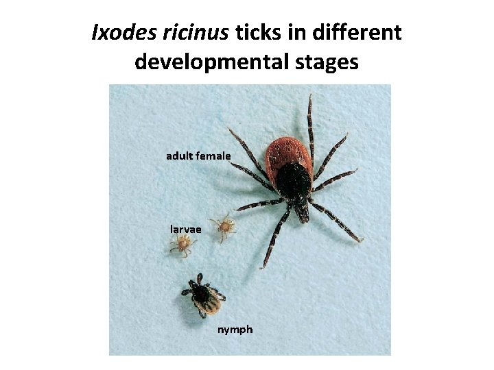
Ixodes ricinus ticks in different developmental stages adult female larvae nymph
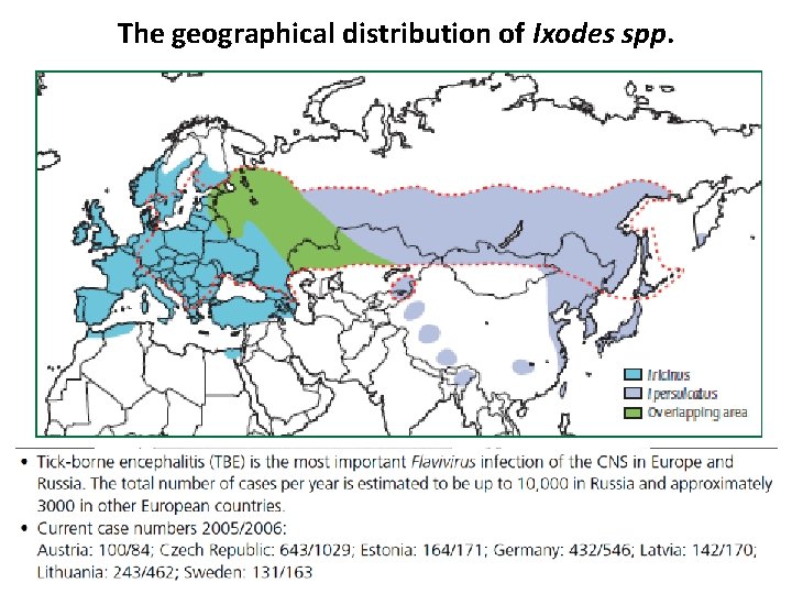
The geographical distribution of Ixodes spp.
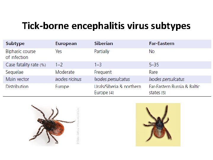
Tick-borne encephalitis virus subtypes
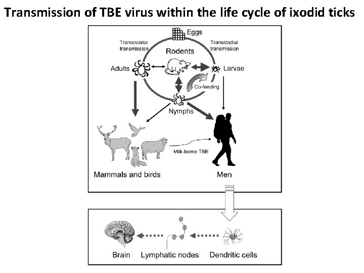
Transmission of TBE virus within the life cycle of ixodid ticks
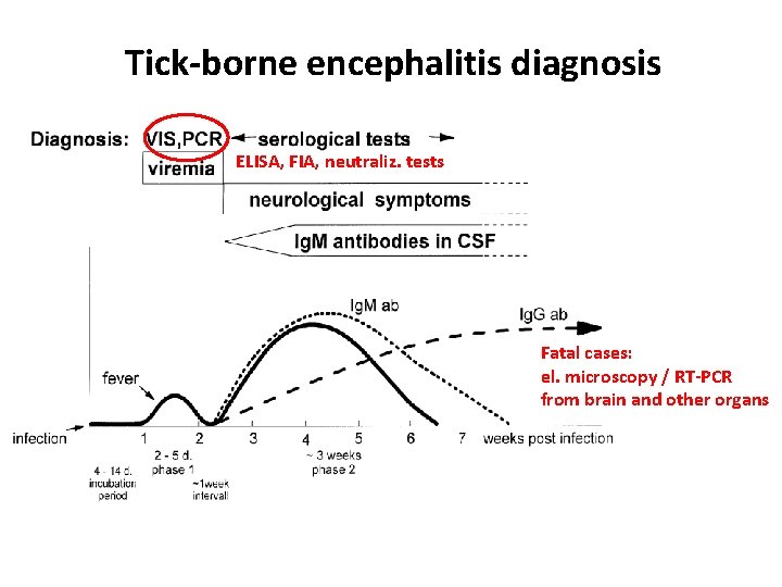
Tick-borne encephalitis diagnosis ELISA, FIA, neutraliz. tests Fatal cases: el. microscopy / RT-PCR from brain and other organs
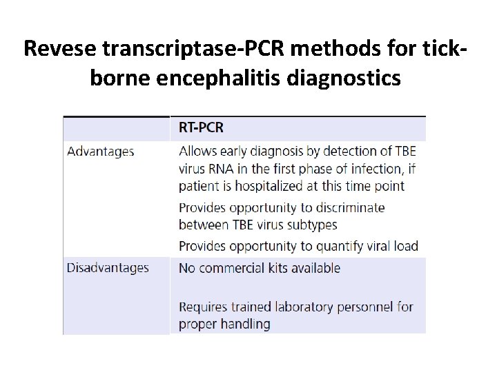
Revese transcriptase-PCR methods for tickborne encephalitis diagnostics
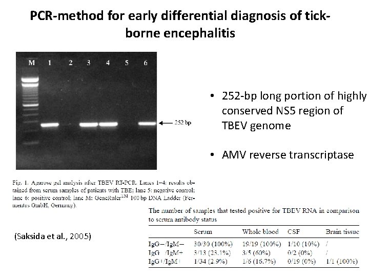
PCR-method for early differential diagnosis of tickborne encephalitis • 252 -bp long portion of highly conserved NS 5 region of TBEV genome • AMV reverse transcriptase (Saksida et al. , 2005)
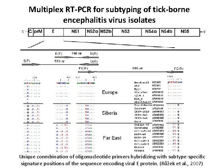
Multiplex RT-PCR for subtyping of tick-borne encephalitis virus isolates Unique conmbination of oligonucleotide primers hybridizing with subtype-specific signature positions of the sequence encoding viral E protein. (Růžek et al. , 2007)
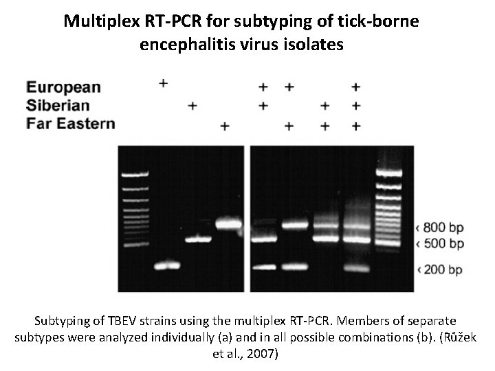
Multiplex RT-PCR for subtyping of tick-borne encephalitis virus isolates Subtyping of TBEV strains using the multiplex RT-PCR. Members of separate subtypes were analyzed individually (a) and in all possible combinations (b). (Růžek et al. , 2007)
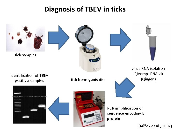
Diagnosis of TBEV in ticks tick samples identification of TBEV positive samples tick homogenisation virus RNA isolation QIAamp RNA kit (Qiagen) PCR amplification of sequence encoding E protein (Růžek et al. , 2007)
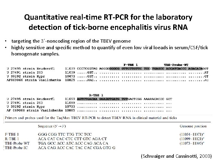
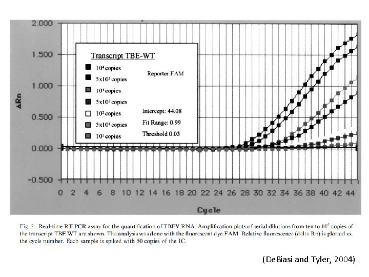
Quantitative real-time RT-PCR for the laboratory detection of tick-borne encephalitis virus RNA • targeting the 3´-noncoding region of the TBEV genome • highly sensitive and specific method to quantify of even low viral loeads in serum/CSF/tick homogenate samples. (Schwaiger and Cassinotti, 2003)
(De. Biasi and Tyler, 2004)
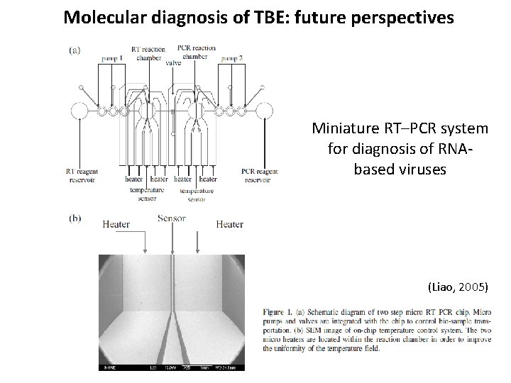
Molecular diagnosis of TBE: future perspectives Miniature RT–PCR system for diagnosis of RNAbased viruses (Liao, 2005)
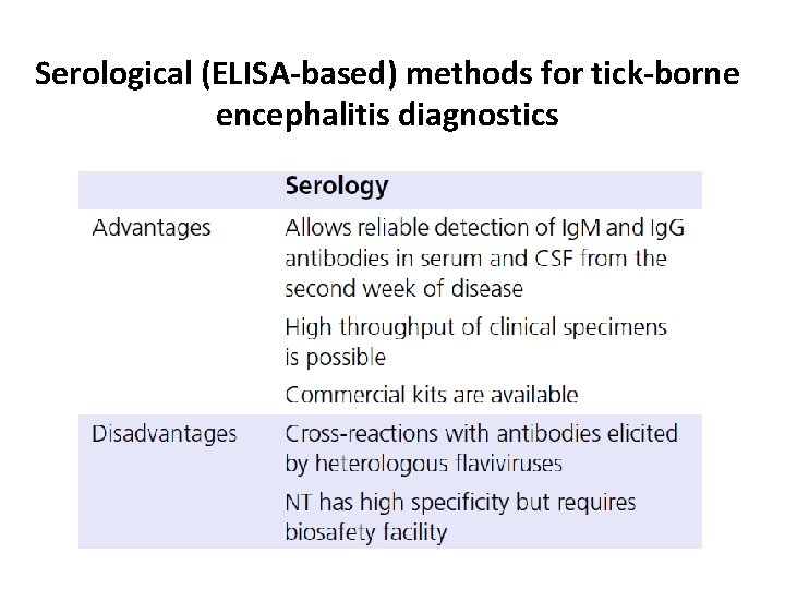
Serological (ELISA-based) methods for tick-borne encephalitis diagnostics
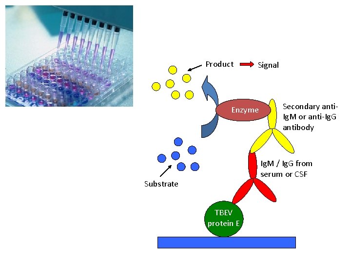
Product Signal Enzyme Secondary anti. Ig. M or anti-Ig. G antibody Ig. M / Ig. G from serum or CSF Substrate TBEV protein E
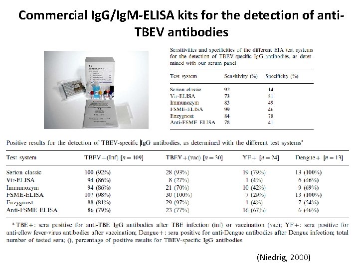
Commercial Ig. G/Ig. M-ELISA kits for the detection of anti. TBEV antibodies (Niedrig, 2000)
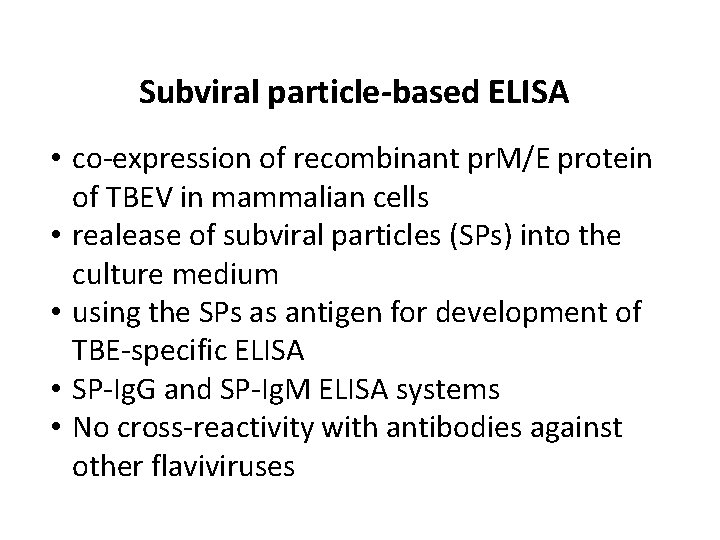
Subviral particle-based ELISA • co-expression of recombinant pr. M/E protein of TBEV in mammalian cells • realease of subviral particles (SPs) into the culture medium • using the SPs as antigen for development of TBE-specific ELISA • SP-Ig. G and SP-Ig. M ELISA systems • No cross-reactivity with antibodies against other flaviviruses
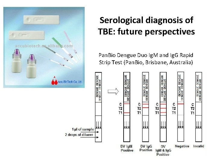
Serological diagnosis of TBE: future perspectives Pan. Bio Dengue Duo Ig. M and Ig. G Rapid Strip Test (Pan. Bio, Brisbane, Australia)
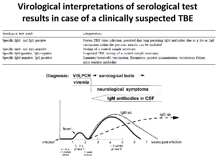
Virological interpretations of serological test results in case of a clinically suspected TBE
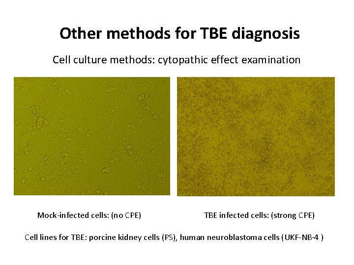
Other methods for TBE diagnosis Cell culture methods: cytopathic effect examination Mock-infected cells: (no CPE) TBE infected cells: (strong CPE) Cell lines for TBE: porcine kidney cells (PS), human neuroblastoma cells (UKF-NB-4 )
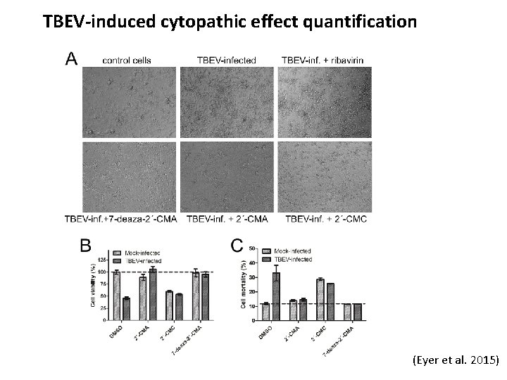
TBEV-induced cytopathic effect quantification (Eyer et al. 2015)
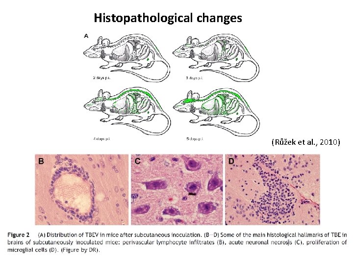
Histopathological changes (Růžek et al. , 2010)
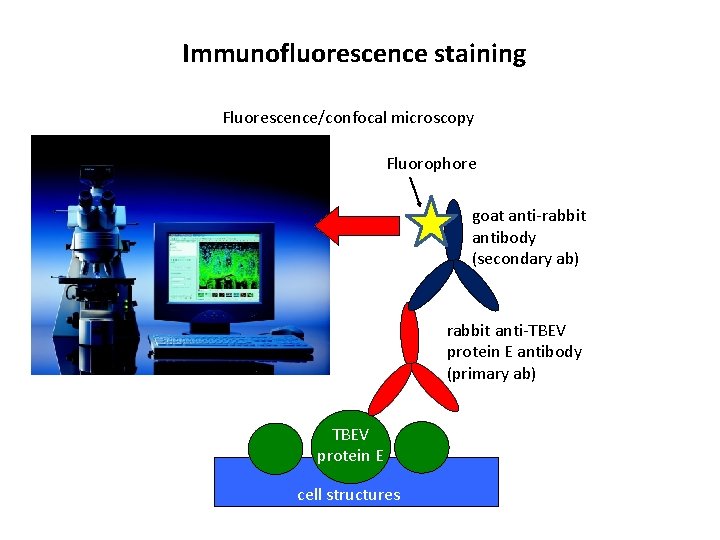
Immunofluorescence staining Fluorescence/confocal microscopy Fluorophore goat anti-rabbit antibody (secondary ab) rabbit anti-TBEV protein E antibody (primary ab) TBEV protein E cell structures
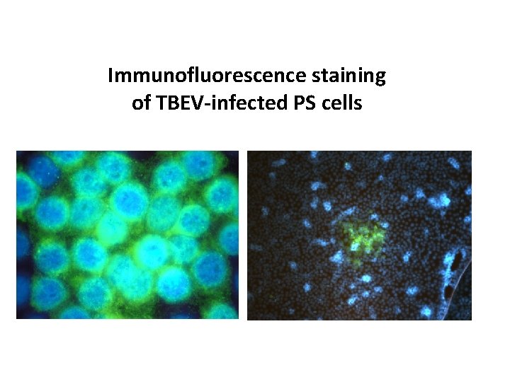
Immunofluorescence staining of TBEV-infected PS cells
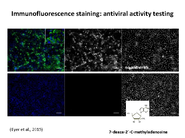
Immunofluorescence staining: antiviral activity testing no antivirals (Eyer et al. , 2015) 7 -deaza-2´-C-methyladenosine
Tick-borne encephalitis diagnosis: conclusion • Currently the diagnosis of TBE is based on serological methods (detection of specific antibodies in serum/whole blood)/CSF from the second week of the disease onwards • Disadvantages: antibody cross-reactivity with other flaviviruses • Molecular detection by PCR could be valuable for the early diagnosis of a specific etiological agent. • Early detection: improving prognosis, introduction of the earlier appropriate therapy