CHRONIC SPECIFIC RHINITIS Dr S Jaya Sandeep NASAL

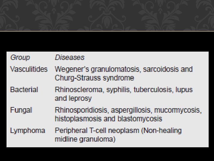


 described this condition as a granulomatous disorder of the nose extending")


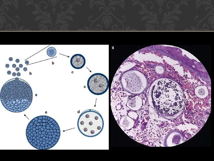
 Development of sporangium. The mature trophocyte then develops into sporangium. A sporangium is")
 Production of endospores. Sporangia filled with endospores develop a high internal pressure and")


- Slides: 38
CHRONIC SPECIFIC RHINITIS Dr S Jaya Sandeep
NASAL SYPHILIS It may manifest in the form of primary, secondary, tertiary or congenital syphilis. Acute primary nasal syphilis It involves the vestibule of nose showing hard painless nodule 3 to 4 weeks after the contact and may disappear spontaneously in 8 to 10 weeks. Venereal diseases research laboratory (VDRL) tests may confirm the diagnosis.
Secondary syphilis of nose It appears 8 to 10 weeks after inoculation and may manifest in the form of simple rhinitis along with crusting and fissuring of nasal vestibule and is suspected when mucous patches appear in pharynx along with lymph nodes. Wassermann’s reaction becomes positive.
Tertiary syphilis It is commonly seen in the nose and lesion found is gumma of the nose invading mucous membrane periosteum and bone leading to sagging of bony bridge of nose and secondary atrophic rhinitis follows. Tenderness of bridge of nose is quite characteristic. Bony septum may show perforation.
Congenital syphilis In infants, snuffles is the most common lesion in the 3 rd week of intrauterine life beginning with catarrhal form of rhinitis and becomes purulent in a short time with excoriation of nasal vestibule ultimately leading to destruction of mucous membrane, periosteum and bone.
Treatment • Antisyphilitic treatment, benzathine penicillin— 2. 4 mega units IM weekly for 3 weeks • Discharge in the nose is removed by nasal toilet or alkaline douche.
TUBERCULOSIS OF NOSE Tuberculosis of nose may be in the form of ulcer or a nodule and affects the cartilaginous part of septum. Examination shows a bright red nodule or ulcer on the septum with rapid course of destruction. Examination of discharge shows tubercle bacilli and biopsy will show typical tubercular lesion. Treatment It is routine antitubercular therapy.
LUPUS VULGARIS Lupus vulgaris is a chronic form of tubercular infection affecting skin and mucous membrane of nose. Female to male ratio is 2 : 1. Symptoms vary from crusting, nose bleed and ulceration with foetor followed by slow progressive fibrosis and contraction causing disfigurement of alae nasi. The 3 B’s are important in diagnosis, i. e. blanching, bacterial examination and biopsy. Apple jelly nodules, on blanching, are typical of lupus vulgaris. Treatment
LEPROSY OF NOSE Leprosy of nose is a granulomatous lesion of nose, which affects mucous membrane, skin and peripheral nerves. It is caused by bacillus Mycobacterium leprae. It is treated with dapsone.
RHINOSCLEROMA Friedmann (1966) described this condition as a granulomatous disorder of the nose extending to nasopharynx and oropharynx It is seen mostly in poor socioeconomic conditions Pathology The disease is caused by Klebsiella rhinoscleromatis (Frisch bacillus). It mainly affects nose, nasopharynx,
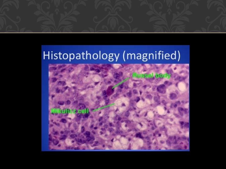
Histological appearance of the biopsy material shows plasma cells, lymphocytes and eosinophils. Mikulicz cells or foam cells and Russell bodies are the characteristic features. Plasma cells get transformed into Russell bodies. There is high content of mucopolysaccharides around the walls of bacilli, which protects it against antibiotics and antibodies
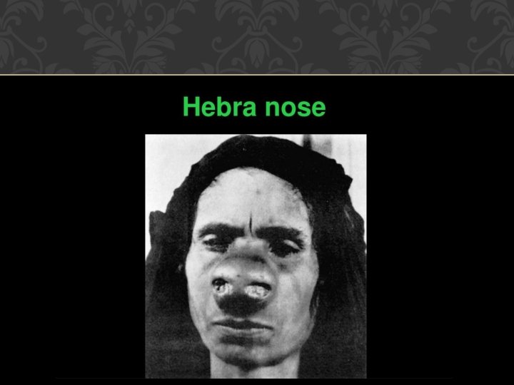
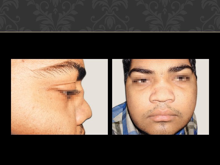
Symptomatology • Stage of congestion and infiltration giving rise to discharge, crusting and foetor • Granulomatous or nodular stage in which bluish red rubbery nodules appear in the anterior part of nose, which later becomes pale and hard • Stage of cicatrization in which there is formation of adhesions, fibrosis, stenosis with distortion of nasal anatomy giving rise to so called Hebra nose. It is also called as Tapir nose. Soft palate deformity, which may
Diagnosis It is confirmed by biopsy and histological features and culture of the bacilli. Treatment i. Ampicillin 2 gm/day for 6 weeks or injection streptomycin or tetracycline or steroids. ii. Radical excision of the mass and repair of the defect
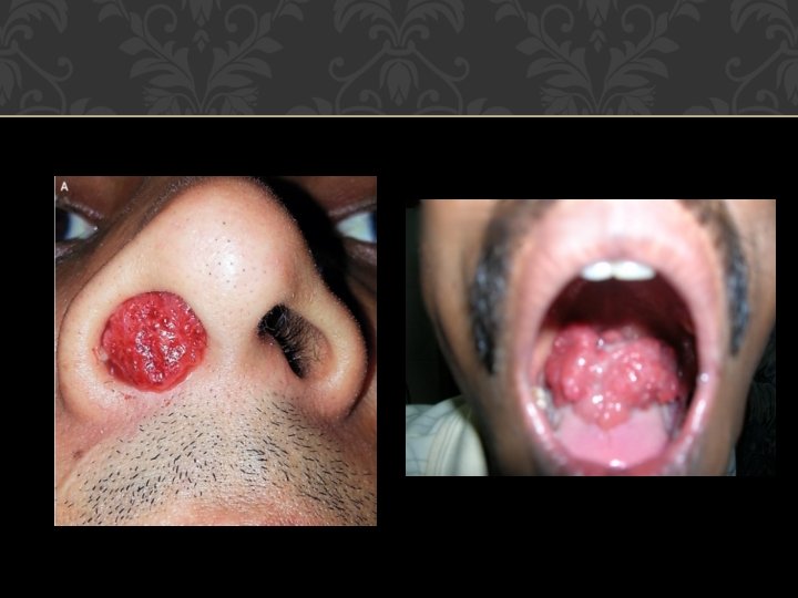
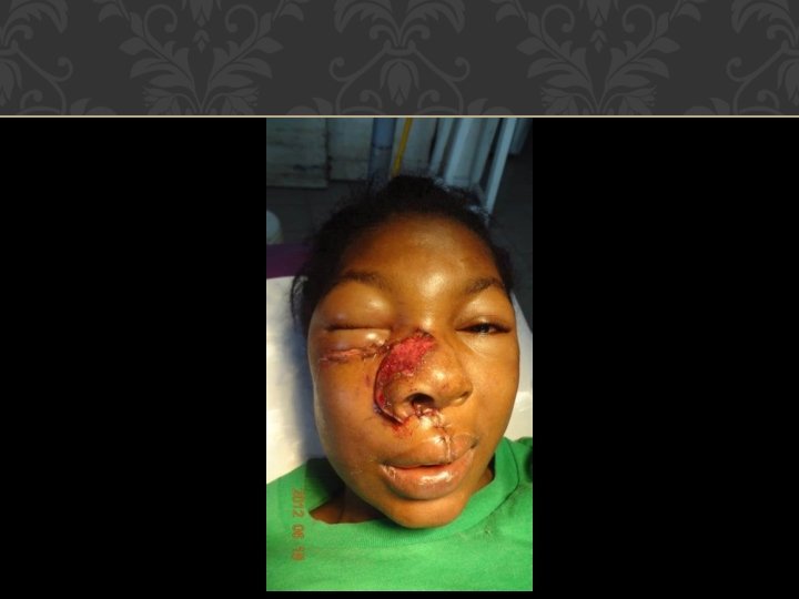
RHINOSPORIDIOSIS Rhinosporidiosis is a fungal infection of the nose caused by Rhinosporidium Seeberi or kinealyi. Young males from South India and Sri Lanka were found to be affected. Mode of infection is by dust or dung of animals, which carries the spores of the fungus. Clinical features The disease mostly affects nose and nasopharynx; other sites such as lip, palate, conjunctiva, epiglottis, larynx, trachea, bronchi, skin, vulva and vagina may also be affected
In the nose, the disease presents as a leafy, polypoidal mass, pink to purple in colour and attached to nasal septum or lateral wall. Sometimes, it extends into the nasopharynx and may hang behind the soft palate. The mass is very vascular and bleeds easily on touch. Its surface is studded with white dots representing the sporangia. In early stages, the patient may complain of nasal discharge which is often blood tinged and nasal stuffiness. Sometimes, frank epistaxis is the only presenting complaint.
Life Cycle Three stages have been recognized in the life cycle of the organism: trophic stage, development of sporangia and production of endospores. (a) Trophic stage. The endospore is oval or rounded, 6– 8 μm in size, clear cytoplasm, vesicular nucleus with a nucleolus and a covering of chitin. It gradually increases in size, begins to divide cytoplasm and nucleus forming small endospores by several divisions. Trophocyte becomes large filled with young endospores.
(b) Development of sporangium. The mature trophocyte then develops into sporangium. A sporangium is 200– 250 μm in diameter, contains 12, 000– 16, 000 endospores. It has a thick wall consisting of two layers: outer chitinous and inner cellulose layer. Endospores mature with the formation of mucoid and chitinous wall. Sporangium filled with thousands of endospores develops a germinal pore ready to burst and liberate the endospores.
(c) Production of endospores. Sporangia filled with endospores develop a high internal pressure and rupture, liberating endospores into the surrounding tissue. If internal pressure is not high, spores are liberated one by one without breaking the wall. After liberation endospores start their life as trophic stage. Some endospores are carried by lymphatic channels to the blood stream to cause disseminated form of disease
Diagnosis This is made on biopsy. It shows several sporangia, oval or round in shape and filled with spores which may be seen bursting through its chitinous wall. Growth of nose, histological picture is typical both in growth and fungal infections. Management i. Wide endoscopic excision of the growth with diathermy and cauterization of the base ii. Medical treatment is not of much use, but dapsone and
CANDIDIASIS Also called moniliasis or thrush. It is caused by Candida albicans. Usually, it occurs in the mouth as white patches, but may be seen in the nose of the patients who have been on prolonged antibiotics, anticancer drugs or marasmic patients. Treatment It is local cleaning and application of nystatin locally or clotrimazole solution.
ACTINOMYCOSIS Actinomycosis is a chronic granulomatous infestation due to Actinomyces or ray fungus and is seen in farmers. Macroscopically, it looks like sulfur granules and microscopically polymorphs, endothelial cells and foreign body giant cells with branching acidophilic fibers radiating out are seen. Nose is usually not involved, but may be infected secondarily from the maxilla or sinuses. Treatment It is with large doses of penicillin for 6 to 8 weeks.
MUCORMYCOSIS Mostly seen in patients with uncontrolled diabetes and it is caused by Mucor family and differs from other fungus by the invasion of walls of blood vessels causing necrosis, ulceration and thrombosis. The patient presents with black necrotic mass in the nasal cavity. Treatment It is treated with amphotericin B and surgical debridement
ASPERGILLOSIS It may be of Aspergillus fumigatus or of niger type and is seen mostly in persons handling small birds. Clinical features A black or greyish membrane is seen in the nasal mucosa. Exploration of maxillary sinus reveals a fungus ball containing semisolid cheesy-white or blackish material. The organisms can be seen on special staining for fungus. Treatment Surgical debridement of the involved tissues and antifungal drugs, e. g. Amphotericin B
SARCOIDOSIS Sarcoidosis is a granulomatous disorder resembling tuberculosis but without caseation of the tubercles. The patient presents with nasal obstruction, nasal bleeding and nasal discharge with lymphadenopathy. Kveim test and biopsy are diagnostic. Treatment It is with steroids, vitamin D and ultraviolet (UV) rays.
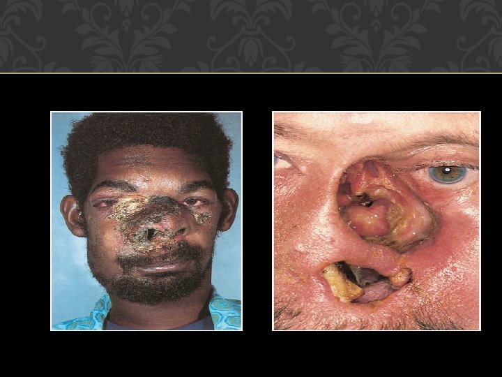
WEGENER’S GRANULOMA Wegener’s granuloma is a systemic condition of unknown etiology, which differs from Stewart’s type by involvement of upper respiratory tract, lungs and kidneys; and in this, destruction of tissues is not so extensive with less tendency of cartilage and bone involvement. Histopathology Multinucleated giant cells closely applied to blood vessels without breakdown of elastic tissue of artery wall unlike giant cell arteritis
Clinical Features Blood-stained discharge, crusting and granulations, cough with hemoptysis, hematoma, saddle nose and perforation of nasal septum may be seen along with fatigue, night sweats and anemia. Investigations • Erythrocyte sedimentation rate (ESR) is raised, eosinophilia may be present • Blood urea and serum creatinine show increased levels • Urine examination shows red cells, casts, albumin • X-ray chest may show single or multiple cavities
Treatment i. Corticosteroids in high doses, they are very effective till healing occurs. 60 mg daily for 8 to 12 weeks. ii. Cytotoxic drugs such as cyclophosphamide and azathioprine have been used with variable success
T-CELL LYMPHOMA Earlier terms used to describe this lesion were midline malignant lesion and polymorphic reticulosis. It is a destructive lesion usually starting on one side of nose involving the upper lip, oral cavity, maxilla and sometimes even extending to orbit. Histologically polymorphic lymphoid tissue with angiocentric and angioinvasive features is seen. There is no vasculitis—a feature typical of Wegener’s granulomatosis.
Unlike Wegener’s granulomatosis, it is rapidly destructive and usually devoid of systemic involvement; there is absence of involvement of lung and kidneys. Immunohistochemical studies of biopsy material are necessary to establish diagnosis of T-cell lymphoma. Localized T-cell lymphoma is treated by radiation while a disseminated disease requires chemotherapy
THANK YOU