
Enterobacteriaceae Klebsiella etc Dr Deepjyoti Kalita MD Ph
Enterobacteriaceae – Klebsiella etc Dr Deepjyoti Kalita MD Ph. D Dept. of Microbiology AIIMS Rishikesh

Learning objective At the end of the session, the students will be able to • Describe morphology and antigens of few important enterobacteriaceae • Describe Pathogenesis & Clinical features • Choose appropriate lab diagnosis and interpret the results • Describe prevention and treatment
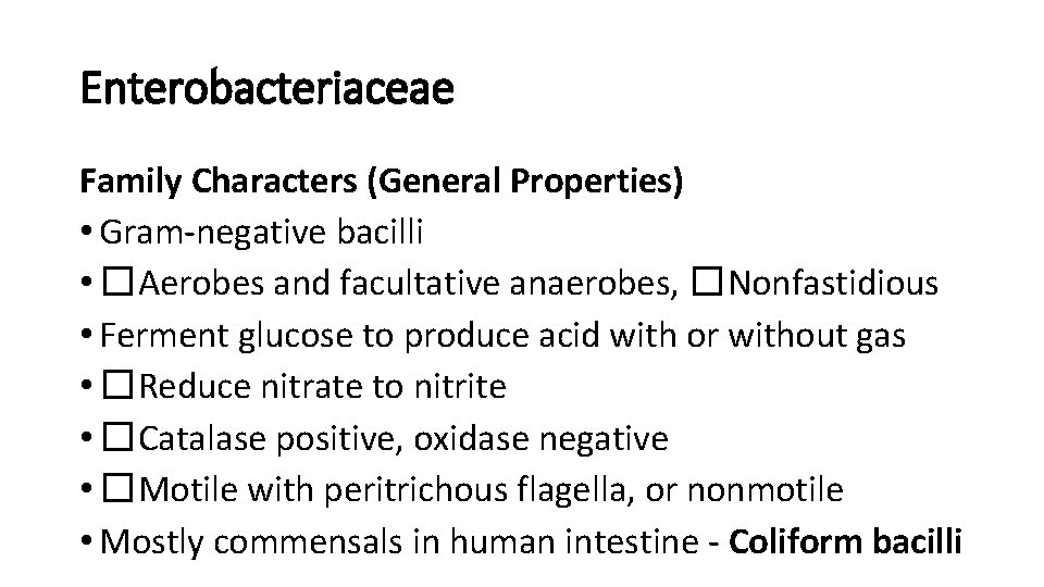
Enterobacteriaceae Family Characters (General Properties) • Gram-negative bacilli • �Aerobes and facultative anaerobes, �Nonfastidious • Ferment glucose to produce acid with or without gas • �Reduce nitrate to nitrite • �Catalase positive, oxidase negative • �Motile with peritrichous flagella, or nonmotile • Mostly commensals in human intestine - Coliform bacilli
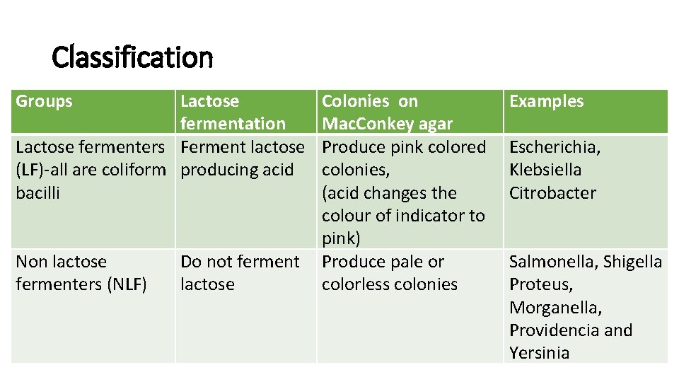
Classification Groups Lactose fermentation Lactose fermenters Ferment lactose (LF)-all are coliform producing acid bacilli Non lactose fermenters (NLF) Do not ferment lactose Colonies on Mac. Conkey agar Produce pink colored colonies, (acid changes the colour of indicator to pink) Produce pale or colorless colonies Examples Escherichia, Klebsiella Citrobacter Salmonella, Shigella Proteus, Morganella, Providencia and Yersinia
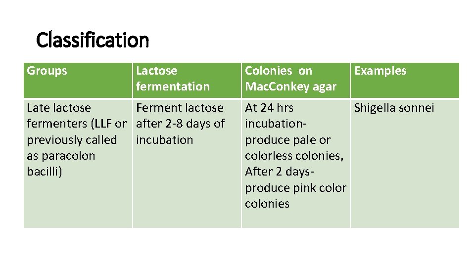
Classification Groups Lactose fermentation Late lactose Ferment lactose fermenters (LLF or after 2 -8 days of previously called incubation as paracolon bacilli) Colonies on Mac. Conkey agar Examples At 24 hrs Shigella sonnei incubationproduce pale or colorless colonies, After 2 daysproduce pink color colonies
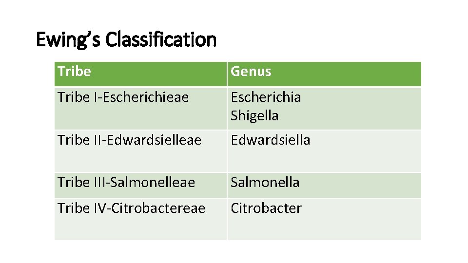
Ewing’s Classification Tribe Genus Tribe I-Escherichieae Escherichia Shigella Tribe II-Edwardsielleae Edwardsiella Tribe III-Salmonelleae Salmonella Tribe IV-Citrobactereae Citrobacter
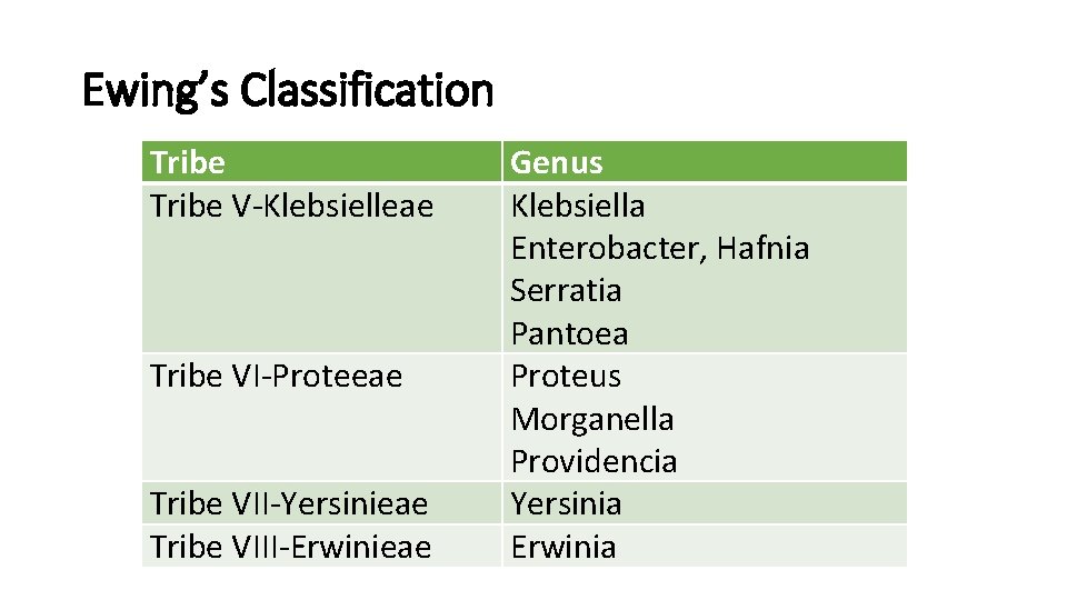
Ewing’s Classification Tribe V-Klebsielleae Tribe VI-Proteeae Tribe VII-Yersinieae Tribe VIII-Erwinieae Genus Klebsiella Enterobacter, Hafnia Serratia Pantoea Proteus Morganella Providencia Yersinia Erwinia
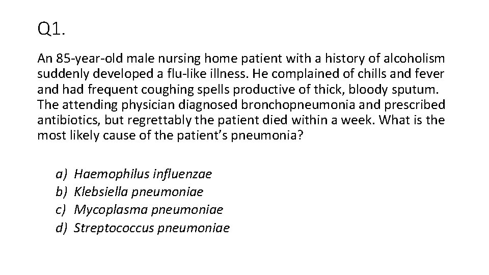
Q 1. An 85 -year-old male nursing home patient with a history of alcoholism suddenly developed a flu-like illness. He complained of chills and fever and had frequent coughing spells productive of thick, bloody sputum. The attending physician diagnosed bronchopneumonia and prescribed antibiotics, but regrettably the patient died within a week. What is the most likely cause of the patient’s pneumonia? a) b) c) d) Haemophilus influenzae Klebsiella pneumoniae Mycoplasma pneumoniae Streptococcus pneumoniae
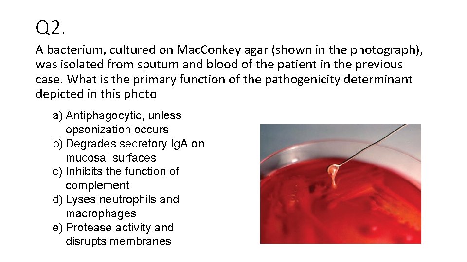
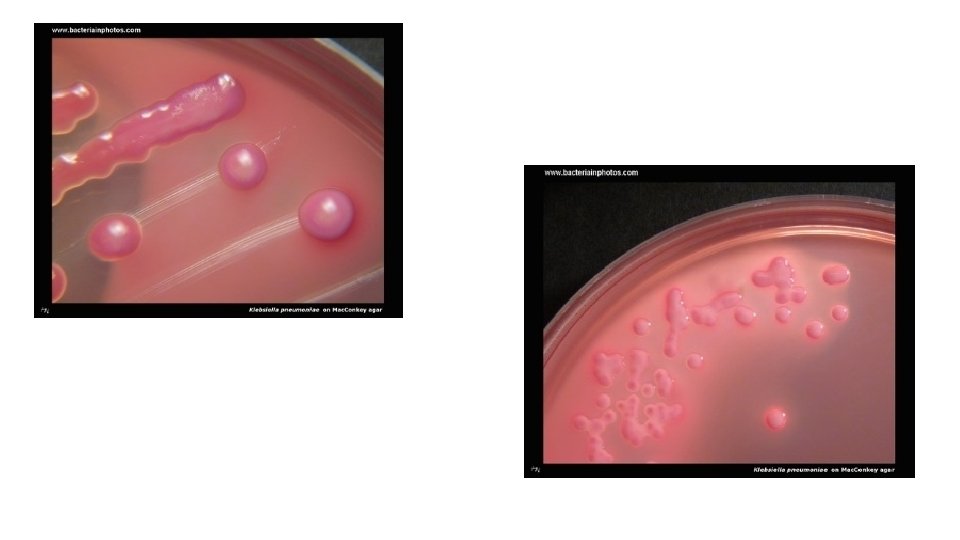
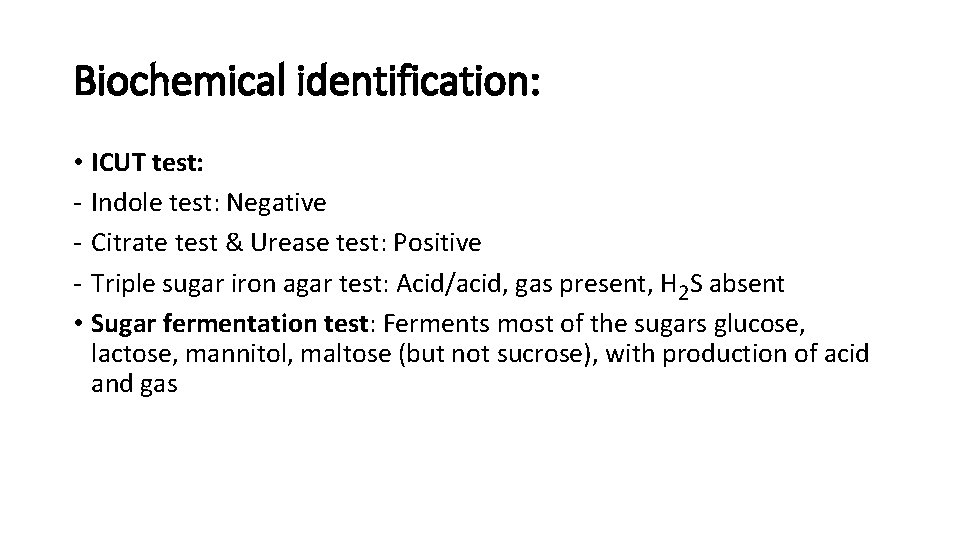
Q 2. A bacterium, cultured on Mac. Conkey agar (shown in the photograph), was isolated from sputum and blood of the patient in the previous case. What is the primary function of the pathogenicity determinant depicted in this photo a) Antiphagocytic, unless opsonization occurs b) Degrades secretory Ig. A on mucosal surfaces c) Inhibits the function of complement d) Lyses neutrophils and macrophages e) Protease activity and disrupts membranes
Q 3 What is the O antigen of Enterobacteriaceae? a) Cell surface polysaccharide b) A channel controlling substance taken into the organism c) A flagella protein d) Cell wall lipopolysaccharide
Q 4. A 23 -year-old woman comes to your office because, for 3 days, she has experienced burning with urination, increased frequency of urination, and a continual feeling that she needs to urinate. She does not have vaginal discharge, fever, or flank pain. Rapid ‘dipstick’ urine tests are consistent with uncomplicated cystitis. Culture of urine on standard media produces a lactose-fermenting Gram negative rod. In situations such as this, what are the likely pathogen? a) Klebsiella pneumoniae b) E Coli c) Enterobacter d) CONS e) Salmonella f) Pseudomonas
Q 5. Isolate from previous Q was tested further with following results • Indole-Negative, Citrate –Positive, Urease – positive, Sucrose ferments with gas • Nonmotile & • Sensitive to 3 rd gen cephalosporines and quinolones Hence the isolate is a) b) c) d) Klebsiella pneumoniae E Coli Enterobacter Pseudomonas

KLEBSIELLEAE Essentials of Medical Microbiology © 2018, Jaypee Brothers Medical Publishers
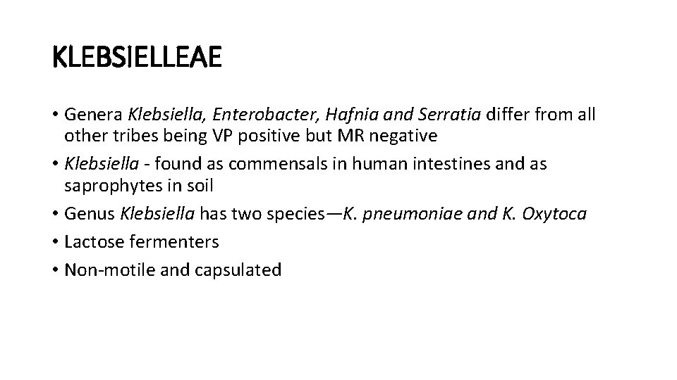
KLEBSIELLEAE • Genera Klebsiella, Enterobacter, Hafnia and Serratia differ from all other tribes being VP positive but MR negative • Klebsiella - found as commensals in human intestines and as saprophytes in soil • Genus Klebsiella has two species—K. pneumoniae and K. Oxytoca • Lactose fermenters • Non-motile and capsulated
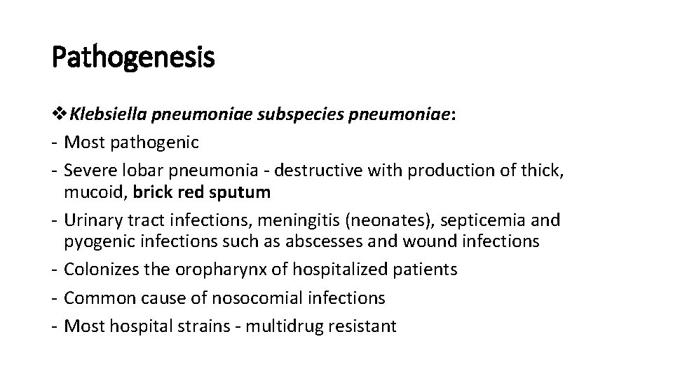
Pathogenesis v. Klebsiella pneumoniae subspecies pneumoniae: - Most pathogenic - Severe lobar pneumonia - destructive with production of thick, mucoid, brick red sputum - Urinary tract infections, meningitis (neonates), septicemia and pyogenic infections such as abscesses and wound infections - Colonizes the oropharynx of hospitalized patients - Common cause of nosocomial infections - Most hospital strains - multidrug resistant
Pathogenesis v. K. pneumoniae subspecies ozaenae - Atrophic rhinitis (or ozena) -foul smelling nasal discharge - Biochemically inactive v K. pneumoniae subspecies rhinoscleromatis - Rhinoscleroma - chronic granulomatous hypertrophy of the nose - South-eastern Europe, India and in Central America - Biochemically inactive
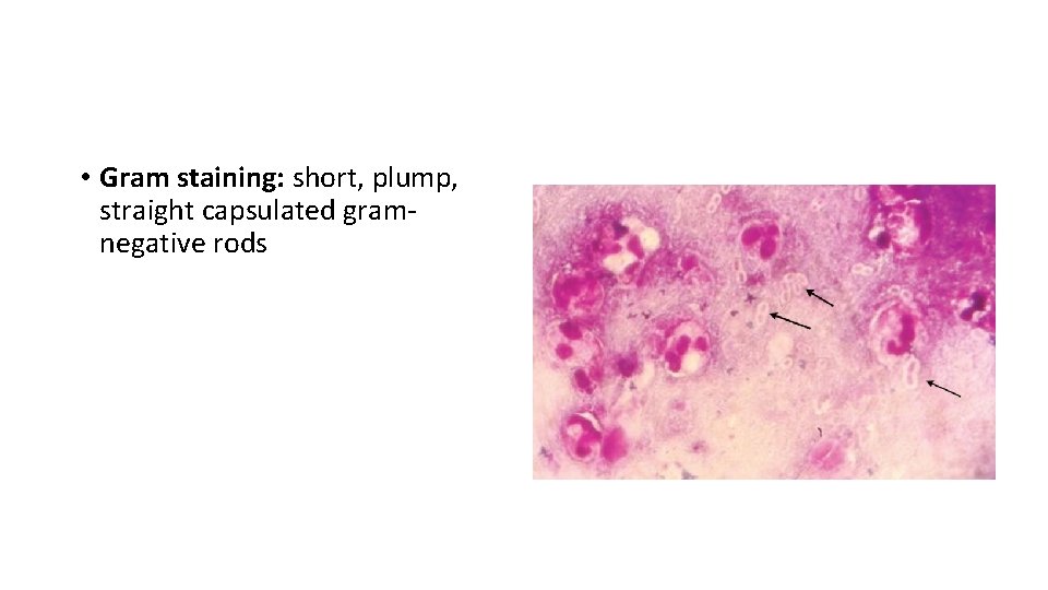
Laboratory Diagnosis • Gram staining: short, plump, straight capsulated gramnegative rods
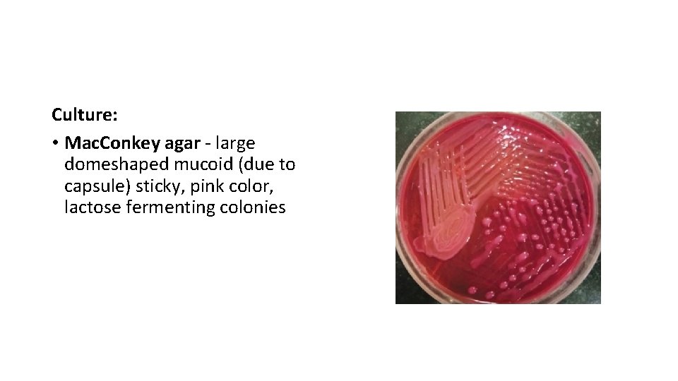
Laboratory Diagnosis Culture: • Mac. Conkey agar - large domeshaped mucoid (due to capsule) sticky, pink color, lactose fermenting colonies
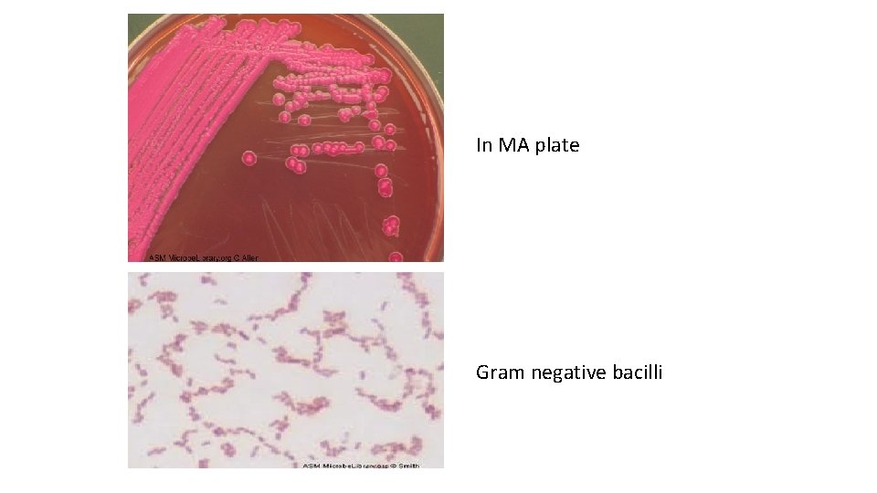
In MA plate Gram negative bacilli
Biochemical identification: • ICUT test: - Indole test: Negative - Citrate test & Urease test: Positive - Triple sugar iron agar test: Acid/acid, gas present, H 2 S absent • Sugar fermentation test: Ferments most of the sugars glucose, lactose, mannitol, maltose (but not sucrose), with production of acid and gas
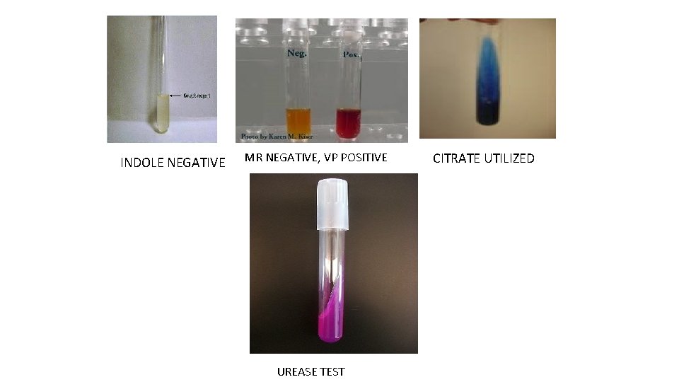
INDOLE NEGATIVE MR NEGATIVE, VP POSITIVE UREASE TEST CITRATE UTILIZED
Klebsiella • VP (Voges-Proskauer) test: Positive • MR (methyl red) test: Negative • K. oxytoca is biochemically similar to K. pneumoniae, but differs from the latter by being indole positive • Most clinical isolates are MDR
Treatment • Based upon antimicrobial susceptibility test report • Hospital strains mostly MDR. Often produce ESBLs or Amp. C β-lactamases resistant to most β-lactams except carbapenems • Carbapenems, amikacin or BL/BLIs - agents of choice for hospital acquired MDR isolates
Treatment • Carbapenem resistant isolates - Polymyxins, fosfomycin or tigecycline
ENTEROBACTER Essentials of Medical Microbiology © 2018, Jaypee Brothers Medical Publishers
ENTEROBACTER • Similar to Klebsiella in most biochemical reactions differs in being motile and ornithine decarboxylase positive • E. aerogenes and E. Cloacae - common isolated species from the clinical specimens • Widely distributed in water, sewage, soil and feces of healthy persons
ENTEROBACTER • Opportunistic pathogens • Wound infection, urinary and respiratory tract infections • Occasionally septicemia and meningitis • Most Enterobacter isolated are MDR • Guideline for treatment is same as that for E. coli or Klebsiella
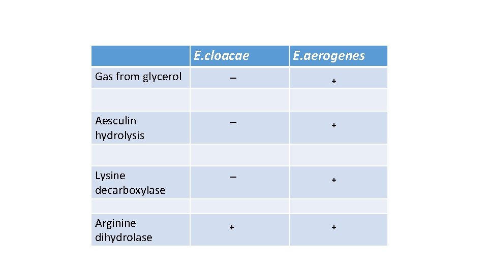
E. cloacae E. aerogenes Gas from glycerol ‒ ₊ Aesculin hydrolysis ‒ ₊ Lysine decarboxylase ‒ ₊ Arginine dihydrolase ₊ ₊
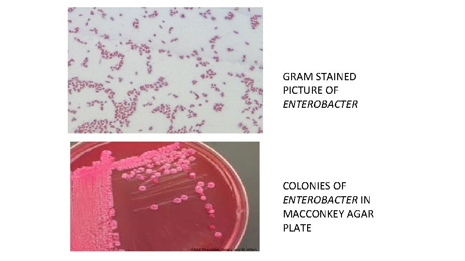
GRAM STAINED PICTURE OF ENTEROBACTER COLONIES OF ENTEROBACTER IN MACCONKEY AGAR PLATE
SERRATIA • Saprophyte found in water, soil and food • May grow in sputum after collection and sputum red (Pseudohemoptysis) • Nosocomial infections - meningitis, endocarditis, septicemia, urinary, respiratory and wound infections • Hospital strains - often non-pigmented and multiple drug resistant (produce Amp. C β-lactamases)
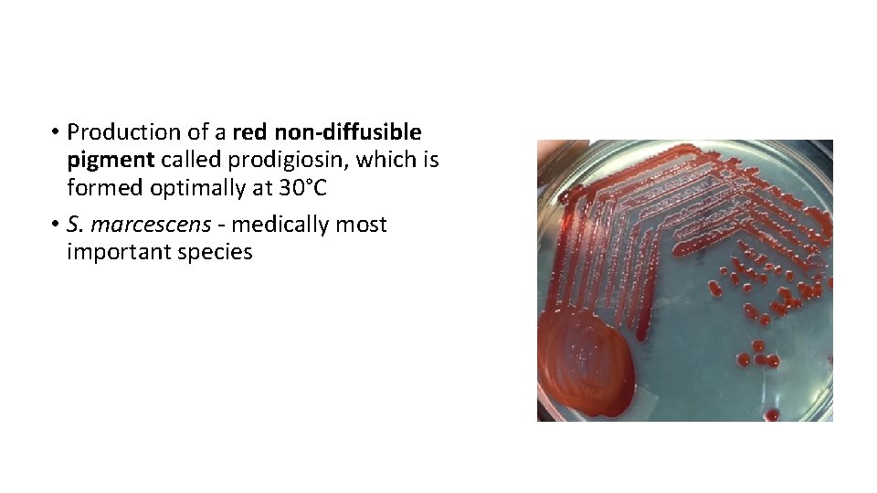
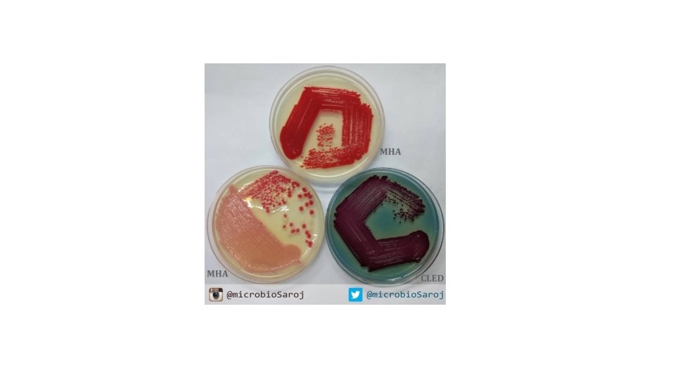
SERRATIA • Production of a red non-diffusible pigment called prodigiosin, which is formed optimally at 30°C • S. marcescens - medically most important species
SERRATIA • Biochemical properties: - Production of lipase, gelatinase and Dnase - Resistant to colistin and cephalothin • Treatment: Most are MDR - Guideline for treatment is same as that for E. coli except - Intrinsically resistant to polymyxins and nitrofurantoin
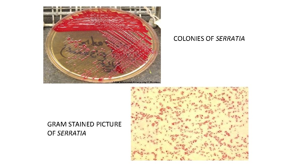
COLONIES OF SERRATIA GRAM STAINED PICTURE OF SERRATIA
HAFNIA • H. alvei - only species • Rarely isolated from wounds, abscess, sputum, urine and blood • Lactose non-fermenter • Positive for lysine and ornithine decarboxylase • Similar to Serratia, the biochemical reactions are most reliable when tested at 30°C.
Q 1. An 85 -year-old male nursing home patient with a history of alcoholism suddenly developed a flu-like illness. He complained of chills and fever and had frequent coughing spells productive of thick, bloody sputum. The attending physician diagnosed bronchopneumonia and prescribed antibiotics, but regrettably the patient died within a week. What is the most likely cause of the patient’s pneumonia? a) b) c) d) Haemophilus influenzae Klebsiella pneumoniae Mycoplasma pneumoniae Streptococcus pneumoniae
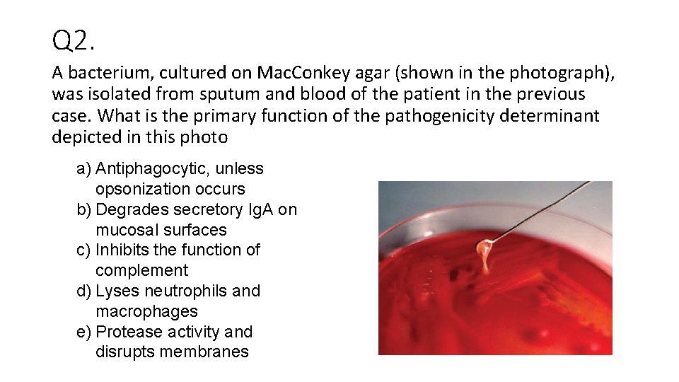
Q 2. A bacterium, cultured on Mac. Conkey agar (shown in the photograph), was isolated from sputum and blood of the patient in the previous case. What is the primary function of the pathogenicity determinant depicted in this photo a) Antiphagocytic, unless opsonization occurs b) Degrades secretory Ig. A on mucosal surfaces c) Inhibits the function of complement d) Lyses neutrophils and macrophages e) Protease activity and disrupts membranes
Q 3 What is the O antigen of Enterobacteriaceae? a) Cell surface polysaccharide b) A channel controlling substance taken into the organism c) A flagella protein d) Cell wall lipopolysaccharide
Q 4. A 23 -year-old woman comes to your office because, for 3 days, she has experienced burning with urination, increased frequency of urination, and a continual feeling that she needs to urinate. She does not have vaginal discharge, fever, or flank pain. Rapid ‘dipstick’ urine tests are consistent with uncomplicated cystitis. Culture of urine on standard media produces a lactose-fermenting Gram negative rod. In situations such as this, what are the likely pathogen? a) Klebsiella pneumoniae b) E Coli c) Enterobacter d) CONS e) Salmonella f) Pseudomonas
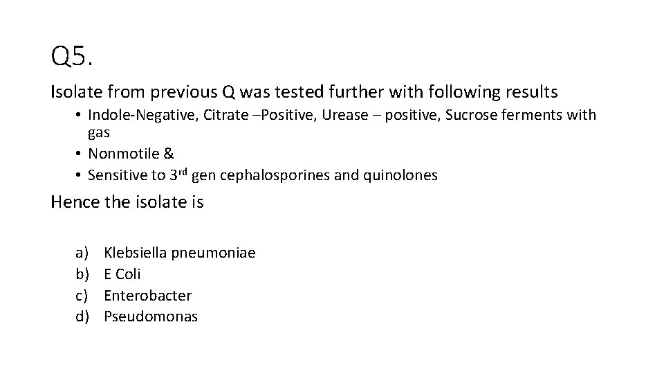
Q 5. Isolate from previous Q was tested further with following results • Indole-Negative, Citrate –Positive, Urease – positive, Sucrose ferments with gas • Nonmotile & • Sensitive to 3 rd gen cephalosporines and quinolones Hence the isolate is a) b) c) d) Klebsiella pneumoniae E Coli Enterobacter Pseudomonas

- Slides: 48