
RHINITIS Dr S Jaya Sandeep CLASSIFICATION Rhinitis Chronic

- Slides: 38
RHINITIS Dr S Jaya Sandeep
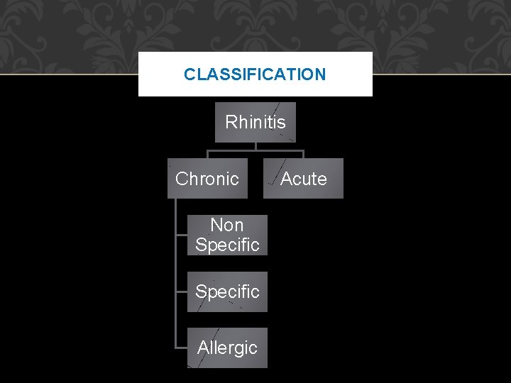
CLASSIFICATION Rhinitis Chronic Non Specific Allergic Acute
ACUTE INFLAMMATORY CONDITIONS Acute Rhinitis or Coryza Incubation period varies from 1 to 3 days and usually transmission occurs by droplet infection such as sneezing, coughing, talking or schooling years due to lack of resistance to infection. Predisposing factors may be change of climate, temperature, humidity, poor nutrition, chronic sinusitis, fatigue, diabetes, tuberculosis and dysfunction of thyroid.
Etiology Causative agents are the viruses such as rhinovirus, influenza and parainfluenza virus, syncytial virus, enteric cytopathogenic human orphan (ECHO) virus, coxsackie, adeno and reoviruses. Secondary invading agents are Streptococcus, Moraxella catarrhalis, Haemophilus influenzae, Staphylococcus and pneumococci. It spreads by kissing and through food, finger and fomites.
PATHOLOGY Ischemia and vasoconstriction Vasodilation Edema Increased activity of nasal glands and goblet cells Leukocytosis and purulent discharge
CLINICAL FEATURES 1. Firstly, there is dryness and ticklish spot in the throat or nasopharynx. 2. sneezing, discharge from the nose and nasal obstruction. 3. Mucous membrane of nose and throat is swollen and congested. 4. Fever appears due to toxemia. 5. On 2 nd or 3 rd day, there is thick discharge, which is mucopurulent, nasal obstruction increases and on 5 th day onwards, symptoms start decreasing and recovery occurs.
COMPLICATIONS Nasopharyngitis Sinusitis Eustachian tube (ET) catarrh Otitis media Lymphadenitis Respiratory tract infection Tonsillitis Nephritis Rheumatism
MANAGEMENT Avoidance of dusty and crowded places Complete isolation of patients Balanced and nutritious diet Sudden fatigue, change of temperature and humidity should be avoided Proper sterilization of fomites is necessary Role of cold (polyvalent) vaccine has not been proved.
TREATMENT Complete bed rest Hot water bath is quite helpful There is role of vitamin C, 500 mg bid for 10 days Antibiotics to prevent complications and secondary infection Antihistaminics and anti-inflammatory drugs are given symptomatically 2 to 3 times a day Smoking and hard drinks are avoided
ACUTE NASAL DIPHTHERIA Causative organism is Corynebacterium diphtheriae. It may be primary or secondary due to faucial diphtheria. The patient presents with pyrexia, general toxemic features, lymphadenitis and palsies. Treatment is with penicillin and antitoxin serum. Erythromycin is given in the patients with allergy to penicillin.
IRRITATIVE RHINITIS Caused by exposure to dust, smoke or irritating gases such as ammonia, formaline, acid fumes, etc. or it may result from trauma inflicted on the nasal mucosa during intranasal manipulation, e. g. removal of a foreign body. There is an immediate catarrhal reaction with sneezing, rhinorrhoea and nasal congestion. The symptoms may pass off rapidly with removal of the offending agent or may persist for some days if nasal epithelium has been damaged.
CHRONIC NONSPECIFIC RHINITIS Atrophic rhinitis Chronic simple rhinitis Chronic hypertrophic rhinitis Rhinitis sicca Rhinitis caseosa Rhinitis medicamentosa
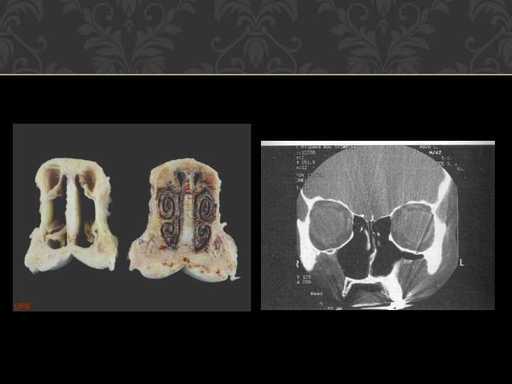
ATROPHIC RHINITIS Atrophic rhinitis is a chronic inflammatory disease, which is characterized by progressive atrophy of mucous membrane of nose and turbinates. There is also associated excessive crusting and abnormal patency of nasal passages It is termed as ozena, if there is associated fetor The disease usually starts at puberty, commonly seen in females.
TYPES 1. Primary atrophic rhinitis type 2. Secondary atrophic rhinitis which may be due to – Syphilis – Tuberculosis – Excessive surgery – Chronic sinusitis – Trauma – Postradiation therapy
CAUSES Heredity Endocrinal Racial Nutrition Immunocompromised Autoimmune
Other Causes Bacterial: Such as Coccobacillus foetidus, Bacillus mucosus, diphtheroid bacilli, Klebsiella ozaenae may be responsible for atrophic rhinitis Empty nose syndrome: It is an iatrogenic condition caused by over enthusiastic removal of nasal turbinates during nasal surgery Syphilis (tertiary) Wide nasal cavity Lupus, leprosy and tubercular Extensive surgery on the nose
PATHOGENESIS There occurs metaplastic changes in columnar ciliated to cuboidal or stratified squamous epithelium Capillaries are dilated and size and number of glands decrease Periarteritis and endarteritis may be present There may be atrophy of bones of the turbinate along with atrophy of nerves In type-I, endarteritis or periarteritis is present In type-II, dilatation of capillaries is seen.
CLINICAL FEATURES Symptoms It is a bilateral condition mostly in females present with: • Nasal obstruction • Nose bleed • Headache • Merciful anosmia—foul smell, which the patient herself is unable to perceive due to atrophy of olfactory nerves and excessive crusting of nose
Signs • Grayish black crusts in the nose • Nasal passages are roomy • Turbinates are shrivelled and detachment of crusts leave a bleeding and ulcerated mucosa • In late cases, septal perforation and saddle nose may be present • Eustachian tube catarrh is present and paranasal sinuses and pharynx may be affected.
TREATMENT Conservative Regular nasal cleaning: By nasal douching with alkaline solution prepared by dissolving 280 ml of warm water in 28. 4 gm of sodabiborate and 56. 7 gm of sodium chloride. Application of 25 percent glucose in glycerine, which inhibits the growth of proteolytic organisms, i. e. saprophytes Estradiol in arachis oil for its vasodilation effect. Antibiotic in antiozene solution—local application in the nose to control secondary infection. Placental extract by submucosal injections. High protein diet, Vitamin A
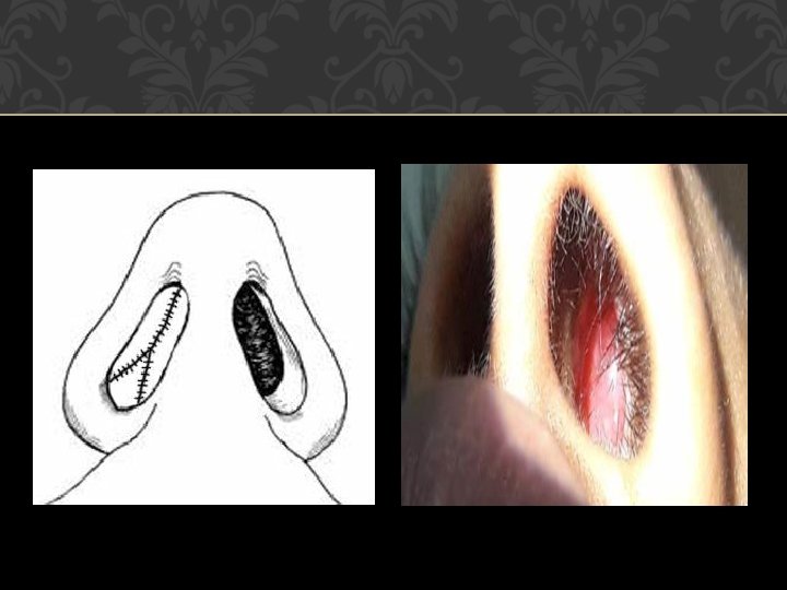
Surgical Submucosal injection of paraffin or teflon paste in glycerine 50 percent decreases the nasal passages. Young’s or modified Young’s operation Folds of skin inside the nostril are raised and are sutured together with the object of complete interruption of air flow for a period of months to years. It helps by giving rest to nasal mucosa and by causing local hypoxia. In modified operation, partial interruption of the air flow is done
Eyries operation (1953): Placement of implants under the nasal mucosa to decrease the size of nasal cavity. Insertion of fat and cartilage or Teflon strips have been inserted after lifting the flaps of mucoperiosteum on the septum or lateral wall. Lautenslager operation is also done for atrophic rhinitis (medialization of lateral wall).
CHRONIC SIMPLE RHINITIS Recurrent attacks of acute rhinitis in the presence of predisposing factors leads to chronicity Persistence of nasal infection due to sinusitis, tonsillitis and adenoids Chronic irritation from dust, smoke, cigarette smoking, sniff. Nasal obstruction due to deviated nasal septum, synechia leading to persistence of discharge in the nose
PATHOLOGY Simple chronic rhinitis is an early stage of hypertrophic rhinitis. There is hyperaemia and oedema of mucous membrane with hypertrophy of seromucinous glands and increase in goblet cells. Blood sinusoids particularly those over the turbinates are distended.
CLINICAL FEATURES Nasal obstruction Nasal discharge Headache Swollen turbinates Post nasal discharge
TREATMENT Treat the underlying cause sinuses, tonsils, adenoids, allergy, personal habits (smoking or alcohol indulgence), environment or work situation (smoky or dusty surroundings). Nasal irrigations with alkaline solution Nasal decongestants Antihistamines Antibiotics
CHRONIC HYPERTROPHIC RHINITIS In this condition, there is hypertrophy of epithelial lining, glands, and turbinates. Important causes leading to this condition are chronic infections of nose, PNS, chronic irritation of nose due to smoke, fumes, dust or local use of drugs such as nasal drops.
Clinical features Nasal obstruction, nasal discharge, anosmia and feeling of heaviness of head. Examination shows thick mucosa with enlarged turbinate, which does not shrink with local drops. Treatment Size of turbinates can be reduced by electrocautery, submucosal diathermy, partial resection of enlarged turbinates or by
Compensatory Hypertrophic Rhinitis This is seen in cases of marked deviation of septum to one side. The roomier side of the nose shows hypertrophy of inferior and middle turbinates. This is an attempt on the part of nature to reduce the wide space to overcome the ill effects of drying and crusting that always attend wider nasal space. Hypertrophic changes in these cases are not reversible with the correction of nasal septum and often require reduction of turbinates at the time of septal surgery.
RHINITIS SICCA Rhinitis sicca is a milder form of atrophic rhinitis, which occurs in alcoholics, anemia, nutritional deficiencies and persons working in hot, dry and dusty occupation. There is inactivity of seromucinous glands along with metaplasia of columnar ciliated epithelium to cuboidal or squamous epithelium.
Clinical Features Irritation, dryness, crusting, bleeding some time in the anterior part of nose only, no fetor. Examination shows a glazed mucosa with thin crusts. Treatment • Removal of the cause whatsoever • Lubrication of the nose with vaseline • Alkaline nasal douche, if required.
RHINITIS CASEOSA Rhinitis caseosa is also called nasal cholesteatoma It is a chronic inflammatory condition with formation of granulations and presence of offensive cheesy material It is seen more in males at any age group. It may be seen in chronic sinusitis infection due to streptothrix alba or the presence of a foreign body
Microscopically, it shows caseous debris with keratinous material, organisms and sometimes cholesterol crystals Clinically, nose shows white debris, bone may be involved along with sinus infection. Treatment Thorough removal of caseous debris from nose by douching or surgical means.
RHINITIS MEDICAMENTOSA Rhinitis medicamentosa is seen more after prolong use of nasal drops and sprays which results in rebound phenomenon causing vasodilatation and engorgement of nasal mucosa. On examination nasal mucosa is boggy and congested. Treatment a. Stop use of decongestants b. Use of oral or topical steroids
THANK YOU