ANATOMY OF NASAL SEPTUM Dr S Jaya Sandeep


 Sphenopalatine artery (Br of maxillary-Br of external carotid) -Posteroinferior septum 2)")


- Slides: 22
ANATOMY OF NASAL SEPTUM Dr S Jaya Sandeep
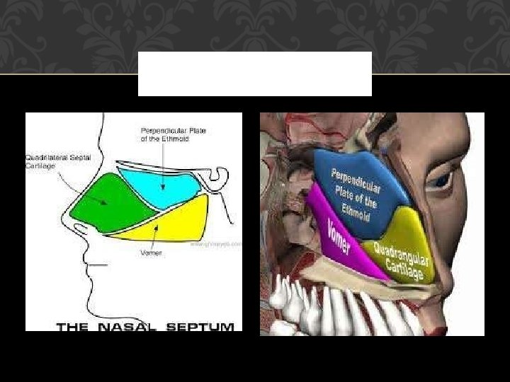
EMBRYOLOGY The primitive septum is initially made entirely of cartilage. The superior part ossifies to form the perpendicular plate of the ethmoid and the vomer in the poster inferior portion, leaving an antero inferior quadrilateral cartilaginous plate
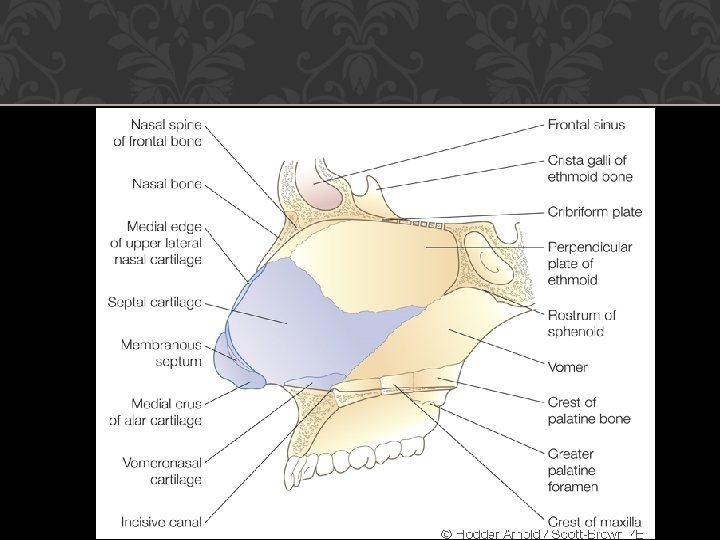
NASAL SEPTUM The nasal septum is composed of 1. Anterior membranous portion 2. Cartilaginous portion : Quadrilateral cartilage, contribution from upper and lower lateral alar cartilages. 3. Bony portion : perpendicular plate of ethmoid, vomer and two bony crests of maxilla and palatine. Quadrilateral cartilage is crucial in development of middle 1/3 of face
CARTILAGINOUS PORTION 3 -4 mm thick in center and 4 -8 mm anterior inferiorly – Footplate Bounded firmly by collagen fibers to bones all around Sits inferiorly in the nasal crest of palatine process of maxilla Anteriorly attached by thin membranous septum to medial crura of lower lateral cartilages
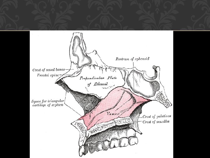
BONY PORTION The perpendicular plate forms the superior and anterior bony septum, is continuous above with the cribriform plate and crista galli. The vomer forms the posterior and inferior nasal septum and articulates by its two alae with the rostrum of the sphenoid.
VOMER BONE Inferiorly – Articulates with nasal crest formed by maxilla and palatine bones Anteriorly – Articulates with perpendicular plate of ethmoid and quadrilateral cartilage Posteriorly – Forms free edge of the septum
CLINICAL IMPORTANCE Deflections may develop at any of the septal articulations and spurs may also be found where the quadrilateral cartilage sends small processes between the ethmoid and vomer. Such deflections are far commoner in men than women, they are most likely to be acquired due to trauma than be Congenital.
HISTOLOGY The mucous membrane is predominantly respiratory with a small area of olfactory epithelium superiorly adjacent to the cribriform plate. Respiratory epithelium is composed of ciliated and non-ciliated pseudo stratified columnar cells, basal pluripotential stem cells and goblet cells.
HISTOLOGY Micro villi – Cytoplasmic extensions which increase surface area and prevent drying Seromucinous glands are present in submucosa and are more important in mucus production in the nasal cavity then the goblet cells which are more numerous in paranasal sinuses.
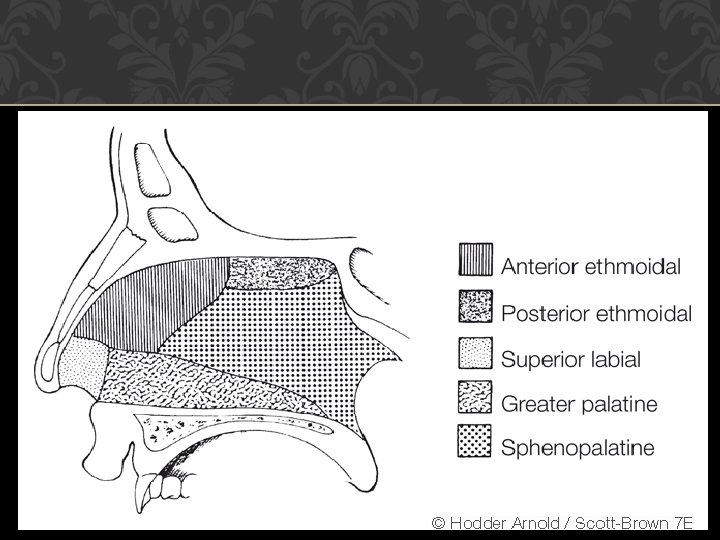
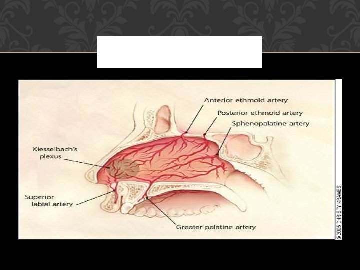
BLOOD SUPPLY 1) Sphenopalatine artery (Br of maxillary-Br of external carotid) -Posteroinferior septum 2) Greater palatine artery (Br of Maxillary) -Anteroinferior portion after entering via incisive canal 3) Superior labial artery (Br of Facial) –Anteriorly, in particular to Kiesselbach's plexus which is situated in Little's area on the anterior septum – a source of epistaxis 4) Ant and Post Ethmoid Arteries (Br of ICA)-Superiorly,
LITTLE’S AREA Kisslebacks plexus is formed by Anterior ethmoidal artery Septal branch of superior labial artery Septal branch of sphenopalatine artery Septal branch of greater palatine artery
VENOUS DRAINAGE The cavernous venous system drains via the sphenopalatine vessels into the pterygoid plexus posteriorly and into the facial veins anteriorly. Superiorly the ethmoidal veins communicate with the superior ophthalmic system and there may be direct intracranial connections through the foramen caecum into the superior sagittal sinus.
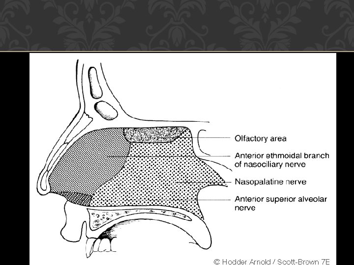
NERVE SUPPLY The maxillary division of the trigeminal nerve provides the sensory supply to the majority of the nasal septum. The nasopalatine nerve supplies the bulk of the bony septum, entering the nasal cavity via the sphenopalatine foramen.
NERVE SUPPLY The anterosuperior part of the septum is supplied by the anterior ethmoidal branch of the nasociliary nerve and a smaller anteroinferior portion receives a branch from the anterior superior alveolar nerve. The posteroinferior septum also receives a small supply from the nerve to the pterygoid canal and a posterior inferior nasal branch of the anterior palatine nerve.
LYMPHATIC DRAINAGE The anterior septum drains with the external nose to the submandibular nodes while drainage is to the retropharyngeal and anterior deep cervical nodes posteriorly.
THANK YOU