Chapter 7 Cell communication and signaling u Signaling


 Contact-dependent: don’t need secreted molecule (C) Neuronal signaling")
 Papacrine: to nearby neighbor (D) Endocrine: to long distance Signal can act over")

")
")


Signaling by phosphorylation : signaling proteins")

 inactivate the protein by stimulating")


 is the largest class of enzyme-coupled")


 and STATs : ØJaks: A class of cytoplasmic tyrosine")


 phosphorylate themselves and then recruit and activate cytoplasmic")


 Protein kinases in both pathways phosphorylate many")


 are specific types of large macromolecular")
 ü Focal adhesions concentrate and direct")


- Slides: 63
Chapter 7 Cell communication and signaling u Signaling Components u The Role of Intracellular Receptor u G-Protein–Coupled Cell-Surface Receptors u Enzyme-Coupled Cell-Surface Receptors u Signal Network System u Cell Signaling And The Cytoskeleton u Cell Communication in Plants 2 nd Edition
SIGNALING COMPONENTS u Four Types of Cell Communication u Cell-Surface Receptors Fall into Three Main Classes u The Basic Cell Signaling Process
Four Types of Cell Communication (A) Contact-dependent: don’t need secreted molecule (C) Neuronal signaling Signal can act over long or short range. From Bruce Alberts et al. , Molecular Biology of The Cell (5 th edition))
(B) Papacrine: to nearby neighbor (D) Endocrine: to long distance Signal can act over long or short range. From Bruce Alberts et al. , Molecular Biology of The Cell (5 th edition)
Contact-dependent signaling controls nerve-cell production: Each future neuron delivers an inhibitory signal to the cells next to it to deter them from specializing as neurons too From Essential Cell Biology (© Garland Science 2010)
From Essential Cell Biology (© Garland Science 2010)
From Essential Cell Biology (© Garland Science 2010)
Some extracellular signal molecule is large and hydrophilic, it needs receptor on cell surface to deliver signal to cell From Essential Cell Biology (© Garland Science 2010)
Cell surface receptors fall into three basic classes 1. An ion-channel-coupled opens or closes in response to binding of its extracellular signal molecule. These channels are also called ‘transmitter-gated ion channels From Bruce Alberts et al. , Molecular Biology of The Cell (5 th edition)
2. G-protein-coupled receptor: When a G-coupled receptor binds its extracellular signal molecule, the signal is passed first to a GTP-binding protein (G protein) that is associated with the receptor. The activated G protein then leaves the receptor and turn on a target enzyme (or ion channel) in the membrane From Bruce Alberts et al. , Molecular Biology of The Cell (5 th edition)
3. Enzyme-coupled receptors: An enzyme-linked receptor binds its extracellular signal molecule, switching on an enzyme activity, either at the other end of the enzyme or other associated enzyme From Bruce Alberts et al. , Molecular Biology of The Cell (5 th edition)
Extracellular signals can act slowly or rapidly From Essential Cell Biology (© Garland Science 2010)
The Basic Cell Signaling Process ü Two different types of signal transduction pathways: one in which a signaling pathway is activated by a diffusible second messenger and another in which a signaling pathway is activated by recruitment of proteins to the plasma membrane. ü Most signal transduction pathways involve a combination of these mechanisms. ü signaling pathways are not typically linear tracks as depicted here, but are branched and interconnected to form a complex web. From Gerald Karp, Cell and Molecular Biology- concepts and experiments (6 th edition)
Many intracellular signaling proteins act as molecular switches: (A)Signaling by phosphorylation : signaling proteins are activated by the addition of a phosphate group by protein kinase, and inactivated by removal of the phosphate by protein phosphatase. Protein kinase phosphates serine/threonine (called ser/thr kinase) or tyrosine (called tyrosine kinase) (B) Signaling by GTP-binding protein: a GTP-binding signaling protein is induced to exchange its bound GDT for GTP (i. e. adds a phosphate to the protein). The hydrolysis of the bound GTP to GDP Then switches the protein off
Signal transduction pathway consisting of protein kinases and protein phosphatases Protein kinase 2 is activated by protein kinase 1. Once activated, protein kinase 2 phosphorylates protein kinase 3, activating the enzyme. Protein kinase 3 then phosphorylates a transcription factor, increasing its affinity for a site on the DNA. Binding of a transcription factor to the DNA affects the transcription of the gene in question. Each of these activation steps in the pathway is reversed by a phosphatase. From Gerald Karp, Cell and Molecular Biology- concepts and experiments (6 th edition)
The regulation of a monomeric GTPases. GTPase-activating proteins (GAPs) inactivate the protein by stimulating it to hydrolyze its bound GTP to GDP. Guanine nucleotide exchange factors (GEFs ) activate the inactive protein by stimulating it to release its GDP; Note that the concentration of GTP in cytosol is 10 times greater than the concentration of GDP, the protein rapidly binds GTP once it ejects GDP and is thereby activated From Bruce Alberts et al. , Molecular Biology of The Cell (5 th edition)
The Role of Intracellular Receptor Extracellular signal molecules bind either to cell-surface receptors or to intracellular enzymes or receptors Some small, hydrophobic extracellular signal can diffuse across the membrane and binds to intracellular receptor in either the cytosol or the nucleus From Essential Cell Biology (© Garland Science 2010)
Some small hydrophobic hormones binds to intracellular receptors that act as transcription regulators From Essential Cell Biology (© Garland Science 2010)
The steroid hormone cortical acts by activating a transcription regulator. Cortisol is one of the hormones produced by the adrenal glands in response to stress From Essential Cell Biology (© Garland Science 2010)
Some Dissolved Gases Cross the Plasma Membrane and Activate Intracellular Enzymes Directly 1. Acetylcholine released by nerve terminal in the blood-vessel wall stimulates endothelial cells lining the blood vessel to make and release NO 2. NO diffuse from endothelial cell to adjacent smooth muscle cells causing muscle cell relaxation From Essential Cell Biology (© Garland Science 2010)
One target protein that can be activated by NO is guanyl cyclase. The activated cyclase catalyzes the production of c. GTP from GTP signal transduction pathway that operates by means of NO and cyclic GMP that leads to the dilation of blood vessels From Gerald Karp, Cell and Molecular Biology-concepts and experiments (6 th edition)
Signaling through G-Protein-Coupled Cell-Surface Receptors u The G protein and G-Protein-Coupled Receptors u G-protein Regulation of Ion Channels u GPCR-Initiated Cyclic AMP Pathway u GPCR-Initiated Inositol Phospholipid Pathway
The G protein and G-Protein-Coupled Receptors G-protein-coupled receptor From Essential Cell Biology (© Garland Science 2010)
ü Binding of signal molecule to receptor causes the conformation change of this receptor, which in turn alters the conformation of the G protein ü The alteration of the α subunit of G protein allows it to exchange its GDP to GTP ü This causes the G protein to break up into two activate components- α subunit and a βγ complex, both can regulate the activity of target proteins in the plasma membrane From Essential Cell Biology (© Garland Science 2010)
The desensitization of G protein ü The phosphorylation of the cytoplasmic domain of the activated GPCR by a G protein-coupled receptor kinase (GRK). ü Arrestins binding to the phosphorylated GPCRs prevents the further activation of G proteins. ü Desensitization is an important mechanism that allows a cell to respond to changes in its environment rather than continue to be in a state of activation endlessly in the presence of an unchanging environment. From Gerald Karp, Cell and Molecular Biology-concepts and experiments (6 th edition)
G-protein-coupled receptor desensitization depends on receptor phosphorylation by PKA, PKC, Ca. MK 2 or G-protein-coupled receptor kinases(GRKs) From Essential Cell Biology (© Garland Science 2010)
G-protein Regulation of Ion Channels Some G-protein regulate ion channels: e. g. G protein couple receptor activation to opening of K+ channels in the plasma membrane of heart muscle cells activated bg complex binds to and opens a K+ channel inactivation of the a subunit by hydrolysis of its bound GTP returns the G protein to its inactive state, allowing the K+ channel to close From Essential Cell Biology (© Garland Science 2010)
GPCR-Initiated Cyclic AMP Pathway ü Some G proteins activate membrane -bound enzymes: enzyme activated by G proteins catalyze the synthesis of intracellular second-messenger molecules ü Second messenger: small molecule formed in or released into the cytosol in response to an extracellular signal (first messenger) that helps to relay the signal to the interior of the cell: c. AMP, IP 3 and Ca 2+ From Essential Cell Biology (© Garland Science 2010)
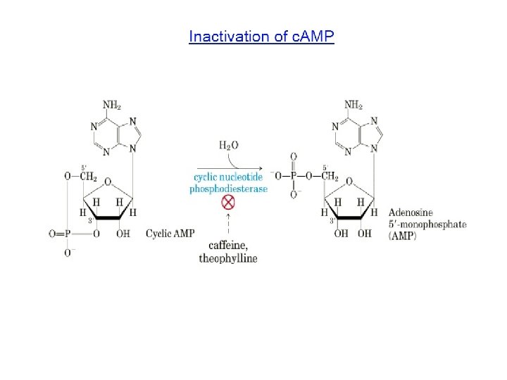
A rise in intracellular cyclic AMP can activate gene transcription ü c-AMP is synthesized by adenylyl cyclase and degraded by c-AMP phosphodiesterase ü c-AMP exerts various effects mainly by activating the enzyme cyclic-AMP-dependent protein kinase (PKA) ü The binding of c-AMP causes the conformation change and activate PKA. The activated PKA then phosphorates certain intracellular proteins on its serine or threonine sites, thus altering their activity From Bruce Alberts et al. , Molecular Biology of The Cell (5 th edition)
GPCR-Initiated Inositol Phospholipid Pathway
The pathway through phospholipase C results in a rise in intracellular Ca+ Signals GPLR GP PLC IP 3 and DAG (twin signals). IP 3 receptor(Ca 2+ channel, located at the surface of s. ER) Elevation of cytosolic Ca 2+; DAG activates PKC to phosphoralate Ser and Thr on target proteins. Calcium binds to calcium-binding proteins(Ca. M) which affects other proteins. From Bruce Alberts et al. , Molecular Biology of The Cell (5 th edition)
Signaling through Enzyme-Coupled Cell-Surface Receptors u Receptor Tyrosine Kinases u Tyrosine-kinase-associated receptor u Receptor Serine/Threonine Kinases
Receptor Tyrosine Kinases ü Receptor tyrosine kinases (RTKs) is the largest class of enzyme-coupled receptors in made up those with a cytoplasmic domain that functions as a tyrosine protein kinase ü Activated receptor tyrosine kinase phosphorylating specific tyrosine on selected intracellular proteins, and assemble a complex of intracellular signaling proteins From Bruce Alberts et al. , Essential cell biology (2 nd edition)
RTKs activate Ras in the fly eye Ras: a small protein that is bound by a lipid tail to the cytoplasmic face of the plasma membrane. The binding of Ras-activating protein makes Ras exchange its bound GDP to GTP. The activated Ras then stimulates the next steps in the signaling pathway. Sev is activated by the Boss protein. The adaptor protein Drk docks on a particular phosphotyrosine on the activated receptor. Drk recruits and stimulates a protein accomplice that functions as a Ras-activating protein Sos stimulates the inactive Ras protein to relay the signal downstream, inducing the R 7 precursor cell to differentiate into a UV-sensing photoreceptor cell. From Bruce Alberts et al. , Molecular Biology of The Cell (5 th edition)
Ras activates a MAP-kinase Phosphorylation cascade MAP kinase: protein kinase that performs a crucial step in relaying signals from cell-surface receptors to the nucleus. It is the final kinase in a 3 -kinase sequence (mitogen-activated protein kinase) From Bruce Alberts et al. , Molecular Biology of The Cell (5 th edition)
RTKs can activate the PI-3 -Akt signaling pathway 1. An extracellular survival signal, such as IGF (insulin-like growth factor) family, activate an RTK, which recruits and activates PI-3 -kinase pathway to promote cell growth and survival 2. PI 3 -kinase (phosphoinositide 3 -kinase) is an enzyme which can hoshphorylate inositol phospholipid in the plasma membrane 3. These phosphorylated lipids become docking sites for specific intracellular signaling proteins, which relocate from cytosol to membrane, where they can activate one another From Essential Cell Biology (© Garland Science 2010)
Activated Akt promotes cell survival : 1. One of the relocated signaling proteins in the PI-3 -kinase pathway is the serine/threonine protein kinase Akt (also called protein kinase B (PKB)). This pathway called PI-3 -kinase-Akt singaling pathway 2. Akt promotes the growth and survival of may cell types often by inactivating the signaling proteins it phosphorylates. It also stimulate cells to grow in size 3. Mechanism of PI-3 -kinase-Akt singaling to promote cell survival: Akt phosphorylates and inactivates a cytosolic protein, Bad, to inhibits apoptosis in cells From Essential Cell Biology (© Garland Science 2010)
Tyrosine-kinase-associated receptor The signaling pathway of the cytokines: ü Unlike the receptor tyrosine kinases, cytokine receptors have no intrinsic enzyme activity but are associated with cytoplasmic tyrosine kinases called JAKs. ü Binding of a cytokine to its receptor causes associated tyrosine kinase JAKs to cross phosphorylate and activate one another , thereby increasing the activity of their tyrosine kinase domain. ü JAKs then phosphorylate the receptor protein on tyrosine. They activate cytoplasmic gene regulatory proteins called STATs migrate to the nucleus where they stimulate transcription of specific target genes.
Jak-STAT signaling pathway Janus kinases(Jaks) and STATs : ØJaks: A class of cytoplasmic tyrosine kinases, including Jak 1, Jak 2, Jak 3, and Tyk 2, and each is associated with particular cytokine receptors; ØSTATs: Jaks then phosphorylate and activate a set of latent gene regulatory proteins called STATs(signal transducers and activators of transcription, which have an SH 2 domain), which move into the nucleus and stimulate the transcription of specific genes. Signaling ligands and cytokine receptors: ØLigands: more than 30 cytokines and hormones activate the Jak-STAT pathway by binding to cytokine receptors. ØCytokine receptors: they are composed of two or more poly peptide chains. All receptor are associated with one or more Jaks.
The Jak-STAT signaling pathway providing a fast track to the nucleus, a more direct route to control gene expression From Bruce Alberts et al. , Molecular Biology of The Cell (5 th edition)
Receptor Serine/Threonine Kinases ü An even more direct signaling pathway; When activated, they directly phosphorylate and activate cytoplasmic gene regulatory proteins called SMADs. ü The hormones and local mediators that activate these receptors belong to the TGF-β (transforming growth factor-β) superfamily of extracellular proteins, which are essential in animal development, tissue repair and in immune regulation, as well as in many other processes. ü Members of the TGF-β superfamily act through receptor serine/threonine kinases that are single-pass transmembrane proteins with a serine/threonine kinase domain on the cytosolic side of the plasma membrane.
TGF-β receptors (receptor serine/ threonine kinases) phosphorylate themselves and then recruit and activate cytoplasmic gene regulatory proteins (called SMADs). The SMADs then dissociate from the receptors and bind to other SMADs, and the complexes then migrate to the nucleus, where they stimulate transcription of specific target genes. From Bruce Alberts et al. , Molecular Biology of The Cell (5 th edition)
SIGNAL NETWORK SYSTEM u Convergence u Divergence u Cross-talk u Signal termination: desensitization
Convergence: Signals from a variety of unrelated receptors can lead to the formation of phosphotyrosine docking sites for the SH 2 domain of the adaptor protein (converge to activate a common effector) From Gerald Karp, Cell and Molecular Biology-concepts and experiments (6 th edition)
Divergence: Signals from the same ligand can diverge to activate a variety of different effectors From Gerald Karp, Cell and Molecular Biology-concepts and experiments (6 th edition)
Cross-talk between two major signalling pathways From Gerald Karp, Cell and Molecular Biology-concepts and experiments (6 th edition)
Signaling pathways can be highly interconnected (cross-talk) Protein kinases in both pathways phosphorylate many proteins, including proteins belonging to the other pathway From Bruce Alberts et al. , Molecular Biology of The Cell (5 th edition)
Some intracellular signaling proteins serve to integrate incoming signals From Bruce Alberts et al. , Molecular Biology of The Cell (5 th edition)
The target cells can become desensitized to a signal molecule by five ways. Sequestration; down-regulation; protein inactivation; Three general ways of the desensitization: 1. Receptor inactivation by alteration; 2. Receptor sequestration by internalization; 3. Receptor down-regulation by destroying in Ls. inhibitory
The mechanisms that shut off a signal are as important as the mechanisms that turn it on Toxins produced by bacteria that cause cholera (affecting Gsa) and whooping cough (pertussis) (affecting Gia)
In actual fact, signaling pathways in the cell are much more complex.
Cell signaling and the cytoskeleton u Function of focal adhesion u Integrin signaling depends on focal adhesion kinase (FAK) u Rho family regulation of the actin cytoskeleton
Function of focal adhesion Focal adhesions (cell–matrix adhesions) are specific types of large macromolecular assemblies. It is the mechanical linkages to the ECM. ü The mechanical-structural function of focal adhesion, which is carried out by actin filaments and associated proteins ü Signaling function from extracellular surface nucleus (where they stimulate the transcription of genes involved in cell growth and proliferation), which is carried out by tyrosine kinase (Src and FAK) When integrins cluster at cell-matrix contacts, FAK is recruited by intracellular anchor protines such as talin or paxillin
Integrin signaling depends on focal adhesion kinase (FAK) ü Focal adhesions concentrate and direct numerous signaling proteins at sites of integrin binding and clustering. ü Signal pathway that lead to the assembly of the fibers of a focal adhesion. ü Rho is involved in regulating the organization of the cell’s actin cytoskeleton.
Model for signaling from the FAK protein-tyrosine kinase. Binding of integrins to the extracellular matrix stimulates FAK activity, leading to its autophosphorylation. Src then binds to the FAK autophosphorylation site and phosphorylates FAK on additional tyrosine residues. These phosphotyrosines serve as binding sites for the Grb-2 -Sos complex, leading to activation of Ras and the MAP kinase cascade, as well as for additional downstream signaling molecules, including PI 3 -kinase. From Geoffrey M. Cooper, The Cell –A Molecular Approach (2 nd edition)
Rho family regulation of the actin cytoskeleton Targets of Rho family members. From Geoffrey M. Cooper, The Cell –A Molecular Approach (2 nd edition)
Cell communication in plant u Plant hormones u Ethylene signaling pathway u Light receptor pathway u Regulating calcium concentrations
Plant hormones Structure of plant hormones From Geoffrey M. Cooper, The Cell –A Molecular Approach (2 nd edition)
1. Plants and animals have been evolving independently for more than a billion years 2. Like animals, plants make extensive use of transmembrane cell-surface receptors, especially enzyme-coupled receptors. e. g. the spindly weed Arabidopsis Thaliana has hundreds of genes encoding receptor serine/threonine kinase, but their serine/threonine kinase receptor is very different to animal’s 3. Plant receptors trigger a large variety of cell signaling prosess involves in plant growth, development and disease resistance 4. Plant don’t use RTKs, steroid-hormone-type nuclear receptors, or c-AMP, they seem to use few GPCRs
Ethylene signaling pathway One of the best-studied signaling pathways in plant mediates the response of cells to ethylene- a gaseous hormone that regulates a diverse array of development processes, including seed germination and fruit ripening. A current view of the ethylene two-component signaling pathway. (A) In the absence of ethylene, the receptors and the MAP-kinase module are active, leading to inhibition of the gene regulatory proteins in the nucleus that are responsible for the transcription of ethylene-responsive genes. (B) In the presence of ethylene, the receptors and the MAP-kinase module are inactive, so the ethylene-responsive genes are transcribed. From Bruce Alberts et al. , Molecular Biology of The Cell (5 th edition)
Light receptor pathway A current view of one way in which phytochromes mediate a light response in plant cells. When activated by light, the phytochrome, which is a dimmer, phosphorylates itself and then moves into the nucleus, where it activates gene regulatory proteins to stimulate the transcription of specific genes. From Bruce Alberts et al. , Molecular Biology of The Cell (5 th edition)
Regulating calcium concentrations in plant cells Cytosolic calcium changes in response to several stimuli, including light, pressure, gravity, and hormones. 脱落酸 Calcium signaling aids in decreasing turgor pressure in guard cells.