CEREBELLUM histology neural connections cerebellar syndromes gnes Nemeskri

CEREBELLUM histology – neural connections – cerebellar syndromes Ágnes Nemeskéri 2018 Semmelweis University Department of Anatomy, Histology and Embryology nemeskeri. agnes@med. semmelweis-univ. hu

Function of cerebellum Cerebellum is classically thought to control movement coordination (Flourens, 1824; Luciani, 1891) motor learning (Marr, 1969; Albus, 1972) BUT recent experimental evidence suggests: it may play a key role in cognition and emotion (Schmahmann, 2004; Schmahmann and Caplan, 2006; Ito, 2008)

„the human cerebellum increased three- to fourfold in last million years. At this huge increase in size of the cerebellum was linked by two-way nerve tracks (20 million on each side of the brain) to the cerebral cortex, including the parietal and prefrontal areas for planning and language functions” (Leiner, Leiner & Dow) Lent et. al. 2012
: „the cerebellum operates as a general co-processor, whose effect")
Functions of cerebellum D’Angelo (2013): „the cerebellum operates as a general co-processor, whose effect depends on the centers to which different modules are connected, affecting cognitive functions as well as sensory-motor processing. ” Modular organization of cerebellum. - majority of connections between neurons and interneurons in the cerebellar cortex occur: within individual modules

Message low-level functions timing - seems the most fundamental prediction learning - cerebellum is primarily involved when unknown problems or circumstances are encountered working memory, error/novelty detection, mental object manipulation high-level functions motor control attention switching language processing imagery visuospatial processing decision-making reasoning unconsciously learning feedforward models of behavior - culture

Histology of the cerebellum Cerebellar cortex - striking, regular, 3 -layered uniform structure - all regions perform same fundamental operation
: stellate cells, basket cells")
Layers of cerebellar cortex Molecular layer - inhibitory interneurons (GABA-erg): stellate cells, basket cells - flattened dendritic trees of Purkinje cells - fibers: parallel fibers originating from granule cells Purkinje cell layer - cell bodies of Purkinje cells and Bergmann glial cells - underneath the Purkinje layer: Lugaro cells whose long dendrites travel along the boundary between Purkinje and the granular layers Granular layer - densely packed with granule cells - interneurons: mainly Golgi cells unipolar brush cells excitatory interneurons (only in flocculo-nodular l. ) relay feedforward input from mossy fibers, and their ON and OFF responses have opposing polarities, they may play a role in aiding this bidirectional response of granule cells to encode direction of motion Lugaro cells-inhibitory interneuron-end on

Granule cells - total number around 50 billion - about 75 % of the brain's neurons: cerebellar granule cells granular layer - axons rises vertically to molecular layer - split in two, traveling horizontally to form: parallel fiber (a distinctive "T" shape) - parallel fibers pass through the dendritic trees of Purkinje cells - making a total of 80 -100 synaptic connections - granule cells use glutamate exerting excitatory effects dendritic glomerulus nucleus rat - granule cell emits only four to five dendrites - end in an enlargement called: dendritic glomerulus -enlargements: sites of excitatory input from mossy fibers inhibitory input from Golgi cells

Parallel fibers and Purkinje dendritic tree in the molecular layer - parallel fibers activate: parallel fibers -on Purkinje cell dendrites – Glutamate receptor δ 2 -stellate and basket cells, which in turn inhibit Purkinje cells

Cerebellar glomerulus - granule cell emits only four to five dendrites-excitatory - one mossy fibre axon terminal sends its messages about sensory stimuli - to about 50 granule cells - dendrites - Golgi cell terminal - inhibitory - glial coat first "processing station" for afferent nerve fibers entering the cerebellum mossy fiber terminal - excitatory from pons vestibular nuclei spinal cord https: //slideplayer. com/slide/4280523/14/images/34/Cerebellar+glomerulus. jpg
 (1787 – 1869) Czech anatomist and physiologist http:")
Purkinje cell Jan Evangelista Purkyně (Czech) (1787 – 1869) Czech anatomist and physiologist http: //blog. nervousencounter. com/wp-content/ uploads/2012/08/Kouichi-Purkinjecellproj 146. 25. jpg

Purkinje cell layer

Purkinje cell dendritic arbor: form nearly two-dimensional, planar in the molecular layer - oriented in a plane, transverse to long axis - dendritic spines: Glutamate receptor delta 2 (Glu. Rδ 2) is selectively expressed in Purkinje cells Glu. Rδ 2 is localised at parallel fiber-Purkinje cell synapses, (60 000 spine/Pcell) but not at climbing fiber-Purkinje cell synapses (smooth, primary, secondary d) Glu. Rδ 2 plays crucial roles in the cerebellar function, plays key roles in parallel fiber (PF) synapse formation, (elimination of surplus climbing fibers , motor coordination, and motor learning. http: //www. cell. ucalgary. ca/h_ marzban/Hassan. Marzbanwebsite/untitled. gif Dendritic spines http: //synapses. clm. utexas. edu/anatomy/compare/fig 4. jp Purkinje cell axon: - the only exit route for information leaving the cerebellum (through the deep cerebellar nuclei) http: //www. scielo. org. ar/img/ revistas/biocell/v 36 n 1/a 01 f 28. gif

Golgi cell http: //www. neuroscience. cam. ac. uk/ uploaded. Files/mostofi_phpt. Taihc. jpg Golgi cell - inhibitory interneuron in the granular layer - short axon synapses onto the dendrite of granule cells - Golgi cell acts by altering the mossy fibre - granule cell synapse - receive excitatory input from mossy fibers, also synapsing on granule cells - dendrites synapses on parallel fibers, which are long granule cell axons - thereby this circuitry allows for feed-forward and feed-back inhibition of granule cells

Basket, stellate cells http: //www. neuro. duke. edu/sites/www. neuro. duke. edu/files/pictur es/2 photon_pair. jpg - in molecular layers: inhibitory interneuron - terminal axons run horizontally collaterals form a basket-like nest in which Purkinje cells rest -synases at axon hillock Stellate cells -synapses on ~12 Purkinje c. dendrites -synapses on Purkinje cell body -inhibitory interneuron -are activated by the mossy fiber-granule cell-parallel fiber pathway inhibition excitation inhibition https: //encrypted-tbn 3. gstatic. com/images? q=tbn : ANd 9 Gc. QKCt. F 98 i. JZO 4 K 0 r. SRVBe. A 4 e. Kq. Iu 03 -5 VTLv 01 RRkp. S 0 x. J 5 -nt 4 SA J. Szentágothai lateral inhibition to allow for generation of reciprocal signals from the same mossy fiber synaptic input.

Deep nuclei of cerebellum J. Nolte: Human Brain - 2009 MRI - deep cerebellar nuclei receive the output from the cerebellar cortex via Purkinje cells (inhibition) - cerebellar nuclei receive afferent projections: from inferior olive, lateral reticular nucl. , upper cervical and lumbar spinal segments, pontine nucle -deep cerebellar nuclei form a functional unit --cerebellar cortex controls the cerebellar output --cerebellar nuclei generate spontaneous action potentials despite the continuous Purkinje inhibition

Dentate nucleus Human dentate nucleus: - dorsal part projects – n. rubrr - VL/VA to motor cortex - ventral part projects through thalamus: VL/VA dorsomedial nucl. prefrontal cortex during purely cognitive tasks there is increased blood floow in different parts of the cerebellum J. Nolte: Human Brain - 2009 MRI Atlas of the Human Cerebellar Nuclei A. Dimitrova, J. Weber, C. Redies, K. Kindsvater, M. Maschke, F. P. Kolb, § M. Forsting, H. C. Diener, and D. Timmann. 2002
 from: - pontocerebellum - cerebral")
Inputs to cerebellum- mossy fibers 1. Mossy fibers (excitatory) from: - pontocerebellum - cerebral cortex via the pontocerebellar pathway pontocerebellum cerebro-ponto-cerebellar system – cerebrocerebellum – - primary and secondary motor cortex, primary sensory cortex - spinocerebellum - spinocerebellar pathways – to spinocerebellum + interposed nucleus spinocerebellum - vestibulocerebellum + fastigial, interposed nuclei - primary and secondary vestibular afferents vestibulocerebellum brainstem reticular formation - all input to cerebellum except olivocerebellar climbing fibers end on dendrites of granule cells › parallel fibers › Purkinje cell Spinocerebellum -several areas contain somatotopic representation of the ipsilateral half of body Vestibulocerebellum contains an additional cell type: unipolar brush cell http: //cobocards. s 3. amazonaws. com/card/480_300/7/7234826. jpg

Inputs to cerebellum- climbing fibers 2. Climbing fibers from the contralateral inferior olivary nucleus - axons pass through pons - enter cerebellum via inf. cer. ped. - they form synapses with the deep cer. nuclei and a single Purkinje cell - each climbing fiber forms synapses with 1 -10 Purkinje cells - very powerful, excitatory input!!!! - central role in motor behaviors Inferior olivary nucleus receives afferents from: Inferior olivary nucleus receives afferents spinal cord vestibular system red nucleus superior colliculus reticular formation sensory and motor cortices gabaergic neurons from the contralateral dentate and interposed n. Inferior olivary nucleus is involved in control and coordination of movements, sensory processing and cognitive tas likely by encoding the timing of sensory input independently of attention or awareness Destruction of inf. olivary nucl. = persistent deficit in motor learning inf. olivary nucl Golgi cells, basket cells, stellate cells are coexcited by every incoming impulse.

Which information to which cerebellar region? http: //what-when-how. com/wpcontent/uploads/2012/04/tmp 15 F 111_thumb 2. jpg Flocculus: visual and auditory information arrive (control of eye movements) Vermis midway: - simple detection of visual and auditory stimuli – activation - area that receives somatosensory information of head Posterior lobe lateral parts: - activation in discriminating among different stimuli - activation even if no movement is involved!!

Connections of the cerebellum J. Nolte: Human Brain - 2009

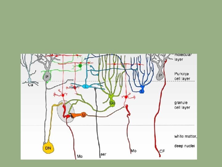
Neural connections of the cerebellum I. cerebellar circuit consists: cortical sections subcortical sections climbing fiber E. D’Angelo and S. Casali - 2013 Cortical circuit is organized as a feedforward excitatory chain assisted by inhibitory loops: mossy fibers excite granule cells, granule cells activate all the other cortical elements: Purkinje, Basket, Stellate - in granular layer, inhibition is provided by Golgi Cells, - in molecular layer inhibition : Stellate Cells and Basket Cells - finally, Purkinje Cells inhibit Deep Cerebellar Nucl. - Inferior Olive, activated by brain stem and spinal cord nuclei, controls Purkinje Cell activity through a single powerful synapse -the whole system can be seen as a complex mechanism controlling the Deep Cerebellar Nuclei output

Neural connections of the cerebellum I. - At subcortical level, the afferent fibers activate Deep Cerebellar nuclei cells (DCN-C) and Inferior Olive cells (IO-C). - Deep Cerebellar Nucl. emits the output and at the same time inhibits the Inferior Olive -two main inputs: - mossy fibers (mf) originating in brain stem and spinal cord nuclei: - diverge to Deep Cerebellar N. and activate the gran. layer (Granule Cells + Golgi Cells) mossy fibers - axon of the Granule Cells bifurcates in the molecular layer: parallel fibers (pf) climbing fibers (cf) originating from the Inferior Olive inferior olive deep cer. nuclei E. D’Angelo and S. Casali - 2013

Modular organization of cerebellum four ideal zones are shown in color each one containing microzones forming a multizonal microcomplex inhibitory interneurons: blue E. D’Angelo and S. Casali - 2013 - a microzone is defined as a group of the order of 1000 Purkinje cells all having same somatotopic receptive field - these Purkinje cells are arranged in a long, narrow strip, oriented perpendicular to cortical folds - Purkinje dendrites flattened in same direction as microzones extend, crossed by parallel fs at right angles - climbing fibers branches (about 10) usually innervate Purkinje cells belonging to same microzone - olivary neurons generating such climbing fibers tend to be coupled by gap junctions !!!!!!! -this helps synchronizing Purkinje cells within a microzone on a millisecond time scale - Purkinje cells in a microzone all send their axons to the same small cluster of output cells of deep cerebellar nuclei - finally, basket cells axons are confined largely to a single microzone - cellular interactions within a microzone are much stronger than those between different microzones

Motor and somatosensory loops cerebellum can modulate motor cortex excitability - in relation to the incoming sensory input Dorsolateral prefrontal cortex (middle frontal gyrus) executive functions: working memory cognitive flexibility planning inhibition abstract reasoning Dorsolateral prefrontal cortex (middle frontal gyrus) E. D’Angelo and S. Casali - 2013 HIGHEST CORTICAL AREA THAT IS INVOLVED IN MOTOR PLANNING, ORGANIZATION AND REGULATION Loops formed with basal ganglia Bidirectional connections 1. From cerebellum: dentate nucleus – thalamus – striatum 2. From basal ganglia: subthalamic nucleus – pontine nuclei - cerebellar cortex Loops formed with limbic system Cerebellum may be connected with: -amygdala, hippocampus, septal nuclei, hypothalamus

From motor control to cognition and emotion Motor control 1. impairment of body balance + eye movement control 2. impairment of gait 3. difficulty executing voluntary, planned movements Lateral cerebellum is involved, through cerebro-cerebellar loops, in the cognitive components of movement planning Functional neuroimaging studies show - activation of the posterior lobe during cognitive planning, working memory, language (verbal memory tasks, verb for noun substitution, synonym generation), mental imagery (“visualizing, ” “seeing in the mind's eye, ” “hearing in the head, ” “imagining the feel of”) sensory discrimination Dysmetria of thoughts or cognitve dysmetria 1. impairment of planning, set shifting, abstract reasoning, working memory, verbal fluency 2. difficulties with spatial cognition, both in visuospatial organization and visuospatial working memory 3. personality change, with blunting of affect and/or disinhibited and inappropriate behavior 4. language deficits including agrammatism cerebellar cognitive affective syndrome Similar to prefrontal syndrome

Neural connections of the cerebellum

Function of the cerebellum • cerebellum is proposed to play a critical role in the learning and execution of both voluntary and certain reflex movements • Motor learning function • Cerebellum utilizes somatosensory information • Cerebellum utilizes vestibular information • Cerebellum utilizes visual information - activates vermis (midway – head area) – participation in movement toward the stimulus - flocculus controls the eye movements • Cerebellum utilizes auditory information - activates vermis (midway – head area) – participation in movement toward the stimulus • Cerebellum utilizes olfactory information • Cerebellum utilizes visceral information • Cerebellum is involved in cognitive functions - fibers from association cortex especially from prefrontal cortex

Anatomical bases of cerebellar syndromes • damage to the cerebellum leads to deficits in motor coordination • damage or loss of the cerebellum: no paralysis, no loss of sensation, no inability to understand a task • but it leads to an inability to perform movements well - difficulty executing voluntary planned movements (dysfunction of cerebrocerebellum) • damage to the cerebellum impairs motor function on the ipsilateral side of the body • If the flocculonodular lobe is damaged, nystagmus (vestibulocerebellum), motor disorders resemble those produced by a lesion of the vestibular apparatus • difficulty in balance and wide based gait, uneven steps – „drunken sailor” (spinocerebellum)

Anatomical bases of cerebellar syndromes Microcephaly, disproportionate pontine and cerebellar hypoplasia syndrome: A clinico-radiologic phenotype linked to calcium/calmodulin-dependent serine protein kinase gene mutation (2 -year-old girl ) http: //www. ijhg. com/articles/2013/19/1/images/Indian. JHum. Genet_2013_19_1_104_112921_u 1. jpg Cerebellar hypoplasia - „Our larger study confirms that the combination of speech delay, ataxia, autistic features, and ocular signs (nystagmus, strabismus, and abnormal ocular movements) can predict the occurrence of cerebellar hypoplasia. ” E Wassmer, P Davies, W P. Whitehouse, S H. Green: Clinical Spectrum Associated With Cerebellar Hypoplasia. http: //www. youtube. com/watch? v=Dox 3_ox 8 C 2 U

Dyslexia and cerebellum Developmental dyslexia a selective difficulty in acquiring reading skills, in spite of normal general intelligence Dyslexics show small right cerebellar anterior lobes on MRI

the essential components of culture are learned and sustained not by the cerebral cortex alone as many traditionally believe, but are learned through repetitious improvements in prediction and control by internal models in the cerebellum. following new explanations of culture are (1) culture can be learned unconsciously but yet be socially in sync with others, (2) the recent evolutionary expansion of the cerebellum was involved in the coevolution of earliest stone tools and language—leading to the cerebellum-driven origin of culture, (3) cerebellar internal models are blended to produce the creative, forward advances in culture, (4) the blending of cerebellar internal models led to human, multi-component, infinitely partitionable and communicable working memory, (5) how excessive television viewing may represent a cultural shift that diminishes the observational learning of internal models of the behavior of others cerebellar cognitive affective syndrome -lowering of intellectual function, - it is now thought that the cerebellum is responsible for monitoring both motor and nonmotor functions. nonmotor deficits are believed to be caused by dysfunction in cerebellar connections to cerebral cortex, limbic system problems with planning, set-shifting, abstract reasoning, verbal fluency, working memory, often perseveration, inattention

Annotation Ataxia = Loss of ability to coordinate voluntary muscular movements ; muscle incoordination and gait unsteadiness

References Egidio D’Angelo and Stefano. Casali Seeking a unified framework for cerebellar function and dysfunction: from circuit operations to cognition. REVIEW ARTICLE published: 10 January 2013 , Frontiers in neuronal circuits http: //www. humpath. com/IMG/jpg_cerebellum_brainstem_1 m_12_2. jpg https: //www. google. hu/url? sa=i&rct=j&q=&esrc=s&source=images&cd=&cad=rja&uact=8&docid=J 5 Ck. RVb. Hr 157 TM&tbnid=JYj. NUSDt. WCu. RHM: &ved=0 CAc. Qj. Rw&url = http%3 A%2 F%2 Fwww. absurdintellectual. com%2 F 2011%2 F 02%2 F 10%2 Ftwo-dimensional-trees-the-technique-of-espalier%2 F&ei=KKQp. VL 7 NG 4 LQyg. Oq 74 G 4 Dg&bvm =bv. 76247554, d. b. GQ&psig=AFQj. CNHw. Yokbp 8 Exc. CBx. Vtlnlfjzlqfr. IA&ust=1412101421702001
- Slides: 35