Development of cerebral hemisphere and cerebellum 2 Embryology

Development of cerebral hemisphere and cerebellum ﴾ ﴿ ﺍ ﺍ ﺍﻹﻧﺴﺎﻥ ﺍ ﻳﺍ ﻳﺍ : 2]. ]ﺍﻹﻧﺴﺎﻥ Embryology 436 MEDICINE KING SAUD UNIVERSITY Important Dr. notes Explanation - We recommend you to study anatomy of cerebrum and cerebellum

OBJECTIVE • Describe the formation of the neural tube. • List the 3 brain vesicles and their derivatives. • Describe the brain flexures. • Describe briefly the development of the cerebrum. • Describe briefly the development of the cerebellum. • Enumerate some congenital anomalies in development of CNS.

Development of Cerebrum And Cerebellum Remember. . By the beginning of the 3 rd week(15 days) of development, three germ cell layers become established The Three Germ Layers: 1 - Ectoderm 2 -Mesoderm 3 -Endoderm (outer) (middle) (inner) - Early devolpment During the middle of the 3 rd week(16 -17 days)the dorsal midline ectoderm undergoes thickening to form the neural plate (neuroectoderm). • The 2 margins of the plate elevate, forming neural folds • a longitudinal, midline depression, called the neural groove is formed. • The 2 neural folds approximate then fuse together, thus sealing the neural groove and creating the neural tube. (complete close) ﻣﺮﺍﺣﻞ 3 ﺗﻜﻮﻥ ﺍﻟﻨﻴﻮﺭﺍﻝ ﺗﻴﻮﺏ ﻣﻦ ﺧﻼﻝ ectoderm neural plate ﺃﻮﻝ ﺷﻴﺀ ﺧﻼﻳﺎ ﺍﻻﻛﺘﻮﺩﻳﺮﻡ)ﻣﻦ ﺟﻬﺔ ﺍﻟﺪﻭﺭﺳﺎﻝ ﻣﻴﺪﻻﻳﻦ( ﺗﺘﻜﺜﻒ ﻭﺗﻜﻮﻥ neural groove ﺑﻌﺪﻳﻦ ﺗﺒﺪﺃ ﺗﺮﺗﻔﻊ ﻃﺒﻘﺔ ﺍﻟﺨﻼﻳﺎ ﻣﻦ ﺍﻟﺠﻬﺘﻴﻦ )ﺍﻻﻃﺮﺍﻑ( ﻭﺗﺼﻴﺮ ﻛﺄﻨﻬﺎ ﺯﺑﺪﻳﺔ )ﺻﺤﻦ ﻋﻤﻴﻖ(ﻭﺗﻜﻮﻥ neural tube ﻭﺗﻠﺘﺤﻢ ﺳﻮﺍ ﻭﺗﻜﻮﻥ ﺍﻝ neural folds آﺨﺮ ﺍﻟﺸﻴﺀ ﺍﻟﺠﻬﺘﻴﻦ ﺍﻟﻤﺮﺗﻔﻌﺔ ﺗﻜﻮﻥ - Formation of neural tube is completed by the middle of 4 th week.

Neural Tube Development Recall: Brain develops from cranial 1/3 of neural tube. ﻳﻌﻨﻲ ﻛﻞ ﺍﻟﻤﺮﺍﺣﻞ ﺍﻟﺠﺎﻳﺔ ﺍﻟﻠﻲ ﺣﺘﺼﻴﺮ ﻓﻲ ﺍﻟﻨﻴﻮﺭﺍﻝ ﺗﻴﻮﺏ ﺭﺡ ﺗﻜﻮﻥ ﻓﻲ ﺍﻟﻜﺮﻳﻨﺎﻝ ﺑﺎﺭﺕ - -: ( ﻧﻘﺴﻢ ﺍﻟﻤﺮﺍﺣﻞ ﺍﻟﺠﺎﻳﺔ ﺣﻠﺘﻴﻦ )ﻣﺮﺣﻠﺔ ﺃﻲ ﻭ ﻣﺮﺣﻠﺔ ﺑﻲ (A)-Three- primary brain vesicles stage (end of 4 th (B)-five secondary brain vesicles stage (5 th week)( )ﺗﻜﻤﻠﺔ ﺍﻧﻘﺴﺎﻣﺎﺕ ﺍﻝ ﺑﺮﺍﻳﻤﺮﻱ ﻓﻴﻨﺘﺮﻛﻠﺰ Neural tube upper end dilates and shows 3 vesicles Prosencephalon divides into: (two lateral parts and the other part in the middle week) (28 days) between them) This 3 vesicles are: (from up to down) 1 - prosencephalon 2 - mesencephalon* 3 -Rhombencephalon (forebrain) (midbrain) (hindbrain) Prosencephalon (forebrain) Cranial 1/3 of neural tube ( )ﻭﺣﺪﺓ ﺑﺎﻟﻴﻤﻴﻦ ﻭﺍﻟﺜﺎﻧﻴﻪ ﺑﺎﻟﻴﺴﺎﺭ 1 -telencephalon it is two lateral 2 -diencephalon ( )ﺗﻜﻮﻥ ﺑﺎﻟﻮﺳﻂ ﺑﻴﻦ ﺍﻟﺠﺰﺋﻴﻦ ﻣﻦ ﺍﻟﺘﻴﻠﻨﺲ ﺳﻴﻔﻠﻮﻥ Rhombencephalon divides into: 1 -metencephalon 2 - myelencephalon telencephalon diencephalon Mesencephalon (midbrain) A Rhombencephalon (hindbrain) Mesencephalon *ﻧﻼﺣﻆ ﺍﻥ ﻫﻲ ﺍﻟﻮﺣﻴﺪﺓ ﺍﻟﻲ ﻣﺎ ﺍﻧﻘﺴﻤﺖ (cerebrum) 2 lateral parts thalamus midbrain ﺯﻱ ﻣﺎﻋﺮﻓﻨﺎ ﺍﻧﻬﺎ ﻣﺎﺭﺡ ﺗﻨﻘﺴﻢ ﺯﻱ ﺑﺎﻗﻲ ﺍﻻﺟﺰﺍﺀ metencephalon myelencephalon (cerebellum+pons) (medulla) don’t confuse : the blue area around the cavities is the one responsible of formation of grey and white matter. (Cavities will latterly form the ventricles of the brain) B

Brain Flexures - By the 4 week, the neural tube grows rapidly and faster than cranial cavity. (This lead to form brain flexures) )ﻫﺬﻱ ﺍﻻﻧﺜﻨﺎﺀﺍﺕ ﺗﺰﻭﺩ ﻣﺴﺎﺣﺔ ﺳﻄﺢ ﺍﻟﺒﺮﻳﻦ -There are 3 brain flexures ﺍﻧﺜﻨﺎﺀﺍﺕ : ( 1 - Cervical flexure (ventral) 2 - Midbrain flexure (ventral) 3 - Pontine flexure (dorsal flexure) - The neural tube grows rapidly and bends ventrally, producing two flexures: 1 -Cervical flexure: Between the hind brain and the spinal cord. 2 - Midbrain flexure: between the prosencephalon and the mesencephalon (midbrain). )( ﻓﻠﻴﻜﺸﺮﺯ ﻳﺘﻜﻮﻧﻮﺍ ﺍﻭﻝ ﺷﻲ)ﻓﻴﻨﺘﺮﺍﻝ( ﻭﺑﻌﺪﻫﻢ ﻳﺘﻜﻮﻥ ﺍﺧﺮ ﻭﺍﺣﺪ)ﺩﻭﺭﺳﺎﻝ 2 ﻋﻨﺪﻱ ﻫﺬﻭﻝ ﺍﻝ 3 - Pontine flexure (dorsal flexure): appears later in the hindbrain, in the opposite direction, thinning of the roof of the hindbrain. . ﻫﺬﺍ ﻣﻌﻨﻰ ﺍﻥ ﺍﻟﺒﻮﻧﺘﺎﺑﻦ ﻓﻠﻴﻜﺸﺮ ﻳﺴﻮﻱ Thinning of the roof

Derivatives of Brain Vesicles *This slide is very impotant * - Its important to know the origin of each derivative Primary Brain Vesicles Secondary Brain Vesicles Derivates In Mature Brain thalamus Prosencephalon (forebrain) 1 -Diencephalon(median part) 2 -Two telencephalon (lateral part) Cerebral hemisphere 1 -mesencephalon midbrain 1 -metencephalon Pons (anterior part) Cerebellum (posterior part) 2 - myelencephalon Medulla ablongata Mesencephalon (midbrain) Rhombencephalon (hindbrain)
 or the forebrain vesicle differentiates into a: 1")
Development of The Cerebrum -The (prosencephalon) or the forebrain vesicle differentiates into a: 1 -Telencephalon (medial part) (it’s cavity forms two lateral ventricles). 2 -Diencephalon (lumen) (it’s cavity forms 3 rd ventricle). -Both cavities communicating with each other through a wide interventricular foramen(foramen of monro) -The cerebral hemispheres first appear on the day 32( ) ﺑﻌﺪ ﺷﻬﺮ ﻭﻳﻮﻣﻴﻦ of pregnancy as a pair of bubble-like outgrowths of the Telencephalon. By 16 weeks( ﺷﻬﻮﺭ 4 )ﺑﻌﺪ , the rapidly growing hemispheres are oval and have expanded back to cover the diencephalon. ( )ﻳﻌﻨﻲ ﻳﺘﻤﺪﺩ ﻣﻦ ﻛﻞ ﺍﺗﺠﺎﻩ ﻭﻳﻐﻄﻲ ﺍﻟﺪﻳﻨﺴﻴﻔﺎﻟﻮﻥ 9 ﺍﻋﺮﻓﻮﺍ ﺍﻥ ﻓﻲ ﺍﻟﺒﺪﺍﻳﺔ ﻳﻜﻮﻥ ﺷﻜﻠﻬﺎ ﺍﻭﻓﺎﻝ ﻻﻥ ﻑ ﺳﻼﻳﺪ ﺣﻴﺘﻐﻴﺮ ﺷﻜﻠﻬﺎ -The cerebral hemispheres expand in all directions. -Its medial wall becomes thin, flat and it is the site of choroid plexus of the lateral ventricle. - By the end of the 3 rd month the surfaces of the cerebral hemispheres are ( smooth and no gyri) - By the 4 th month , grey matter is growing faster than white matter - folding of the cortex > formation of gyri and sulci. - the cortex becomes folded into gyri separated by sulci. -The gyri and sulci effectively increase the surface area of the brain. - The detailed pattern of gyri and sulci varies between individuals ﻭﺑﺘﻜﺒﺮ ﺍﻟﻬﻴﻤﺴﻔﻴﺮ ﻭﺗﻤﺪﺩ ﻭﺭﺍﺡ ﻳﻜﻮﻥ ﻓﻴﻬﺎ ﻣﻨﻄﻘﻪ ﻣﺴﻄﺤﺔ ﻳﺘﻜﻮﻥ ﻓﻴﻬﺎ to produce (CSF) choroid plexus

Development of The Cerebrum -The wall of the telencephalon is formed of 3 layers : 1 - Ependymal(inner): (lining the cavity of the lateral ventricle). 2 - Mantel (middle): nerve cells forming the grey matter. 3 - Marginal (outer): nerve fibers forming the white matter. -As development proceeds, the following changes occur as: - Most of the nerve cells in mantel layer migrate to the marginal layer forming the cerebral cortex. -Some cells do not migrate and remains to form the basal ganglia. - Corpus striatum: (it is nuclei of basal ganglia) - appears in 6 th week ﻓﻴﻨﺘﺮﻳﻜﺎﻝ ﻭﺍﻟﻤﺎﺭﺟﻴﻨﺎﻝ ﺑﺎﻟﺨﺎﺭﺝ ﻭﺗﺤﺘﻮﻱ ﻋﻠﻰ ﻧﻴﺮﻑ ﻭﻫﻲ ﺗﻜﻮﻥ ﺍﻟﻮﺍﻳﺖ ﻣﺎﺗﺮ ﺍﻣﺎ ﺍﻟﻤﻴﻨﺘﺎﻝ ﺗﻜﻮﻥ ﺑﺎﻟﺪﺍﺧﻞ ﻭﻫﻲ ﺍﻟﻠﻲ ﺑﺘﻜﻮﻥ ﻟﻲ ﺍﻟﻘﺮﻱ ﻣﺎﺗﺮ ﺑﻌﺪﻳﻦ ﺭﺍﺡ ﺗﻬﺎﺟﺮ ﺍﻟﻤﺎﻧﺘﺎﻝ ﻟﺒﺮﺍ ﻋﺸﺎﻥ ﺗﻜﻮﻥ ﺍﻟﺴﻴﺮﺑﺮﺍﻝ ﻛﻮﺭﺗﻴﻜﺲ ﻭﻳﺒﻘﻰ ﻣﻨﻬﺎ ﺟﺰﺀ ﺻﻐﻴﺮ ﻣﺎ ﻳﻬﺎﺟﺮ ﻫﺬﺍ ﺍﻟﺠﺰﺀ ﺭﺍﺡ ﻳﻜﻮﻥ ﺍﻟﺒﻴﺴﻞ ﻗﺎﻧﻘﻠﻴﺎ in the floor of each hemisphere. - Cell bodies in the cortex differentiate and their fibers passing (as internal capsule) through (corpus striatum) to divide it into caudate and lentiform nuclei. -This fiber pathway forms( the internal capsule).
 ﺃﺸﻴﺎﺀ")
Development of The Cerebrum Rapid growing (sulci and gyri + C-shape + Insula) ﺃﺸﻴﺎﺀ ﻣﻊ ﺍﻟﺘﻜﻮﻳﻦ ﺭﺡ ﻭﺗﺎﺧﺪ ﺷﻜﻞ ﺍﻝ ﺣﺮﻑ ﺍﻝ 3 ﻓﻲ ﺍﻟﺒﺪﺍﻳﺔ ﻋﻨﺪﻱ : ﺳﻲ ﻛﻮﺩﺍﻝ ﻧﻴﻮﻛﻠﻴﺲ 1 ﻻﺗﺮﺍﻝ ﻓﻴﻨﺘﺮﻳﻜﺎﻝ 2 oval) ﻧﺘﺬﻛﺮ ﻛﺎﻥ ﺷﻜﻠﻪ ﻗﺒﻞ : ﺍﻟﻬﻴﻤﺴﻔﺴﺮ 3 - -Further expansion of hemispheres gives it C- shape as well as its cavity (lateral ventricle). including caudate nucleus which elongates to assume the C- shape - The cortex covering the surface of the corpus striatum grows slowly compering to other area , and that will push this area (called insula) inside to the depth of the lateral sulcus of the brain. - So, the insular lobe is a portion of cerebral cortex that has invaginated to lie deep within the lateral sulcus. ﺍﻟﻜﻮﺭﺗﻴﻜﺲ ﺍﻟﻠﻲ ﺗﻐﻄﻲ ﺍﻟﻜﻮﺭﺑﺲ ﺳﺘﺮﺍﻳﺘﻮﻡ ﻳﻜﻮﻥ ﻧﻤﻮﻫﺎ ﺃﺒﻄﺊ ﻣﻦ ﺃﻲ ﺟﺰﻱ ﺛﺎﻧﻲ ﻣﻦ ﺍﻟﻜﻮﺭﺗﻴﻜﺲ ﻋﺸﺎﻥ ﻛﺬﺍ ﺑﻴﺘﻤﺪﺩ ﻛﻞ ﺍﺟﺰﺍﺀ ﺍﻟﻜﻮﺭﺗﻴﻜﺲ ﻣﺎ ﻋﺪﺍ ﻫﻲ ﺑﺘﻜﻮﻥ ﻣﺘﻜﻮﻧﻪ ﺩﺍﺧﻞ ﻭﺗﻜﻮﻥ ﻟﻲ ﺍﻝ insula Cerebral commissures: -Cerebral cortex develops fibers connect between the corresponding regions in right and left hemisphere. These are : ( )ﺍﻋﺮﻓﻮﻫﻢ ﻛﺄﺴﻤﺎﺀ ﻓﻘﻂ ﻻ ﺗﺪﺧﻠﻮﻥ ﺑﺎﻟﺘﻔﺎﺻﻴﻞ • Optic chiasma • Anterior and posterior commissures. • Hippocampal commissure. • Habenular commissure. • Lamina terminalis. • Corpus callosum: (is a major commissural fibres that connect the two cerebral hemispheres).

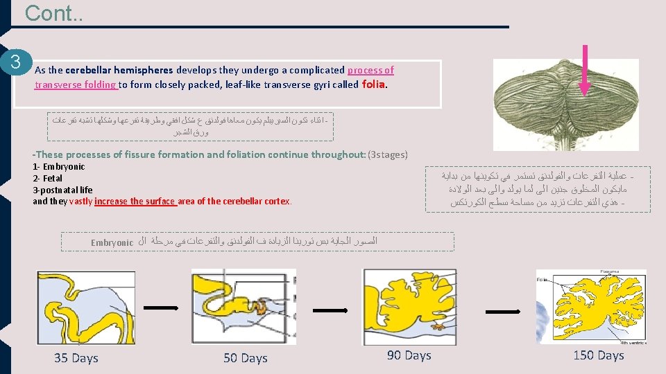
Development of The Cerebellum * *ﺍﻧﺘﺒﻬﻮﺍ ﻫﻨﺎ ﺣﻨﺒﺪﺃ ﻧﺘﻜﻠﻢ ﻋﻦ ﺍﻟﻤﺨﻴﺦ ﺧﻼﺹ ﺧﻠﺼﻨﺎ ﺗﻜﻮﻳﻦ ﺍﻟﻤﺦ *This silde is very important * Extra -The metencephalon develops into: 1 - pons (anterior part) 2 - cerebellum (dorsal part). 1 - Pontine flexure - results in: In the middle of pone -1. 2. 3. 2 Moving 2 the alar plates laterally then pending medially. Stretching and thinning of the roof plate Widening of the cavity to form the 4 th ventricle -The dorsal parts thicken to form Rhombic lips , that will give rise to the cerebellum. Every pontine flexure has (2 basal plate and 2 alar plate). . . ﺍﻻﺭ ﺑﻠﻴﺘﺲ 2 ﺍﺣﻨﺎ ﻳﻬﻤﻨﺎ ﺍﻟﺒﻮﻧﺘﺎﻳﻦ ﻓﻠﻴﻜﺸﺮ ﻳﺘﻜﻮﻥ ﻋﻨﺪﻱ ﻣﻦ ﻧﺺ ﺍﻟﺒﻮﻥ ﻟﻴﺶ ﺍﻟﺒﻮﻥ ؟ ﻻﻥ ﺍﻟﺒﻮﻥ ﻭﺍﻟﺴﻴﺮﻳﺒﻠﻢ ﻳﻄﻠﻌﻮﺍ ﻣﻦ ﻧﻔﺲ ﺍﻟﻤﻜﺎﻥ ﻭﺟﻨﺐ ﺑﻌﺾ ﺍﻻﺍﺭ ﺑﻠﻴﺘﺲ ﺗﺘﺤﺮﻙ ﻻﺗﺮﺍﻝ ﻟﺒﺮﺍ ﻟﻮ ﺗﻼﺣﻈﻮﺍ ﻑ ﺍﻟﺼﻮﺭﺓ ﺍﻟﺜﺎﻧﻴﺔ ﻭﺑﻌﺪﻳﻦ ﺗﻤﻴﻞ ﻣﻴﺪﻳﺎﻟﻲ ﻣﻦ ﺗﺤﺖ 2 ﺍﻟﻤﻬﻢ ﻭﺑﻜﺪﺍ , ﺍﻟﺮﻭﻭﻑ ﻣﻦ ﻓﻮﻕ ﻳﺼﻴﺮﻟﻪ ﺍﺳﺘﺮﻳﺘﺲ ﻭﺑﻜﺬﺍ ﺍﺣﻨﺎ ﻛﺒﺮﻧﺎ ﻣﺴﺎﺣﺔ ﺍﻝ ﻛﺎﻓﺘﻲ ﻑ ﺍﻟﻨﺺ ﻋﺸﺎﻥ ﻳﻜﻮﻧﻠﻲ ﺍﻝ ﻓﻴﻨﺘﺮﻛﺎﻝ ﺍﻟﺮﺍﺑﻊ (very important) -Some neuroblasts migrate from the mantel layer to the marginal layer and form the cerebellar cortex. ﻧﻔﺲ ( )ﻓﻜﺮﺓ ﺍﻟﺴﺮﻳﺒﺮﺍﻝ ﻛﻮﺭﺗﻴﻜﺲ -Others remains in the mantel layer and give rise to the cerebellar nuclei ( )ﻧﻔﺲ ﻓﻜﺮﺓ ﺍﻟﺒﻴﺰﺍﻝ ﻗﺎﻧﻘﻠﻴﺎ ﻱ ﺍﻟﺴﺮﻳﺒﺮﻡ - The cerebellar peduncles develop later on, as the axons of the neurons of the cerebellar nuclei grow out to reach the brain stem

Congenital Anomalies of The Brain *This slide is very important * Extra 1 - Hydrocephalus: Increase secretion of (CSF) and decrease absorption of it. accumulation of cerebrospinal fluid 2 - Cranium bifidum: with or without meningocele (the meninges are exposed) and meningoencephalocele (the meninges with some brain tissue are exposed) ﺑﺎﻟﺘﺎﻟﻲ ﻧﻼﻗﻲ ﺍﻧﻪ ﻓﻲ ﺯﻳﺎﺩﺓ ﻓﻲ ﺣﺠﻢ ﺍﻟﺪﻣﺎﻍ 4 - Arnold-Chiari malformation : (herniated part of cerebellum (tonsils)through the foramen magnum leading to (CSF obstruction , and hydrocephalus ) also in aqueductal stenosis and in brain tumours. )ﻳﺼﻴﺮ ﺗﻮﻧﺴﻼﻳﺘﺲ ﺗﺘﺤﺮﻙ ﻣﻦ ﻣﻜﺎﻧﻬﺎ ﻭﺗﻨﺰﻝ ﻋﻨﺪ ﺍﻟﻔﻮﺭﻣﺎﻥ ﻣﺎﺟﻨﻢ ﻭﺗﺴﻮﻱ ( ﻛﻞ ﺍﻟﻤﻀﺎﻋﻔﺎﺕ ﺍﻟﻲ ﻗﻮﻟﻨﺎ ﻋﻠﻴﻬﺎ 6 -Mental retardation. ( )ﺗﺨﻠﻒ ﻋﻘﻠﻲ 7 - Seizures (changes in electrical activity). 8 - Cerebral palsy. ( )ﺷﻠﻞ ﻛﺎﻣﻞ 9 - Agenesis of corpus callosum. ) ( )ﻣﻦ ﺍﻟﺠﻬﺘﻴﻦ ﻣﺎ ﺍﺭﺗﺒﻄﻮﺍ ﺑﺒﻌﺾ ﺍﻭ ﻣﺎﺗﻜﻮﻥ ﺃﺴﺎﺳﺎ 3 - Microcephaly: ( )ﺗﻜﻮﻳﻦ ﺍﻟﺪﻣﺎﻍ ﻏﻴﺮ ﻣﻜﺘﻤﻞ (abnormal smallness of the head, a congenital condition associated with incomplete brain development). 5 - Anencephaly: (no brain) It is due to failure of closure of the cranial neuropore of the neural tube. the brain and skull are minute and the infant does not usually survive. The frequency of this case 1: 1000. ﻭﻻﺩﺓ 1000 ﻳﺤﺪﺙ ﻓﻲ ﻭﻻﺩﺓ ﻭﺍﺣﺪﺓ ﻣﻦ ﻛﻞ. . ﻫﻮ ﺟﺪﺍ ﻧﺎﺩﺭ ﻣﺎﻳﻘﺪﺭ ﻳﻌﻴﺶ ﺑﻌﺪ ﺍﻟﻮﻻﺩﺓ ﺍﻻ ﻛﻢ ﺳﺎﻋﻪ ﺛﻢ ﻳﻤﻮﺕ Only in boys slide Neural tube has upper and lower opening. -The upper opening close at 23 -26 day , if it not close it will cause anencephaly -Lower will close at 27 dayes , if it not close , it will cause spinal bifid.

SUMMARY Time Changes Beginning of the 3 rd week Formation of 3 germ cell layers (ectoderm, mesoderm, endoderm) Middle of 3 rd week Forming Neural plate (Beginning of neural tube formation) 4 th week Forming brain flexures Middle of the 4 th week End of neural tube formation End of 4 th week Three vesicles stage (3 primary vesicles) 5 th week Five vesicles stage (2 ry brain vesicles) Development of The Cerebrum Day 32 (between 4 th and 5 th week) Cerebral hemispheres appear as pair of bubble-like outgrowths 6 th week Formation of corpus striatum 16 week Cerebral hemispheres are oval and have expanded back to cover the diencephalon The end of 3 rd month Smooth Surfaces of the cerebral hemispheres 4 th month The cortex become folded into gyri separated by sulci ﺗﺄﻜﺪﻭﺍ ﺍﻧﻜﻢ ﻓﺎﻫﻤﻴﻦ ﻛﻮﻳﺲ ﺍﻟﺴﻼﻳﺪﺍﺕ ﺍﻟﻠﻲ ﺟﻨﺒﻬﺎ ﻧﺠﻤﺔ -

MCQ’S 1 - which part of the embryonic ectoderm will thicken to form the neuroectoderm ? a- inner cell layers b- margins of the dorsal ectoderm c- the dorsal midline ectoderm 2 - the 2 cerebral hemispheres will be developed from : a- Prosencephalon by 5 th week b- telencephalon after the end of 5 th week c- diencephalon by the 5 th week 3 - the cerebellar peduncle will be developed from : a- cerebellar cortex reaching brainstem b- cerebellar nuclei reaching brain stem c- cerebral nuclei reaching cerebellum 4 - cerebellar growth will continue: a- before fetal life b- after postnatal life c- until postnatal life 5 - what also can cause hydrocephalus beside herniated cerebellum ? b- aqueductal stenosis c- both a and b are correct 1 - c 2 - B 3 - b 4 - c 5 - c a- brain tumor
. USEFUL • VIDEOS")
References ANY SUGGESTION OR ISSUE • Dr. slides (male and female). USEFUL • VIDEOS https: //www. youtube. com/watch? v=AWYNm. Usf. KWc • https: //www. youtube. com/watch? v=lhape. Oo 6 la. A&feature =youtu. be Embryology @Embryology 436@gmail. com Editing file Your Suggestion here

TEAM MEMBERS § TEAM LEADERS : Yazeed Al-mutairi Nehal Beyari. ▪ BOYS : • Mohammed Almutlaq • Muhanned Alzahrani ▪ GIRLS : • Razan Alotaibi • Thikrayat Omar • Do’aa Walid • Ohood Abdullah • Nouf Aloqili
- Slides: 16