ANEMIAS 2252021 Dr Alka Stoelinga 1 Hemolysis 2252021
- Slides: 45
ANEMIAS 2/25/2021 Dr. Alka Stoelinga 1
Hemolysis 2/25/2021 Dr. Alka Stoelinga 2
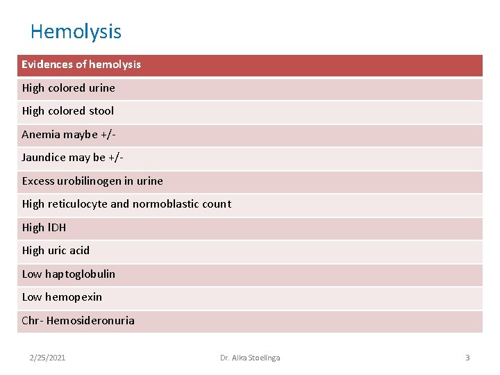
Hemolysis Evidences of hemolysis High colored urine High colored stool Anemia maybe +/Jaundice may be +/Excess urobilinogen in urine High reticulocyte and normoblastic count High l. DH High uric acid Low haptoglobulin Low hemopexin Chr- Hemosideronuria 2/25/2021 Dr. Alka Stoelinga 3
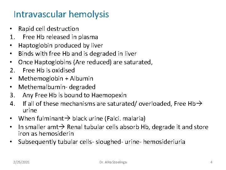
Intravascular hemolysis Rapid cell destruction Free Hb released in plasma Haptoglobin produced by liver Binds with free Hb and is degraded in liver Once Haptoglobins (Are reduced) are saturated, Free Hb is oxidised Methemoglobin + Albumin Methemalbumin- degraded Any Free Hb is bound to Haemopexin If all of these mechanisms are saturated/ overloaded, Free Hb urine • When fulminant black urine (Falci. malaria) • In smaller amt Renal tubular cells absorb Hb, degrade it and store iron as hemosiderin • Subsequently tubular cells- sloughed- urine- hemosideriuria • 1. • • • 2. • • 3. 4. 2/25/2021 Dr. Alka Stoelinga 4
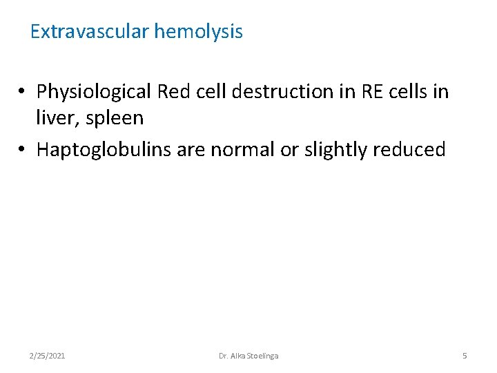
Extravascular hemolysis • Physiological Red cell destruction in RE cells in liver, spleen • Haptoglobulins are normal or slightly reduced 2/25/2021 Dr. Alka Stoelinga 5
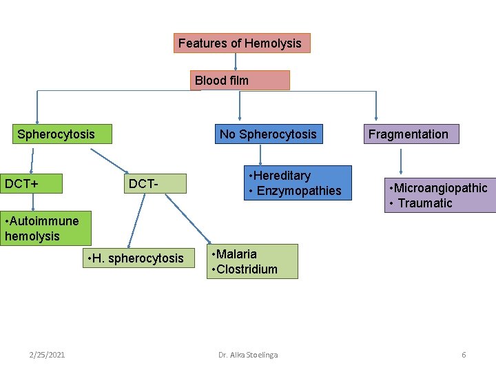
Features of Hemolysis Blood film Spherocytosis DCT+ No Spherocytosis DCT- • Hereditary • Enzymopathies Fragmentation • Microangiopathic • Traumatic • Autoimmune hemolysis • H. spherocytosis 2/25/2021 • Malaria • Clostridium Dr. Alka Stoelinga 6
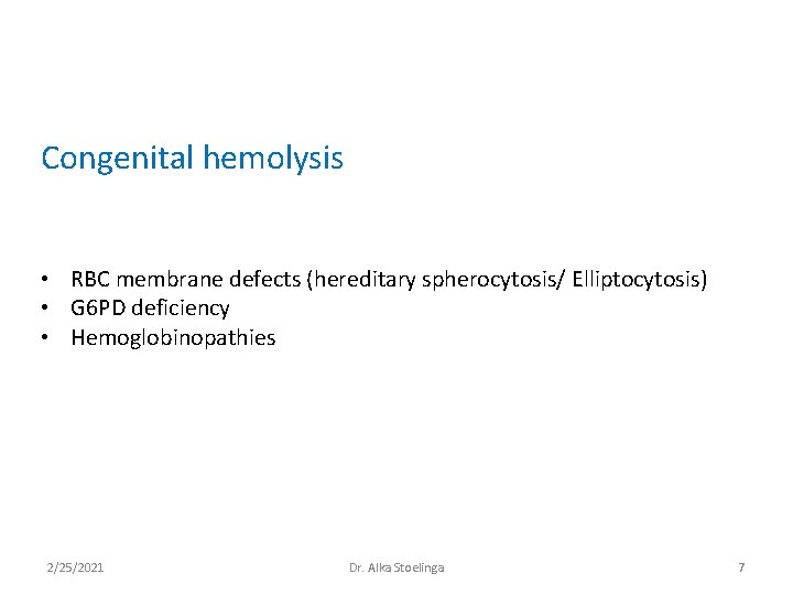
Congenital hemolysis • RBC membrane defects (hereditary spherocytosis/ Elliptocytosis) • G 6 PD deficiency • Hemoglobinopathies 2/25/2021 Dr. Alka Stoelinga 7
Red cell membrane defect • Defect in cytoskeleton • Usually due to quantitative or functional deficiency of one or more proteins in cytoskeleton • Cells loose their normal elasticity • Each time they pass through spleen, they lose membrane relative to their cell volume Raised MCHC Abnormal shape Reduced cell survival (EVH) 2/25/2021 Dr. Alka Stoelinga 8
Hereditary Spherocytosis • Autosomal dominant trait • 25% have no family history • Abnormalities in Beta spectrin and ankyrin Hemolytic crisis- when severity of hemolysis increases Megaloblastic crisis- follows folate deficiency Aplastic crisis- Parvovirus infection • Presents with severe anemia and low reticulocyte counts 2/25/2021 Dr. Alka Stoelinga 9
Investigations • Blood picture- presence of spherocytes • DCT –ve • Osmotic fragility test- Increased sensitivity to lysis in hypotonic saline solution Treatment • Folic acid prophylaxis 5 mg once weekly • Blood transfusion after cross matching • Consider splenectomy – – Growth retardation in children Recurrent severe crisis Death of a family member from the disease Symptomatic cholecystitis 2/25/2021 Dr. Alka Stoelinga 10
G 6 PD deficiency • HMP shunt • NADPH – protects red cells against oxidative stress • G 6 PD deficiency Impairs production of NADPH • Affects male; females are carriers Clinical Features: • Acute drug induced hemolysis • Chronic compensated hemolysis • Infection or acute illnesses • Neonatal jaundice • Favism (Vicia fava/ broad beans) 2/25/2021 Dr. Alka Stoelinga 11
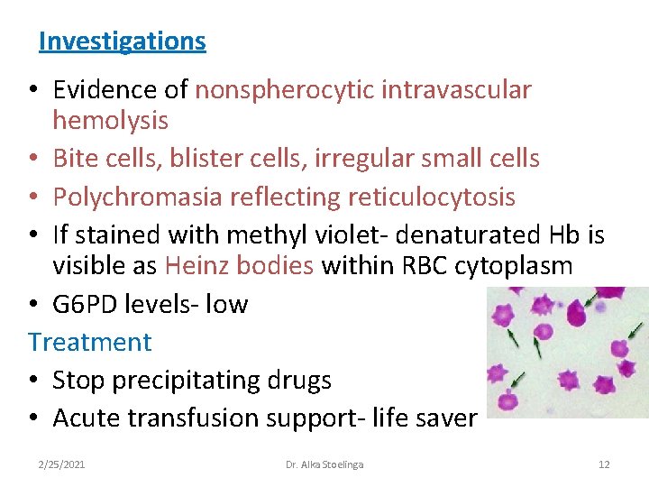
Investigations • Evidence of nonspherocytic intravascular hemolysis • Bite cells, blister cells, irregular small cells • Polychromasia reflecting reticulocytosis • If stained with methyl violet- denaturated Hb is visible as Heinz bodies within RBC cytoplasm • G 6 PD levels- low Treatment • Stop precipitating drugs • Acute transfusion support- life saver 2/25/2021 Dr. Alka Stoelinga 12
Autoimmune Hemolytic Anemia • Red cell autoantibodies RBC destruction • Ig. G/ M, rarely Ig. E or A CLASSIFICATION • Optimal temperature at which the antibody is active is used to classify AHA • 1. Warm antibodies – – bind best at 37⁰C Majority are Ig. G React against Rh antigens 80% of cases • 2. Cold antibodies – Bind best at 4⁰C but can bind upto 37⁰C – Majority are Ig. M and bind compliment – 20% of cases 2/25/2021 Dr. Alka Stoelinga 13
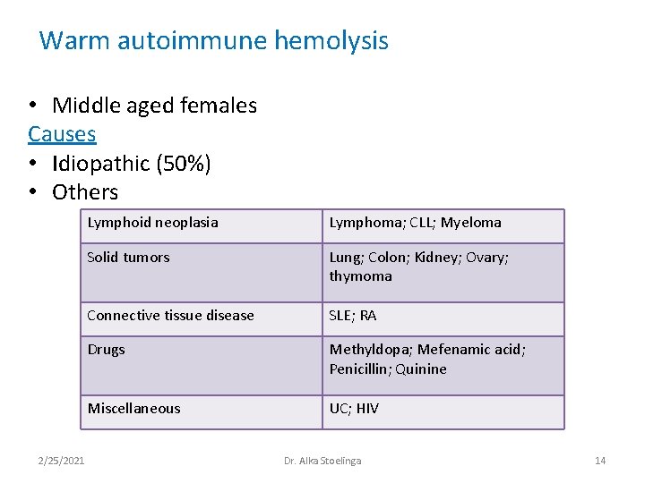
Warm autoimmune hemolysis • Middle aged females Causes • Idiopathic (50%) • Others 2/25/2021 Lymphoid neoplasia Lymphoma; CLL; Myeloma Solid tumors Lung; Colon; Kidney; Ovary; thymoma Connective tissue disease SLE; RA Drugs Methyldopa; Mefenamic acid; Penicillin; Quinine Miscellaneous UC; HIV Dr. Alka Stoelinga 14
Investigations • Hemolysis and spherocytes • DCT/ Antiglobulin test Treatment • Manage underlying condition • Stop offending drugs • Prednisolone 1 mg/kg • Blood transfusion after cross matching • Splenectomy to be considered • Immunosuppressive therapy- Azathioprim/ Cyclophosphamide 2/25/2021 Dr. Alka Stoelinga 15
Cold Agglutinin disease • Ig. M binds red cells at 4⁰C and cause them to agglutinate • Low grade intravascular hemolysis- cold, painful, blue fingers, toes, ears, nose (acrocyanosis) • Blood film- red cell agglutination • MCV- raised • Ig. M Treatment • Keep extremities warm during winter • Steroids • Cross matching and blood transfusion 2/25/2021 Dr. Alka Stoelinga 16
• The Direct Coombs test, is used to test for autoimmune hemolytic anemia. • In certain diseases or conditions an individual's blood may contain Ig. G antibodies that can specifically bind to antigens on the red blood cell (RBC) surface membrane, and their circulating red blood cells (RBCs) can become coated with Ig. G alloantibodies and/or Ig. G autoantibodies • Complement proteins may subsequently bind to the bound antibodies. • The direct Coombs test is used to detect these antibodies or complement proteins that are bound to the surface of red blood cells • Procedure: • A blood sample is taken and the RBCs are washed (removing the patient's own plasma) and then incubated with antihuman globulin (also known as "Coombs reagent"). If this produces agglutination of RBCs, the direct Coombs test is positive, a visual indication that antibodies (and/or complement proteins) are bound to the surface of red blood cells 2/25/2021 Dr. Alka Stoelinga 17
Hemoglobinopathies. Sickle cell anemia 2/25/2021 Dr. Alka Stoelinga 18
2/25/2021 Dr. Alka Stoelinga 19
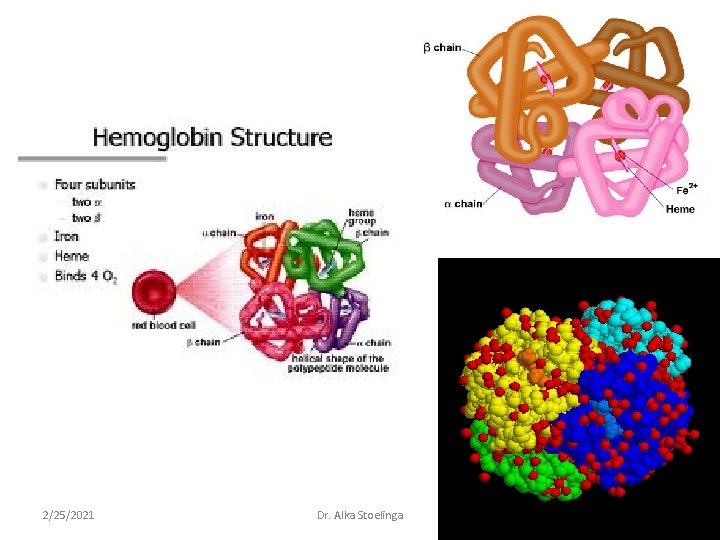
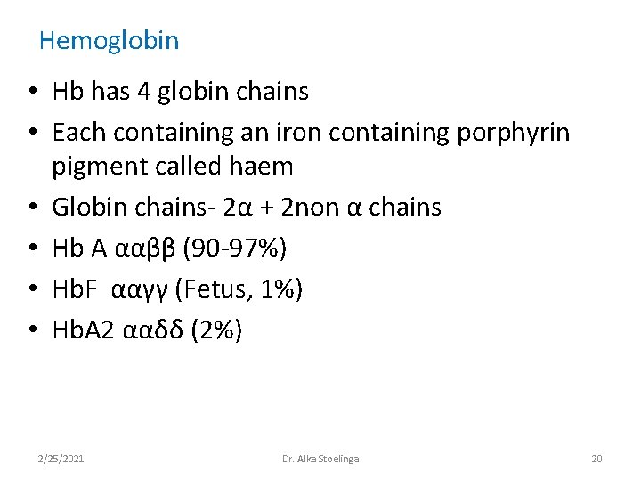
Hemoglobin • Hb has 4 globin chains • Each containing an iron containing porphyrin pigment called haem • Globin chains- 2α + 2 non α chains • Hb A ααββ (90 -97%) • Hb. F ααγγ (Fetus, 1%) • Hb. A 2 ααδδ (2%) 2/25/2021 Dr. Alka Stoelinga 20
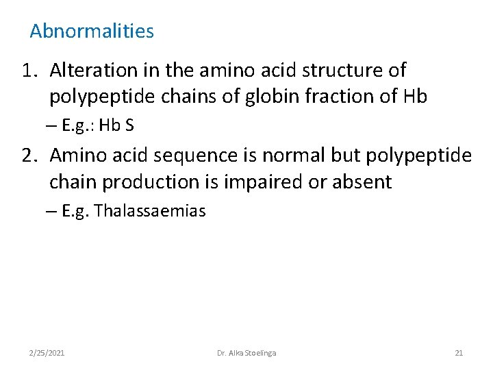
Abnormalities 1. Alteration in the amino acid structure of polypeptide chains of globin fraction of Hb – E. g. : Hb S 2. Amino acid sequence is normal but polypeptide chain production is impaired or absent – E. g. Thalassaemias 2/25/2021 Dr. Alka Stoelinga 21
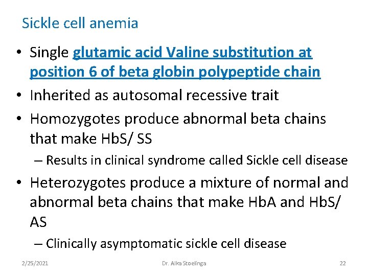
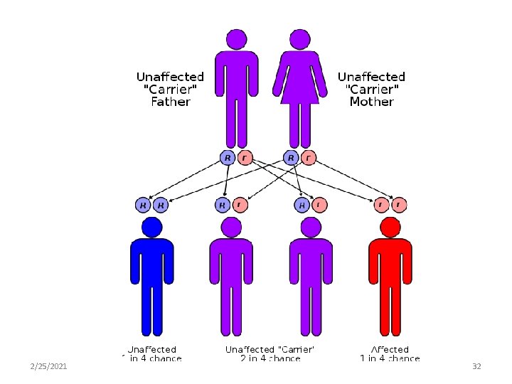
Sickle cell anemia • Single glutamic acid Valine substitution at position 6 of beta globin polypeptide chain • Inherited as autosomal recessive trait • Homozygotes produce abnormal beta chains that make Hb. S/ SS – Results in clinical syndrome called Sickle cell disease • Heterozygotes produce a mixture of normal and abnormal beta chains that make Hb. A and Hb. S/ AS – Clinically asymptomatic sickle cell disease 2/25/2021 Dr. Alka Stoelinga 22
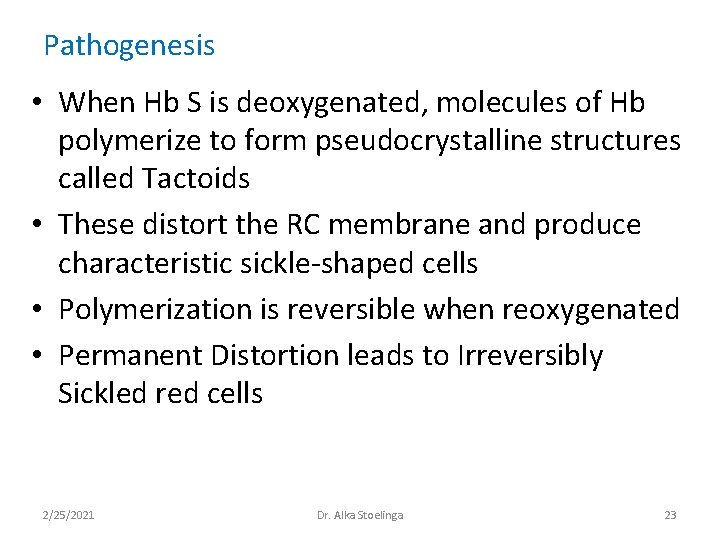
Pathogenesis • When Hb S is deoxygenated, molecules of Hb polymerize to form pseudocrystalline structures called Tactoids • These distort the RC membrane and produce characteristic sickle-shaped cells • Polymerization is reversible when reoxygenated • Permanent Distortion leads to Irreversibly Sickled red cells 2/25/2021 Dr. Alka Stoelinga 23
Note • Sickling is precipitated by: – Hypoxia – Acidosis – Dehydration – Infection • Irreversibly sickled cells have shortened survival • Plugs the vessels in microcirculation • This results in a no. of acute syndromes called crisis and chronic organ damage 2/25/2021 Dr. Alka Stoelinga 24
Clinical Features 1. Vaso Occlusive crisis (Most common) • Acute severe bony pain – Affects areas of active marrow – Hands and feet in children – Femora, humeri, ribs, pelvis and vertebrae in adults • Systemic response: – Tachycardia – Sweating – Fever 2/25/2021 Dr. Alka Stoelinga 25
Clinical Features 2. Sickle chest syndrome: • Follow vaso-occlusive crisis • Most common cause of death in Adult sickle cell disease a) Bone marrow infarction leads to fat emboli to lungs b) Ventilatory failure 2/25/2021 Dr. Alka Stoelinga 26
Clinical Features 3. • • • Sequestration crisis Thrombosis of venous outflow from organ Loss of function Acute pain Spleen (Children) Massive Splenomegaly severe anemia circulatory collapse death • Recurrent Sickling in spleen in children infarction • In adults No functional spleen • Capsular stretching Liver sequestration Severe pain 2/25/2021 Dr. Alka Stoelinga 27
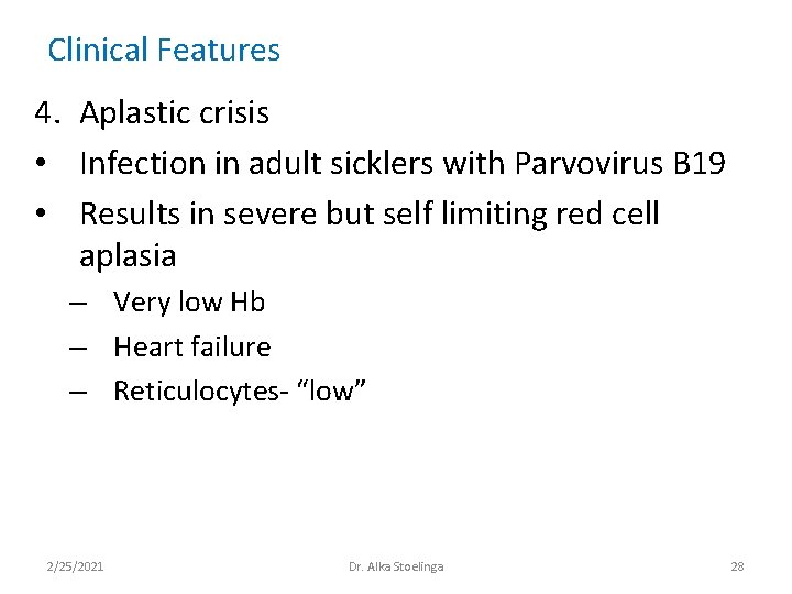
Clinical Features 4. Aplastic crisis • Infection in adult sicklers with Parvovirus B 19 • Results in severe but self limiting red cell aplasia – Very low Hb – Heart failure – Reticulocytes- “low” 2/25/2021 Dr. Alka Stoelinga 28
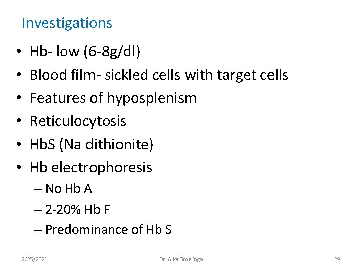
Investigations • • • Hb- low (6 -8 g/dl) Blood film- sickled cells with target cells Features of hyposplenism Reticulocytosis Hb. S (Na dithionite) Hb electrophoresis – No Hb A – 2 -20% Hb F – Predominance of Hb S 2/25/2021 Dr. Alka Stoelinga 29
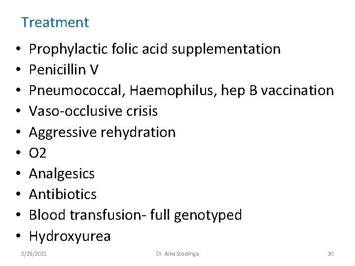
Treatment • • • Prophylactic folic acid supplementation Penicillin V Pneumococcal, Haemophilus, hep B vaccination Vaso-occlusive crisis Aggressive rehydration O 2 Analgesics Antibiotics Blood transfusion- full genotyped Hydroxyurea 2/25/2021 Dr. Alka Stoelinga 30
Hemoglobinopathies. Thalassaemia 2/25/2021 Dr. Alka Stoelinga 31
2/25/2021 Dr. Alka Stoelinga 32
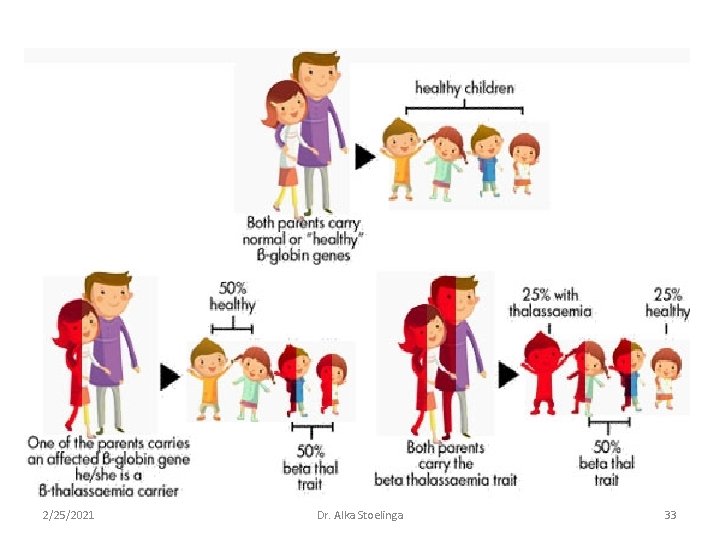
2/25/2021 Dr. Alka Stoelinga 33
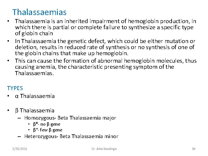
Thalassaemias • Thalassaemia is an inherited impairment of hemoglobin production, in which there is partial or complete failure to synthesize a specific type of globin chain • In Thalassaemia the genetic defect, which could be either mutation or deletion, results in reduced rate of synthesis or no synthesis of one of the globin chains that make up hemoglobin. • This can cause the formation of abnormal hemoglobin molecules, thus causing anemia, the characteristic presenting symptom of the Thalassaemias. TYPES • α Thalassaemia • β Thalassaemia – Homozygous- Beta Thalassaemia major • β⁰- no β gene • β⁺- few β gene – Heterozygous- Beta Thalassaemia minor 2/25/2021 Dr. Alka Stoelinga 34
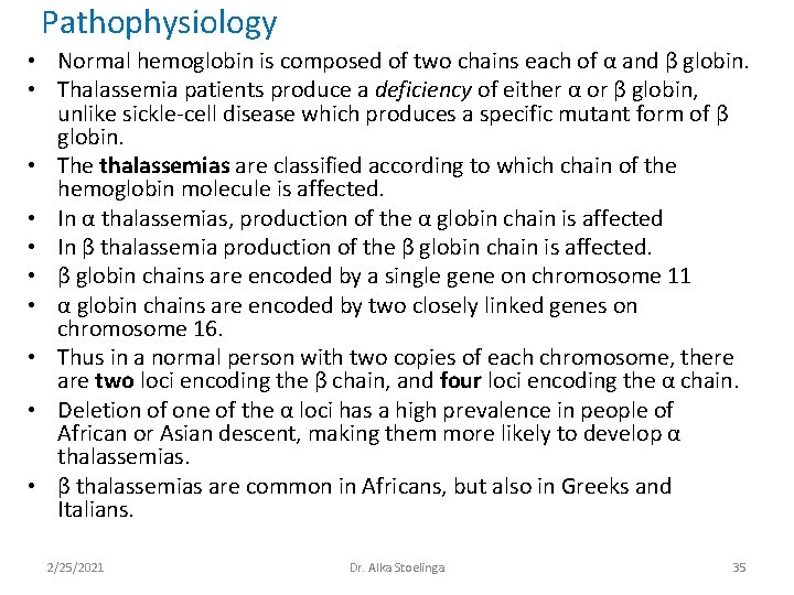
Pathophysiology • Normal hemoglobin is composed of two chains each of α and β globin. • Thalassemia patients produce a deficiency of either α or β globin, unlike sickle-cell disease which produces a specific mutant form of β globin. • The thalassemias are classified according to which chain of the hemoglobin molecule is affected. • In α thalassemias, production of the α globin chain is affected • In β thalassemia production of the β globin chain is affected. • β globin chains are encoded by a single gene on chromosome 11 • α globin chains are encoded by two closely linked genes on chromosome 16. • Thus in a normal person with two copies of each chromosome, there are two loci encoding the β chain, and four loci encoding the α chain. • Deletion of one of the α loci has a high prevalence in people of African or Asian descent, making them more likely to develop α thalassemias. • β thalassemias are common in Africans, but also in Greeks and Italians. 2/25/2021 Dr. Alka Stoelinga 35
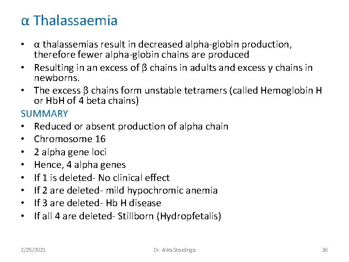
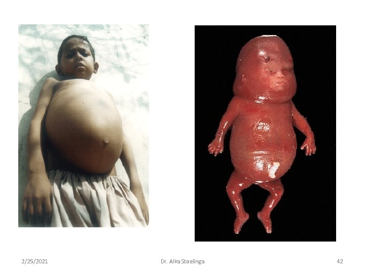
α Thalassaemia • α thalassemias result in decreased alpha-globin production, therefore fewer alpha-globin chains are produced • Resulting in an excess of β chains in adults and excess γ chains in newborns. • The excess β chains form unstable tetramers (called Hemoglobin H or Hb. H of 4 beta chains) SUMMARY • Reduced or absent production of alpha chain • Chromosome 16 • 2 alpha gene loci • Hence, 4 alpha genes • If 1 is deleted- No clinical effect • If 2 are deleted- mild hypochromic anemia • If 3 are deleted- Hb H disease • If all 4 are deleted- Stillborn (Hydropfetalis) 2/25/2021 Dr. Alka Stoelinga 36
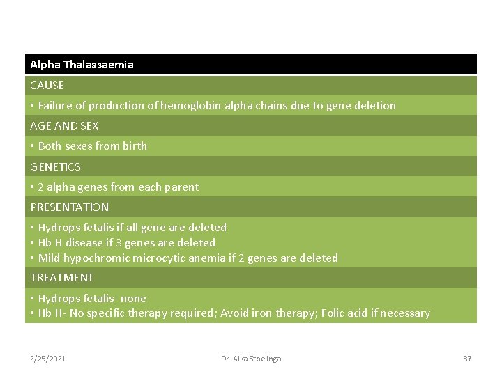
Alpha Thalassaemia CAUSE • Failure of production of hemoglobin alpha chains due to gene deletion AGE AND SEX • Both sexes from birth GENETICS • 2 alpha genes from each parent PRESENTATION • Hydrops fetalis if all gene are deleted • Hb H disease if 3 genes are deleted • Mild hypochromic microcytic anemia if 2 genes are deleted TREATMENT • Hydrops fetalis- none • Hb H- No specific therapy required; Avoid iron therapy; Folic acid if necessary 2/25/2021 Dr. Alka Stoelinga 37
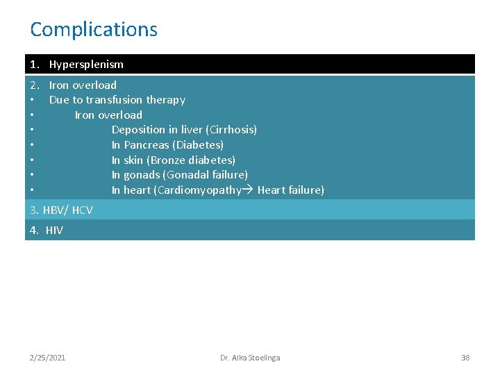
Complications 1. Hypersplenism 2. Iron overload • Due to transfusion therapy • Iron overload • Deposition in liver (Cirrhosis) • In Pancreas (Diabetes) • In skin (Bronze diabetes) • In gonads (Gonadal failure) • In heart (Cardiomyopathy Heart failure) 3. HBV/ HCV 4. HIV 2/25/2021 Dr. Alka Stoelinga 38
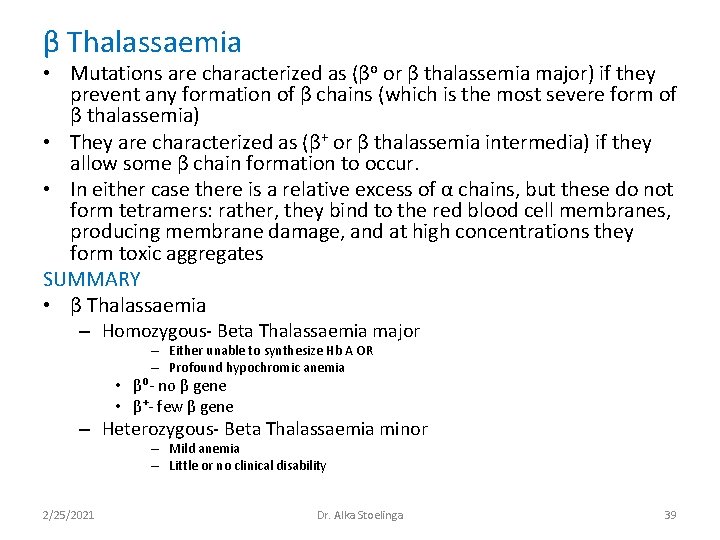
β Thalassaemia • Mutations are characterized as (βo or β thalassemia major) if they prevent any formation of β chains (which is the most severe form of β thalassemia) • They are characterized as (β+ or β thalassemia intermedia) if they allow some β chain formation to occur. • In either case there is a relative excess of α chains, but these do not form tetramers: rather, they bind to the red blood cell membranes, producing membrane damage, and at high concentrations they form toxic aggregates SUMMARY • β Thalassaemia – Homozygous- Beta Thalassaemia major – Either unable to synthesize Hb A OR – Profound hypochromic anemia • β⁰- no β gene • β⁺- few β gene – Heterozygous- Beta Thalassaemia minor – Mild anemia – Little or no clinical disability 2/25/2021 Dr. Alka Stoelinga 39
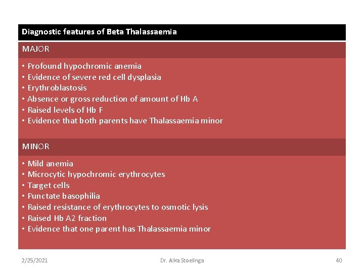
Diagnostic features of Beta Thalassaemia MAJOR • Profound hypochromic anemia • Evidence of severe red cell dysplasia • Erythroblastosis • Absence or gross reduction of amount of Hb A • Raised levels of Hb F • Evidence that both parents have Thalassaemia minor MINOR • Mild anemia • Microcytic hypochromic erythrocytes • Target cells • Punctate basophilia • Raised resistance of erythrocytes to osmotic lysis • Raised Hb A 2 fraction • Evidence that one parent has Thalassaemia minor 2/25/2021 Dr. Alka Stoelinga 40
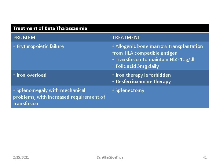
Treatment of Beta Thalassaemia PROBLEM TREATMENT • Erythropoietic failure • Allogenic bone marrow transplantation from HLA compatible antigen • Transfusion to maintain Hb> 10 g/dl • Folic acid 5 mg daily • Iron overload • Iron therapy is forbidden • Desferrioxamine therapy • Splenomegaly with mechanical problems, with increased requirement of transfusion • Splenectomy 2/25/2021 Dr. Alka Stoelinga 41
2/25/2021 Dr. Alka Stoelinga 42
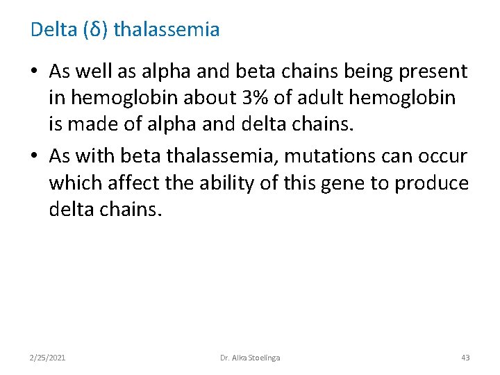
Delta (δ) thalassemia • As well as alpha and beta chains being present in hemoglobin about 3% of adult hemoglobin is made of alpha and delta chains. • As with beta thalassemia, mutations can occur which affect the ability of this gene to produce delta chains. 2/25/2021 Dr. Alka Stoelinga 43
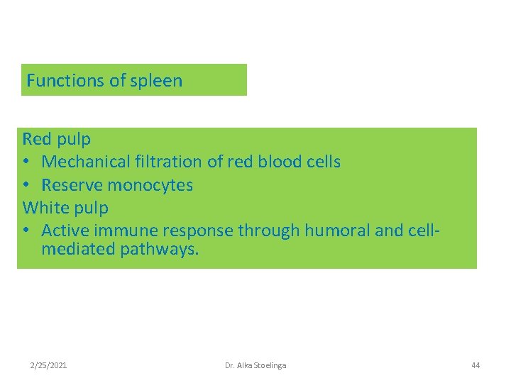
Functions of spleen Red pulp • Mechanical filtration of red blood cells • Reserve monocytes White pulp • Active immune response through humoral and cellmediated pathways. 2/25/2021 Dr. Alka Stoelinga 44
• Hematological malignancies 2/25/2021 Dr. Alka Stoelinga 45