Anal Canal Fissure In Ano Haemorrhoids Dr Amit


 Examination Haemorrhoid directed: • Pain acute/chronic/ cutaneous")


 Non-healed (20%) 0. 2%")


 relaxation via – nitric oxide donation")

- Slides: 37
Anal Canal Fissure In Ano Haemorrhoids Dr. Amit Gupta Associate Professor Dept. of Surgery
Anorectal Anatomy Arterial Supply Inferior rectal A middle rectal A Nerve Supply Sympathetic: Superior hypogastric plexus Parasympathetic: Venous drainage Inferior rectal V middle rectal V S 234 (nerviergentis Pudendal Nerve: Motor and sensory 3 hemorrhoidal complexes L lateral Anal canal R antero-lateral R posterolateral Lymphatic drainage Above dentate: Inf. Mesenteric Below dentate: internal iliac Anal verge
• Pain? -> painless • Bright red bleeding • Prolapse associated with defecation Internal External • Anoderm • Swell, discomfort, difficult hygiene • Pain? -> Thrombosed
Background • • They are part of the normal anoderm cushions They areas of vascular anastamosis in a supporting stroma of subepithelial smooth muscles. The contribute 15 -20% of the normal resting pressure and feed vital sensory information. 3 main cushions are found • • • L lateral R anterior This combination is only in 19% R posterior But can be found anywhere in anus Prevalence is 4% Miss labelling by referring physicians and patients is common
Pathogenesis Abnormal haemorrhoids are dilated cushions of arteriovenous plexus with stretched suspensory fibromuscular stroma with prolapsed rectal mucosa 3 main processes: 1. Increased venous pressure 2. Weakness in supporting fibromuscular stroma 3. Increased internal sphincter tone
Risk factors Habitual 1. 2. 3. 4. 5. 6. 7. 8. Constipation and straining Low fibre high fat/spicy diet Prolonged sitting in toilet Pregnancy Aging Obesity Office work Family tendency Pathological 1. 2. 3. 4. 5. 6. 7. Chronic diarrhea (IBD) Colon malignancy Portal hypertension Spinal cord injury Rectal surgery Episiotomy Anal intercourse
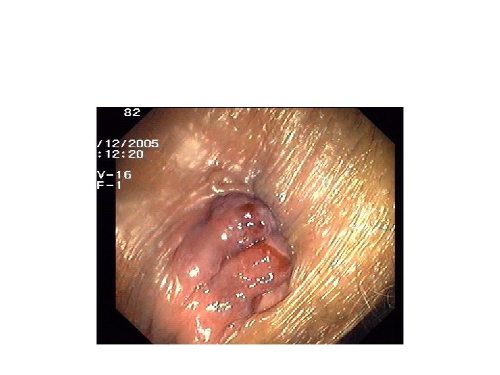
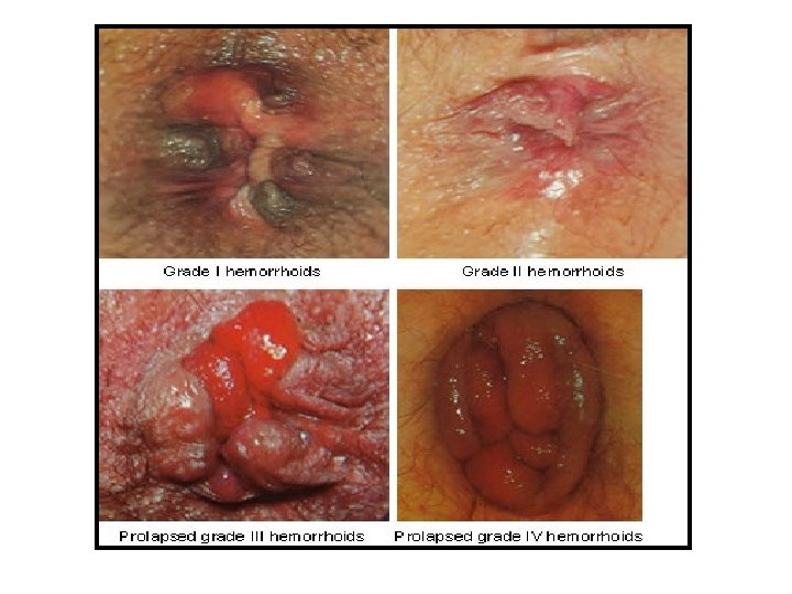
Classification Origin in relation to Dentate line 1. 2. 3. Internal: above DL External: below DL Mixed Degree of prolapse through anus • 1 st: bleed but no prolapse • 2 nd: spontaneous reduction • 3 rd: manual reduction • 4 th: not reducable
Thrombosed external piles
First-degree internal piles viewed through anoscope
Second-degree internal prolapsed piles, reduced spontaneously
Third-degree internal prolapsed piles, requiring manual reduction
Fourth-degree strangulated internal and thrombosed external piles
Clinical assessment History ( Full history required) Examination Haemorrhoid directed: • Pain acute/chronic/ cutaneous • Lump acute/ sub-acute • Prolapse define grade • Bleeding fresh, post defecation • Pruritis and mucus Local General GI: • Digital: • Change in bowel habit • Mucus discharge • Tenasmus/ back pain • Weight loss • Anorexia • Other system inquiry • Inspect for: –Lumps, note colour and reducability –Fissures –Fistulae –Abscess –Masses –Character of blood and mucus • Perform proctoscopy and sigmoidoscopy General abdominal examination
Investigations The diagnosis of haemorrhoids is based on clinical assessment and proctoscopy Further investigations should be based on a clinical index of suspicion • Lab: CBC / Clotting profile/ Group and save • Proctography: if rectal prolpse is suspected • Colonoscopy: if higher colonic or sinister pathology is suspected
Complications 1. Ulceration 2. Thrombosis 3. Sepsis and abscess formation 4. Incontinence Thrombosed internal haemorrhoids Thrombosed external haemorrhoids
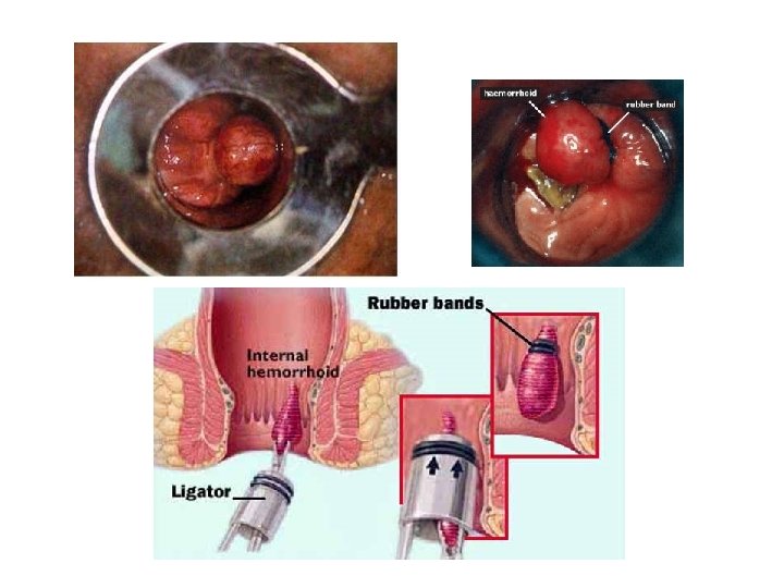
Internal Haemorrhoids Treatment Conservative Measures Grade 1&2 • Dietary modification: high fibre diet • Stool softeners • Bathing in warm water • Topical creams NOT MUCH VALUE Minimally invasive Indicated in failed medical treatment and grades 3&4 • injection sclerotherapy • Rubber band ligation • Laser photocoagulation • Cryotherapy freezing • Stapled haemorrhoidectomy Surgical Indications: 1. Failed other treatments 2. Severely painful grade 3&4 3. Concurrent other anal conditions 4. Patient preference
Excision of thrombosed external hemorrhoid.
Closed hemorrhoidectomy
Grade 4 hemorrhoid before reduction Stapling device Placement of stapling device obturator
External Haemorrhoids Treatment • If presentation less than 72 hours: Enucleate under LA or GA Leave wound open to close by secondary intension Apply pressure dressing for 24 hours post op • If more than 72 hours: • Conservative measures
Anal Fissure Linear tears in the anal mucosa exposing the internal sphincter 90% are posterior
• Young & middle aged adults • Male = Female • Location – posterior midline (most common) Anterior midline fissures – more common in females • In any event, length of each fissure is remarkably constant, extending from the dentate line to the anal verge and corresponding roughly to the lower half of the internal sphincter
Pathology • Acute fissures – heal promptly with conservative treatment • Secondary changes if present, it does not heal readily – Sentinel pile – Hypertrophied anal papilla – Long standing • Fibrous induration in lateral edges of fissure • Fibrosis at the base of ulcer (internal sphincter) – At any stage • Frank suppuration – intersphincteric / perianal abscess
Etiology • Initiation – trauma • Why midline posterior fissures are more common? • Dietary factors – Decreased risk – raw foods, vegetables, whole grain bread – Increased risk – white bread sausages etc. • Secondary fissure – – – Crohn’s disease Previous anal surgery, especially hemorrhoidectomy Fistula-in-ano surgery Anterior fissure in females resulting from childbirth Long standing loose stools with chronic laxative abuse
• Initiation – trauma • Perpetuation of fissure – abnormality of internal anal sphincter • Higher resting pressure within the internal anal sphincter in pts with fissures than in normal control • Rectal distension reflex relaxation of internal anal sphincter overshoot contractions in these patients sphincter spasm and pain • Elevated sphincter pressures cause ischemia of the anal lining resulting in pain and failure to heal • Posterior commissure perfused more poorly than the other portion of the anal canal
Clinical Features • Pain and spasm – Sharp, agonizing during defecation, recurrent, worsens constipation. • Bleeding – In small amounts, – approximately 70% of patients note bright red blood on the toilet paper or stool • Discharge – Irritation and pruritis ani due to malodorous discharge of the pus • Constipation Painless non-healing fissure with occasional bleed – may be a progenitor of IBD
Diagnosis • Inspection – Acute fissure is seen as Linear tear – most important • Palpation • Anoscopy • Sigmoidoscopy • Biopsy
Differential diagnosis • • Anorectal suppuration Pruritus ani Fissure in inflammatory bowel disease Carcinoma Syphilitic fissures Tuberculous ulcer Anal abrasion
Treatment Acute Fissure Stool-bulking agents Warm sitz bath Healed (80%) Non-healed (20%) 0. 2% Nitroglycerin or 2% Nifedipine or Diltiazem 4 -6 wks Repeat treatment with alternate medication Non-healed (30%) Healed (70%) Chronic fissure Botulinum toxin A injection Open LIS 98% healing Closed LIS
Acute anal fissure: • Spontaneous healing, High fiber diet, adequate water intake and warm sitz bath, stool softener/bulk laxative, suppositories • Sodium tetradecyl sulphate
• Chronic anal fissure: – Conservative – Surgical
Pharmacological Sphincterotomy • Pharmacological manipulation of anal sphincter tone as an alternative modality to surgery for the treatment of anal fissure • Shares the same goal as lateral sphincterotomy without its possible long-term side effects • Pharmacological agents lower anal canal resting pressure producing chemical sphincterotomy without causing permanent damage to the anal sphincter mechanism
• By enhancing internal anal sphincter (IAS) relaxation via – nitric oxide donation – intracellular Ca 2+ depletion – muscarinic receptor stimulation – adrenergic inhibition • This improves blood supply at the site of the fissure that would promote healing of anal fissures • Nitric oxide donors and calcium channel blockers, agents that directly reduce resting anal pressure, has now largely replaced traditional surgical methods as first-line treatment for chronic anal fissure
Other Agents • Botulinum Toxin A • L-arginine • Gonyautoxin • Topical sildenafil