
SURGERY OF ANAL CANAL AND RECTUM ANATOMY Anatomic




- Slides: 69
SURGERY OF ANAL CANAL AND RECTUM
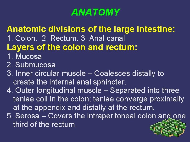
ANATOMY Anatomic divisions of the large intestine: 1. Colon. 2. Rectum. 3. Anal canal Layers of the colon and rectum: 1. Mucosa 2. Submucosa 3. Inner circular muscle – Coalesces distally to create the internal anal sphincter. 4. Outer longitudinal muscle – Separated into three teniae coli in the colon; teniae converge proximally at the appendix and distally at the rectum. 5. Serosa – Covers the intraperitoneal colon and one third of the rectum.
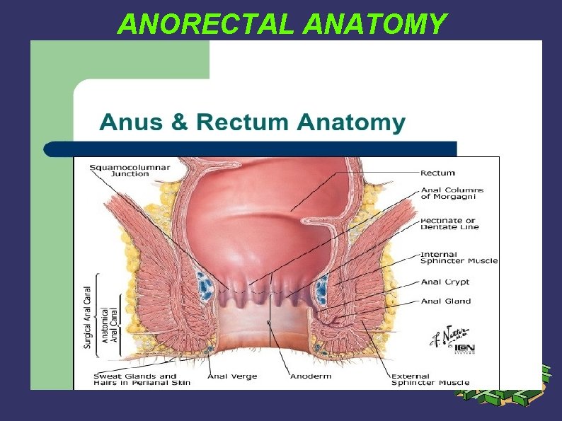
ANORECTAL ANATOMY
Colorectal and Anorectal Vascular Supply The arterial supply to the colon is highly variable. In general, the arterial supply to the colon is as follows: 1. Superior mesenteric artery branches a. Ileocolic artery (absent in up to 20 percent of people) supplies blood flow to the terminal ileum and proximal ascending colon. b. Right colic artery supplies the ascending colon. c. Middle colic artery supplies the transverse colon.
Colorectal and Anorectal Vascular Supply 2 2. Inferior mesenteric artery branches a. Left colic artery supplies the descending colon. b. Sigmoidal branches supply the sigmoid colon. c. Superior rectal art. supplies the proximal rectum. The terminal branches of each artery form anastomoses with the terminal branches of the adjacent artery and communicate via the marginal artery of Drummond 3. Internal iliac artery branches a. Middle rectal artery (variable presence and size). b. Internal pudendal artery branch. c. Inferior rectal artery supplies the lower rectum and anal canal.
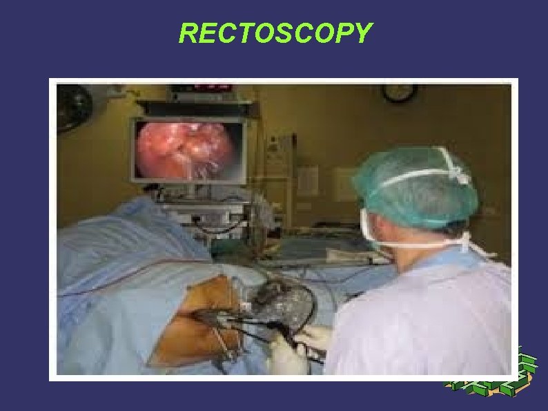
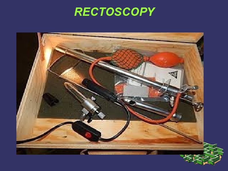
INVESTIGATIONS 1. Anoscopy – The anoscope is useful for examination of the anal canal and can generally allow examination of the distal 6– 8 cm of the anus. Anoscopy can be diagnostic or therapeutic (e. g. , sclerotherapy or rubber band ligation of hemorrhoids). 2. Proctoscopy – The rigid proctoscope is useful for examination of the rectum and distal sigmoid colon and is occasionally used therapeutically (e. g. , polypectomy, electrocoagulation, or detorsion of a sigmoid volvulus).
RECTOSCOPY
RECTOSCOPY
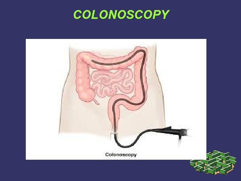
INVESTIGATIONS 2 3. Flexible Sigmoidoscopy and Colonoscopy – provide excellent visualization of the colon and rectum. Sigmoidoscopes measure 60 cm in length and may allow visualization as high as the splenic flexure. Partial preparation with enemas is usually adequate for sigmoidoscopy and most patients can tolerate this procedure without sedation. Colonoscopes measure 100– 160 cm in length and are capable of examining the entire colon and terminal ileum. A complete oral bowel preparation usually is necessary. Both sigmoidoscopy and colonoscopy can be used diagnostically and therapeutically.
COLONOSCOPY
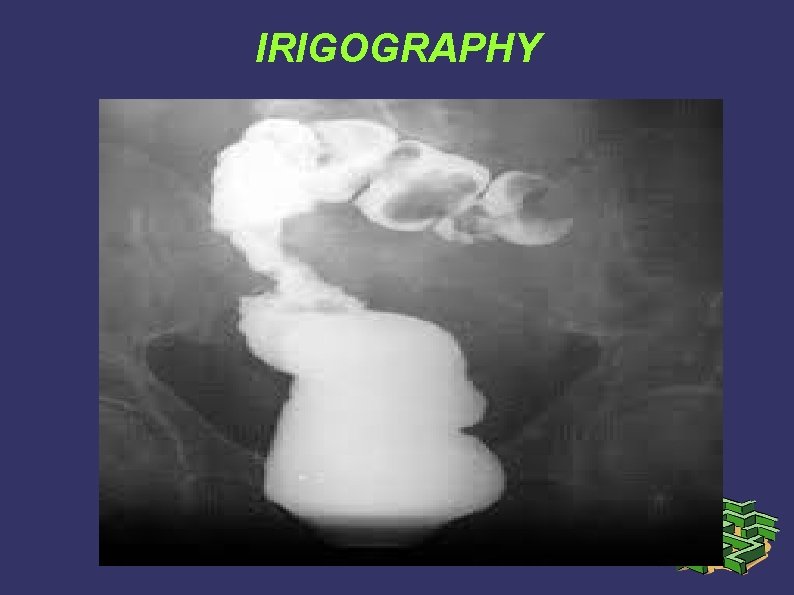
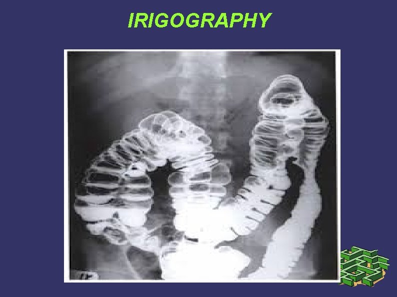
INVESTIGATIONS 3 1. Plain radiograph of the abdomen (supine, upright, and diaphragmatic views) are useful for detecting free intraabdominal air, bowel gas patterns suggestive of small or large bowel obstruction, and volvulus. 2. Contrast studies are useful for evaluating obstructive symptoms, delineating fistulous tracts, and diagnosing small perforations or anastomotic leaks. Gastrografin is recommended if perforation or leak is suspected. Doublecontrast barium enema is more sensitive for the detection of mass lesions greater than 1 cm in
IRIGOGRAPHY
IRIGOGRAPHY
INVESTIGATIONS 4 3. Computed Tomography – (CT) is commonly employed in the evaluation of patients with abdominal complaints. Its utility is primarily in the detection of extraluminal disease, such as intraabdominal abscesses and pericolic inflammation, and in staging colorectal carcinoma. Extravasation of oral or rectal contrast also may confirm the diagnosis of perforation or anastomotic leak. Nonspecific findings such as bowel wall thickening or mesenteric stranding may suggest inflammatory bowel disease, enteritis/colitis, or ischemia. A standard CT scan is relatively insensitive for the detection of intraluminal lesions.
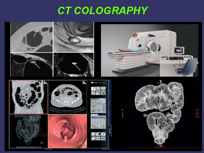
INVESTIGATIONS 5 4. Virtual Colonoscopy (CT colography) – is a technique that is designed to overcome some of the limitations of traditional CT scanning. This technology uses helical CT and three-dimensional reconstruction to detect intraluminal colonic lesions. Oral bowel preparation, oral and rectal contrast, and colon insufflation are used to maximize sensitivity. 5. Magnetic Resonance Imaging (MRI) – The main use of MRI in colorectal disorders is in evaluation of pelvic lesions. MRI is more sensitive than CT for detecting bony involvement or pelvic sidewall extension of rectal tumors. MRI with an endorectal coil can be helpful in the detection and delineation
CT COLOGRAPHY
INVESTIGATIONS 6 6. Positron Emission Tomography (PET) – is used for imaging tissues with high levels of anaerobic glycolysis, such as malignant tumors. PET has been used as an adjunct to CT in the staging of colorectal cancer and may prove useful in discriminating recurrent cancer from fibrosis. 7. Angiography – is occasionally used for the detection of brisk bleeding (approximately 0. 5– 1. 0 m. L per minute) within the colon or small bowel. If extravasation of contrast is identified, infusion of vasopressin or angiographic embolization can be therapeutic.
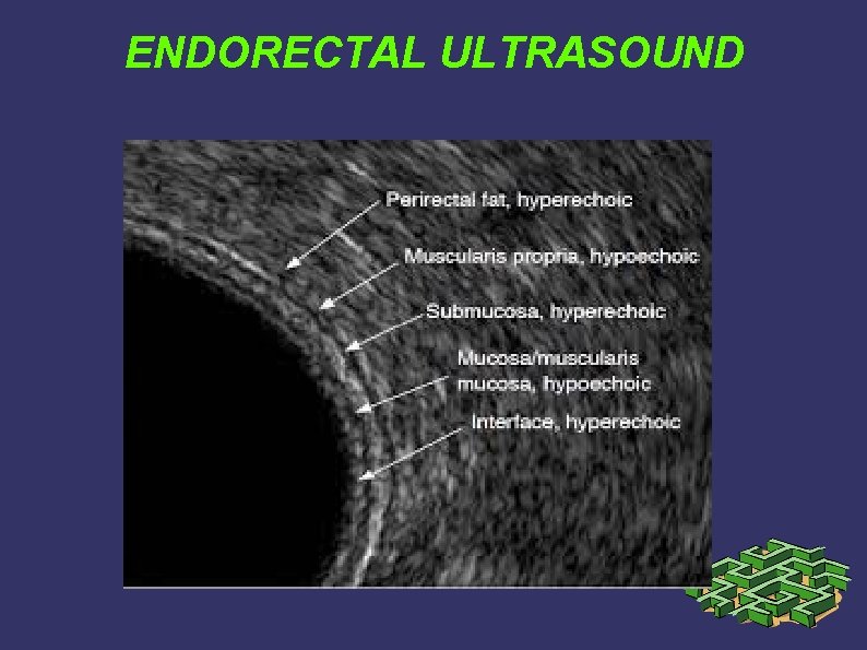
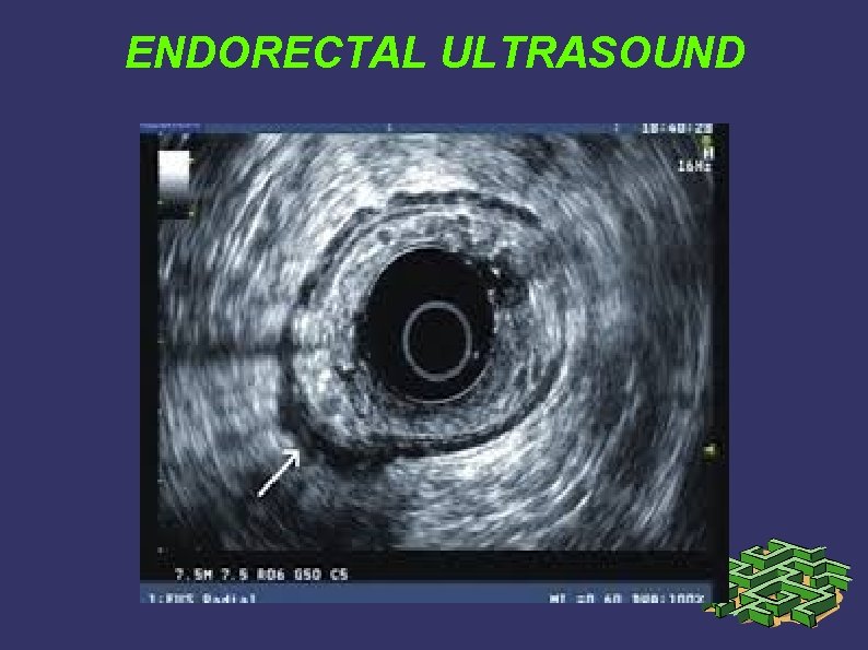
INVESTIGATIONS 7 8. Endorectal and Endoanal Ultrasound – is primarily used to evaluate the depth of invasion of neoplastic lesions in the rectum. Ultrasound can reliably differentiate most benign polyps from invasive tumors and can differentiate superficial (T 1–T 2) from deeper (T 3–T 4) tumors. This modality also can detect enlarged perirectal lymph nodes. Ultrasound may also prove useful for early detection of local recurrence after surgery. Endoanal US is used to evaluate the layers of the anal canal. Internal anal sphincter, external anal sphincter, and puborectalis muscle can be differentiated. Endoanal US is particularly useful for detecting sphincter defects
ENDORECTAL ULTRASOUND
ENDORECTAL ULTRASOUND
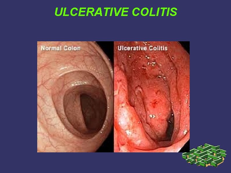
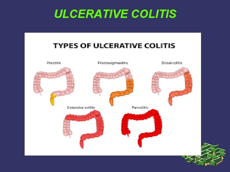
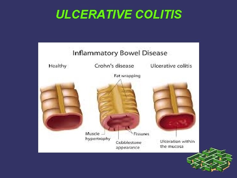
ULCERATIVE COLITIS Ulcerative colitis is a dynamic disease charact. by remissions and exacerbations. The clinical spectrum ranges from an inactive or quiescent phase to low-grade active disease to fulminant disease. The onset of UC may be insidious, with minimal bloody stools, or the onset can be abrupt, with severe diarrhea and bleeding, tenesmus, abdominal pain, and fever. The severity of symptoms depends on the degree and extent of inflammation. Although anemia is common, massive hemorrhage is rare. Physical findings often are nonspecific.
ULCERATIVE COLITIS
ULCERATIVE COLITIS
ULCERATIVE COLITIS
ULCERATIVE COLITIS 2 Diagnosis of UC almost always is made endoscopically. Because the rectum is invariably involved, proctoscopy may be adequate to establish the diagnosis. A complete evaluation with colonoscopy or barium enema during an acute flare is contraindicated because of the risk of perforation. Because the inflammation in ulcerative colitis is purely mucosal, strictures are highly uncommon. Any stricture diagnosed in a patient with ulcerative colitis must be presumed to be malignant until proven
ULCERATIVE COLITIS 3 Indications for Surgery: Emergency surgery is required for patients with lifethreatening hemorrhage, toxic megacolon, or fulminant colitis who fail to respond rapidly to medical therapy. Patients with fulminant colitis should be treated with bowel rest, hydration, broad-spectrum antibiotics, and parenteral corticosteroids. Colonoscopy and barium enema are contraind. and antidiarrheal agents should be avoided. Clinical deterioration or failure to improve within 24 h mandates surgery.
ULCERATIVE COLITIS 4 Indications for elective surgery include: - intractability despite maximal medical therapy, - development of major complications from medical therapy, and - risk of developing colorectal carcinoma. Risk of malignancy increases with pancolonic disease and the duration of symptoms; the risk is approximately 2 percent after 10 years, 8 percent after 20 years, and 18 percent after 30 years.
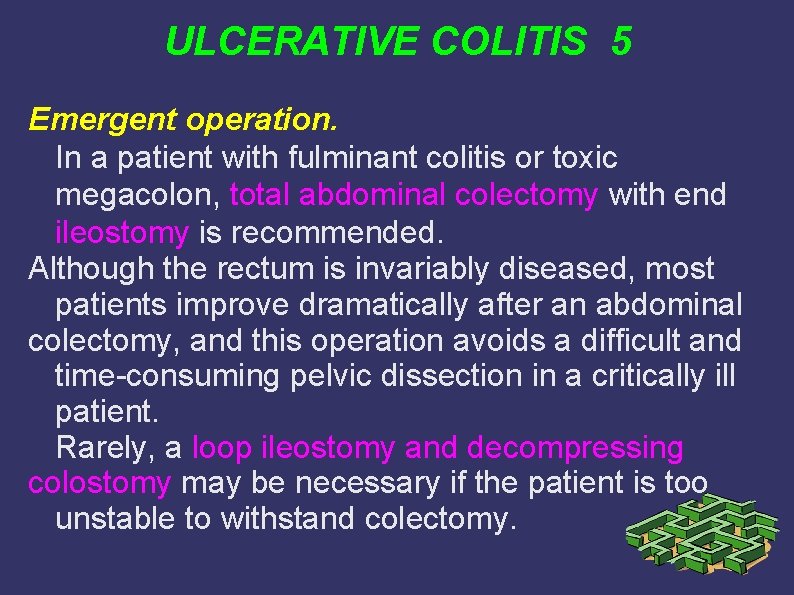
ULCERATIVE COLITIS 5 Emergent operation. In a patient with fulminant colitis or toxic megacolon, total abdominal colectomy with end ileostomy is recommended. Although the rectum is invariably diseased, most patients improve dramatically after an abdominal colectomy, and this operation avoids a difficult and time-consuming pelvic dissection in a critically ill patient. Rarely, a loop ileostomy and decompressing colostomy may be necessary if the patient is too unstable to withstand colectomy.
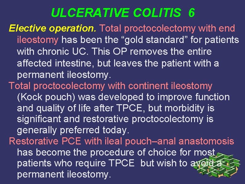
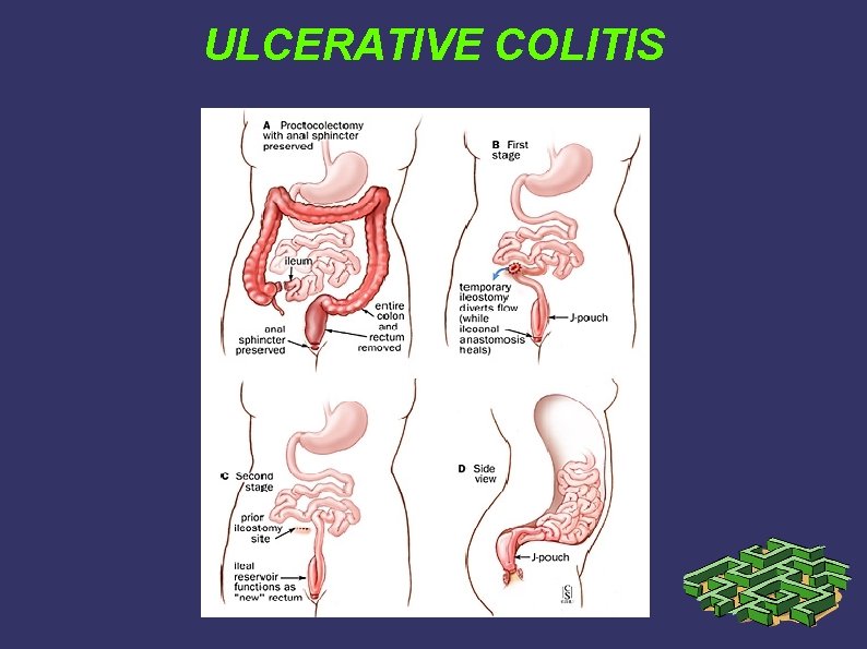
ULCERATIVE COLITIS 6 Elective operation. Total proctocolectomy with end ileostomy has been the “gold standard” for patients with chronic UC. This OP removes the entire affected intestine, but leaves the patient with a permanent ileostomy. Total proctocolectomy with continent ileostomy (Kock pouch) was developed to improve function and quality of life after TPCE, but morbidity is significant and restorative proctocolectomy is generally preferred today. Restorative PCE with ileal pouch–anal anastomosis has become the procedure of choice for most patients who require TPCE but wish to avoid a permanent ileostomy.
ULCERATIVE COLITIS
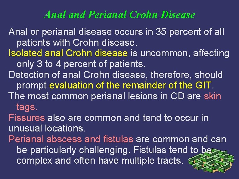
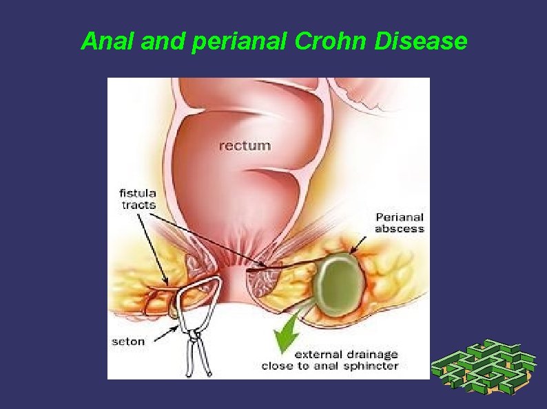
Anal and Perianal Crohn Disease Anal or perianal disease occurs in 35 percent of all patients with Crohn disease. Isolated anal Crohn disease is uncommon, affecting only 3 to 4 percent of patients. Detection of anal Crohn disease, therefore, should prompt evaluation of the remainder of the GIT. The most common perianal lesions in CD are skin tags. Fissures also are common and tend to occur in unusual locations. Perianal abscess and fistulas are common and can be particularly challenging. Fistulas tend to be complex and often have multiple tracts.
Anal and perianal Crohn Disease 2 Treatment of anal and perianal Crohn disease focuses on alleviation of symptoms. In general, skin tags and hemorrhoids should not be excised unless they are extremely symptomatic because of the risk of creating chronic, nonhealing wounds. Fissures may respond to local or systemic therapy; sphincterotomy is relatively contraindicated because of the risk of creating a chronic, nonhealing wound, and because of the increased risk of incontinence in a patient with diarrhea from underlying colitis or small bowel disease. Abscesses should be drained and evacuated
Anal and perianal Crohn Disease 3 Medical treatment of underlying proctitis with salicylate and/or corticosteroid enemas may be helpful; however, control of sepsis is the primary goal of therapy. Metronidazole also has been used with some success in this setting. Infliximab has shown some efficacy in healing chronic fistulas secondary to Crohn disease. However, it is of paramount importance to drain any and all abscesses before initiating immunosuppressive therapy such as corticosteroids or infliximab.
Anal and perianal Crohn Disease
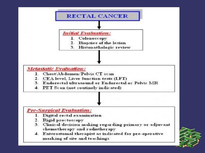
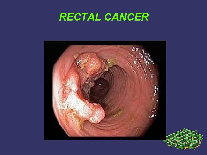
RECTAL CANCER DEF: Rectal cancer is a disease in which cancer cells form in the tissues of the rectum; colorectal cancer occurs in the colon or rectum. HIST: Adenocarcinomas comprise the vast majority (98%) of colon and rectal cancers; more rare rectal cancers include lymphoma (1. 3%), carcinoid (0. 4%), and sarcoma (0. 3%). CHAR: The incidence and epidemiology, etiology, pathogenesis, and screening recommendations are common to both colon cancer and rectal cancer. The image below depicts the staging and workup
RECTAL CANCER
RECTAL CANCER
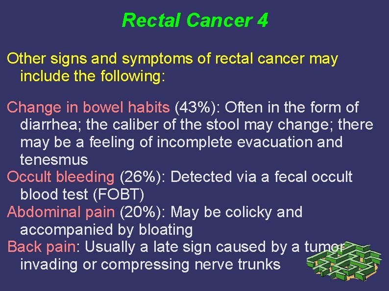
Rectal cancer 3 Signs and symptoms Bleeding is the most common symptom of rectal cancer, occurring in 60% of patients. However, many rectal cancers produce no symptoms and are discovered during digital or proctoscopic screening examinations.
Rectal Cancer 4 Other signs and symptoms of rectal cancer may include the following: Change in bowel habits (43%): Often in the form of diarrhea; the caliber of the stool may change; there may be a feeling of incomplete evacuation and tenesmus Occult bleeding (26%): Detected via a fecal occult blood test (FOBT) Abdominal pain (20%): May be colicky and accompanied by bloating Back pain: Usually a late sign caused by a tumor invading or compressing nerve trunks
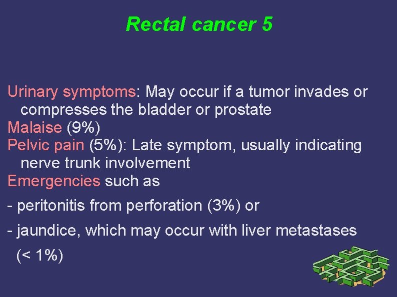
Rectal cancer 5 Urinary symptoms: May occur if a tumor invades or compresses the bladder or prostate Malaise (9%) Pelvic pain (5%): Late symptom, usually indicating nerve trunk involvement Emergencies such as - peritonitis from perforation (3%) or - jaundice, which may occur with liver metastases (< 1%)
Rectal Cancer 6 Diagnosis Perform physical examination with specific attention to the size and location of the rectal tumor in addition to possible metastatic lesions, including enlarged lymph nodes or hepatomegaly. In addition, evaluate the remainder of the colon.
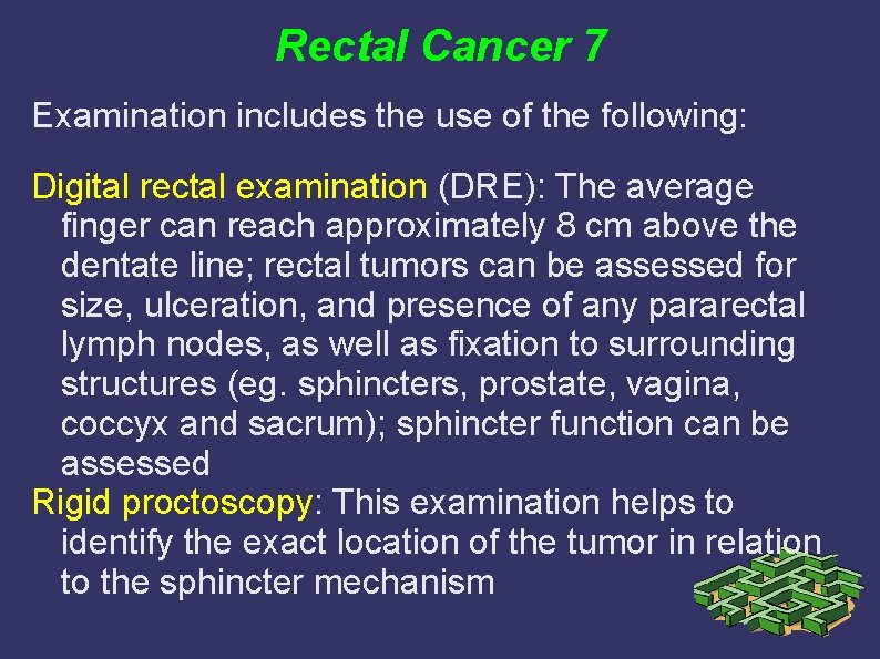
Rectal Cancer 7 Examination includes the use of the following: Digital rectal examination (DRE): The average finger can reach approximately 8 cm above the dentate line; rectal tumors can be assessed for size, ulceration, and presence of any pararectal lymph nodes, as well as fixation to surrounding structures (eg. sphincters, prostate, vagina, coccyx and sacrum); sphincter function can be assessed Rigid proctoscopy: This examination helps to identify the exact location of the tumor in relation to the sphincter mechanism
Rectal Cancer- 8 Laboratory tests Routine laboratory studies in patients with suspected rectal cancer include the following: Complete blood count Serum chemistries Liver and renal function tests Carcinoembryonic antigen (CEA) test Cancer antigen (CA) 19 -9 assay, if available: May be useful for monitoring the disease Histologic examination of tissue specimens
Rectal Cancer 9 Imaging studies If metastatic rectal cancer is suspected, the following radiologic studies may be obtained: CT scanning of the chest, abdomen, and pelvis Endorectal ultrasonography Endorectal or pelvic magnetic resonance imaging (MRI) Positron emission tomography (PET) scanning: Not routinely indicated
Rectal Cancer 10 - management A multidisciplinary approach that includes colorectal surgery, medical oncology, and radiation oncology is required for optimal treatment of patients with rectal cancer. Surgical technique, use of radiotherapy, and method of administering chemotherapy are important factors. Strong considerations should be given to the intent of surgery, possible functional outcome, and preservation of anal continence and genitourinary functions. The first step involves achievement of cure, because the risk of pelvic recurrence is high in patients with rectal cancer, and locally recurrent rectal cancer has a poor prognosis.
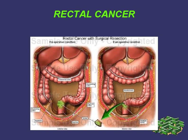
Rectal cancer 11 Radical resection of the rectum is the mainstay of therapy. The timing of surgical resection is dependent on the size, location, extent, and grade of the rectal carcinoma. OP management of rectal cancer may include the following: Transanal excision: For early-stage cancers in a select group of patients Transanal endoscopic microsurgery: Form of local excision that uses a special operating proctoscope that distends the rectum with insufflated carbon dioxide and allows the passage of dissecting instruments Endocavity radiotherapy: Delivered under sedation via a special proctoscope in the operating room Sphincter-sparing procedures: Low anterior resection, coloanal anastomosis, abdominal perineal resection Abdomino – perineal rectum amputation with stoma
Rectal Cancer 12 Adjuvant medical management: Adjuvant medical therapy may include the following: Adjuvant radiation therapy Intraoperative radiation therapy Adjuvant chemoradiation therapy Radioembolization
Rectal Cancer 13 Pharmacotherapy The National Comprehensive Cancer Network guidelines recommend the use of as many chemotherapy drugs as possible to maximize the effect of adjuvant therapies for colon and rectal cancer. The following agents may be used in the management of rectal cancer: Antineoplastic agents (eg. fluorouracil, vincristine, leucovorin, irinotecan, oxaliplatin, cetuximab, bevacizumab, panitumumab) Vaccines (eg. quadrivalent human papillomavirus [HPV] vaccine)
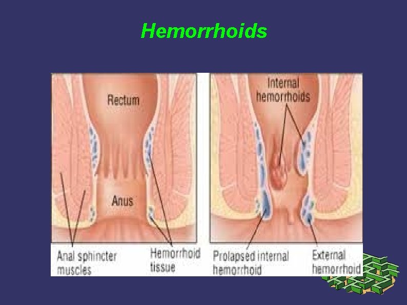
Hemorrhoids are cushions of submucosal tissue containing venules, arterioles, and smoothmuscle fibers that are located in the anal canal. Three hemorrhoidal cushions are found in the left lateral, right anterior, and right posterior positions and are thought to function as part of the continence mechanism. Because hemorrhoids are a normal part of anorectal anatomy, treatment is only indicated if they become symptomatic.
Hemorrhoids 2 1. External hemorrhoids are located distal to the dentate line and are covered with anoderm. Thrombosis of an external hemorrhoid may cause significant pain. Treatment of external hemorrhoids and skin tags are only indicated for symptomatic relief. 2. Internal hemorrhoids are located proximal to the dentate line and covered by insensate anorectal mucosa. Internal hemorrhoids may prolapse or bleed, but rarely become painful unless they develop thrombosis and necrosis.
Hemorrhoids
Hemorrhoids 3 Internal hemorrhoids are graded according to the extent of prolapse: First-degree hemorrhoids—bulge into the anal canal Second-degree hemorrhoids—prolapse through the anus but reduce spontaneously Third-degree hemorrhoids—prolapse through the anal canal and require manual reduction Fourth-degree hemorrhoids—prolapse but cannot be reduced and are at risk for strangulation
Hemorrhoids 4 Treatment: Medical Therapy (dietary fiber, stool softeners, increased fluid intake, and avoidance of straining) – appropriate for bleeding first- and seconddegree hemorrhoids. Rubber Band Ligation, sclerotherapy, infrared photocoagulation – appropriate for bleeding first-, second-, and selected third-degree hemorrhoids. Excision of Thrombosed External Hemorrhoids.
Hemorrhoids 5 Operative hemorrhoidectomy. A number of surgical procedures have been described for elective resection of symptomatic hemorrhoids. All are based on decreasing blood flow to the hemorrhoidal plexuses and excising redundant anoderm and mucosa: 1. Closed Submucosal Hemorrhoidectomy 2. Open Hemorrhoidectomy 3. Whitehead Hemorrhoidectomy
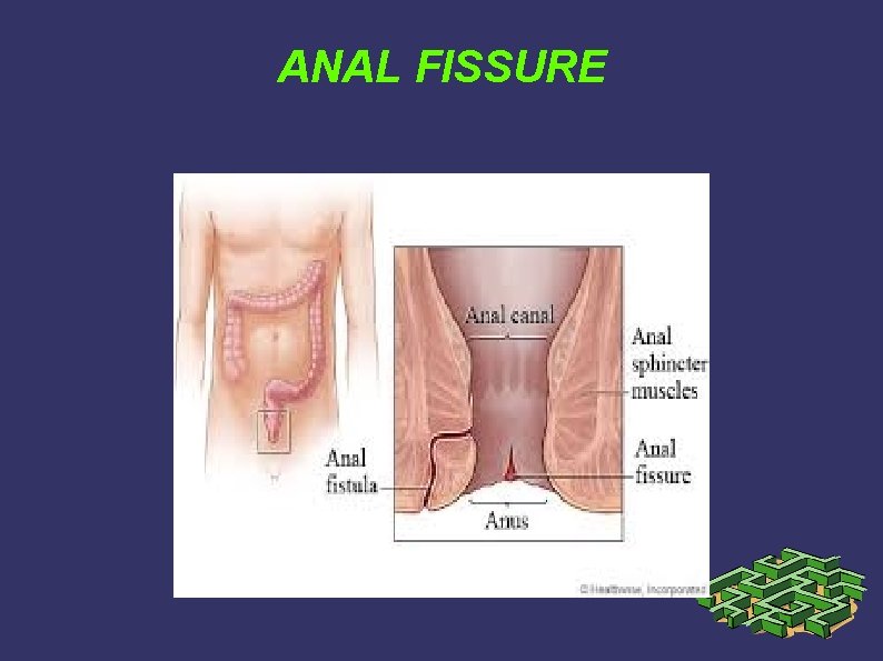
Anal fissure DEF: A fissure in ano is a tear in the anoderm distal to the dentate line. Most anal fissures occur in the posterior midline. Ten to 15 percent occur in the anterior midline. Less than 1 percent of fissures occur off midline. Symptoms and Findings: Characteristic symptoms include tearing pain with defecation and hematochezia. On physical examination, the fissure can often be seen in the anoderm by gently separating the buttocks. Patients are often too tender to tolerate digital rectal examination, anoscopy, or proctoscopy.
ANAL FISSURE
Anal fissure 2 An acute fissure is a superficial tear of the distal anoderm and almost always heals with medical management. Chronic fissures develop ulceration and heaped-up edges with the white fibers of the internal anal sphincter visible at the base of the ulcer.
Anal fissure 3 Therapy focuses on breaking the cycle of pain, spasm, and ischemia: 1. Dietary changes – bulk agents, stool softeners, and warm sitz baths. 2. Topical agents – Lidocaine jelly or other analgesic creams, nitroglycerin ointment 3. Botulinum toxin – has been proposed as an alternative to surgical sphincterotomy for chronic fissure. Although there is limited experience with this approach, results appear to be superior to other medical therapy, and complications such as incontinence are rare.
Anal fissure 4 4. Surgical sphincterotomy – Surgical therapy has been recommended for chronic fissures that have failed medical therapy, and lateral internal sphincterotomy is the procedure of choice for most surgeons. Approximately 30 percent of the internal sphincter fibers are divided. Healing is achieved in more than 95 percent of patients by using this technique and most patients experience immediate pain relief. Recurrence is rare (less than 10 percent), but the risk of minor incontinence ranges from 5 – 15 percent.
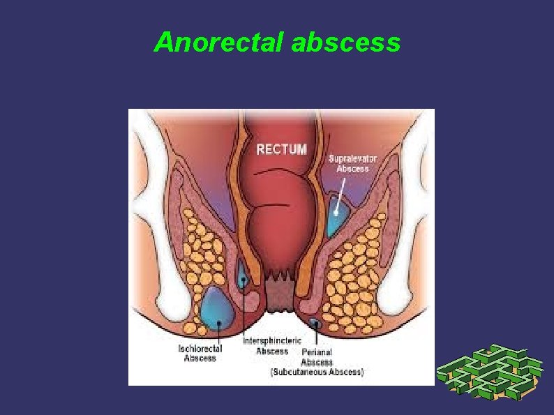
Anorectal abscess DG: Pain is the most common presenting complaint. A palpable mass often is detected by inspection of the perianal area or by digital rectal examination. Occasionally, patients will present with fever, urinary retention, or life-threatening sepsis. The diagnosis of a perianal or ischiorectal abscess can usually be made with physical exam. alone. However, complex or atypical presentations may require imaging studies (CT or MRI) to fully delineate the anatomy of the abscess. Treatment: Anorectal abscesses should be treated by drainage as soon as the diagnosis is established.
Anorectal abscess
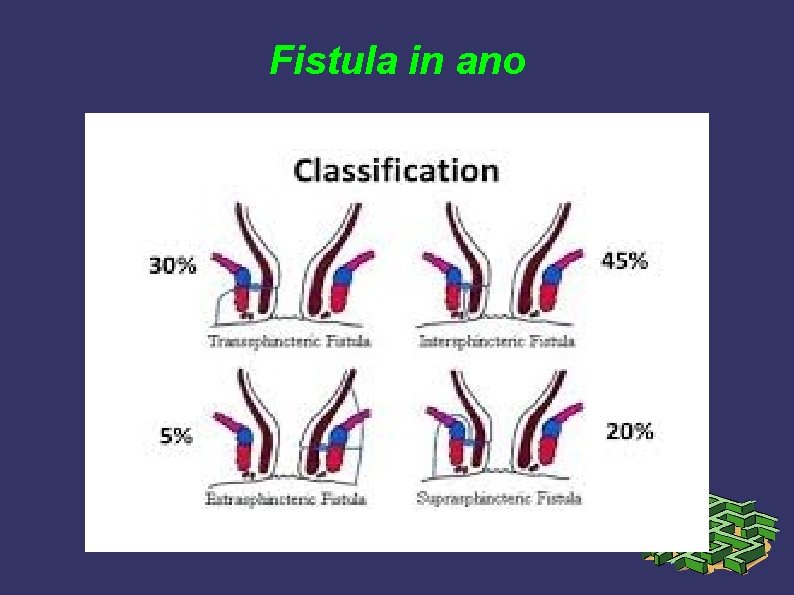
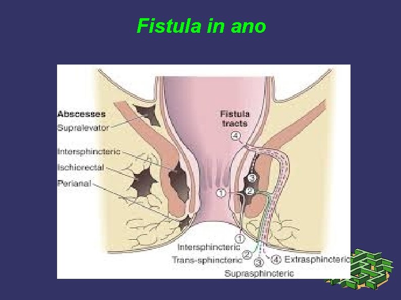
Fistula in ano Drainage of an anorectal abscess results in cure for about 50 % patients. The remaining 50 % develop a persistent fistula in ano. The fistula usually originates in the infected crypt (internal opening) and tracks to the external opening. The course of the fistula often can be predicted by the anatomy of the previous abscess. DG: Patients present with persistent drainage from the internal and/or external openings. Fistulas with an external opening anteriorly connect to the internal opening by a short, radial tract. Fistulas with an external opening posteriorly track in a curvilinear fashion to the posterior midline (Goodsall rule).
Fistula in ano
Fistula in ano
Fistula in ano 2 Fistulas are categorized based on their relationship to the anal sphincter complex and treatment options are based on these classifications. The goal of treatment of fistula in ano is eradication of sepsis without sacrificing continence. Because fistulous tracks encircle variable amounts of the sphincter complex, surgical treatment is dictated by the location of the internal and external openings and the course of the fistula.
Fistula in ano 3 1. Intersphincteric fistula – tracks through the distal internal sphincter to an external opening near the anal verge 2. Transsphincteric fistula – results from an ischiorectal abscess and extends through both the internal and external sphincters. 3. Suprasphincteric fistula– originates in the intersphincteric plane and tracks up and around the entire external sphincter 4. Extrasphincteric fistula – originates in the rectal wall and tracks around both sphincters to exit laterally, usually in the ischiorectal fossa. 5. Complex, nonhealing fistula – may result from Crohn disease, malignancy, radiation proctitis
Trauma of rectum Management of colonic injury depends on the mechanism of injury, the delay between the injury and surgery, the overall condition and stability of the patient, the degree of peritoneal contamination, and the condition of the injured colon. A primary repair may be considered in hemodynamically stable patients with few additional injuries and minimal contamination if the colon appears otherwise healthy. Contraindications to primary repair include shock, injury to more than two other organs, mesenteric vascular damage, and extensive fecal contam. A delay of greater than 6 h between the injury and the operation is a relative contraindication to primary repair. Injuries caused by high-velocity gunshot wounds or blast injuries are usually treated by fecal diversion after debridement of all nonviable tissue. CLASS: Penetrating trauma Blunt trauma
Thanks for Your attention