Anal Canal Surgical anatomy Pilonidal sinus Perianal abscess

Anal Canal – Surgical anatomy, Pilonidal sinus, Perianal abscess, fistula In Ano Dr Amit Gupta Associate Professor Dept Of Surgery

• Measures 2 to 4 cm in length • longer in men than in women. • The dentate or pectinate line marks the transition point between columnar rectal mucosa and squamous anoderm. • The 1 to 2 cm of mucosa just proximal to the dentate line shares histologic characteristics of columnar, cuboidal, and squamous epithelium and is referred to as the anal transition zone. • The dentate line is surrounded by longitudinal mucosal folds, known as the columns of Morgagni, into which the anal crypts empty. • These crypts are the source of cryptoglandular abscesses

Lining of the anal canal
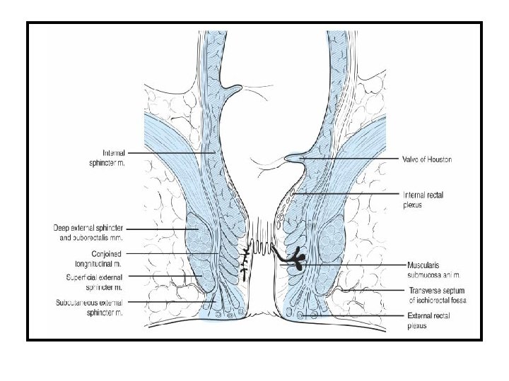

Anorectal Anatomy Arterial Supply Inferior rectal A middle rectal A Nerve Supply Sympathetic: Superior hypogastric plexus Parasympathetic: Venous drainage Inferior rectal V middle rectal V S 234 (nerviergentis Pudendal Nerve: Motor and sensory 3 hemorrhoidal complexes L lateral Anal canal R antero-lateral R posterolateral Lymphatic drainage Above dentate: Inf. Mesenteric Below dentate: internal iliac Anal verge

Pilonidal sinus Pathogenesis: A sinus tract at natal cleft resulting from: • Blockage of hair follicle • Folliculitis • Abscess followed by sinus formation. • Hair trapping • Foreign body reaction • The sinus tract is cephald Associated with: • Caucasians • Hirsute • Sedentary occupations • Obese • Poor hygeine

Presentation & Treatment Acute abscess Incision and drainage Recurrence: 40% Chronic Pain and discharge Wide local excision • with primary closure or • closure by secondary intension Recurrence: 8 -15%

Perianal Abscess • Infection originates in the intersphincteric plane, most likely in one of the anal glands. • This may result in – – simple intersphincteric abscess extend vertically either upward downwards horizontally circumferentially resulting in varied clinical presentations.
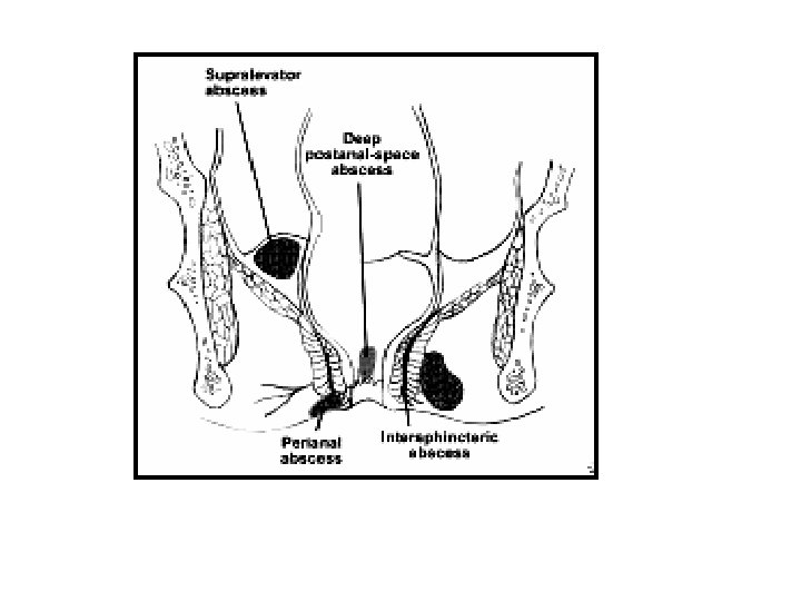

Aetiology & Pathogenesis: • 4 -10 glands at dentate line. • Infection of the cryptglandular epithelium resulting from obstruction of the glands. • Ascending infection into the intersphincteric space and other potential spaces. • Bacteria implicated: E. Coli. , Enterococci, bacteroides Other causes: • Crohn • TB • Carcinoma, Lymphoma and Leukaemia • Trauma • Inflammatory pelvic conditions (appendicitis)

Pathophysiology Glandular secretion stasis Infection & suppuration Anal crypts obstruction abscess formation
 and fever •")
Clinical presentation Abscess Clinical presentation Perianal • Perianal pain, discharge (pus) and fever • Tender, fluctuant, erythematous subcutaneous lump Ischio-rectal • Chills, fever, ischiorectal pain • Indurated, erythematous mss, tender Intersphincteric Supralevator • Rectal pain, chills and fever, discharge • PR tender. Difficult to identify are. EUA needed

Treatment • Abscesses should be drained when diagnosed. • Simple and superficial abscesses can most often be drained under local anesthesia • Patients who manifest systemic symptoms, immunocompromised and those with complex, complicated abscesses are best treated in a hospital setting. • An intersphincteric abscess is drained by dividing the internal sphincter at the level of the abscess

Incision and drainage of anorectal abscess Modification of Hanley's technique for incision and drainage of horseshoe abscess

Fistula in Ano • In anorectal abscess 50% develop a persistent fistula in ano. • The fistula usually originates in the infected crypt (internal opening) and tracks to the external opening, usually the site of prior drainage. The course of the fistula can often be predicted by the anatomy of the previous abscess. • Majority of fistulas are cryptoglandular in origin, trauma, Crohn's disease, malignancy, radiation, or unusual infections (tuberculosis, actinomycosis, and chlamydia) may also produce fistulas. • A complex, recurrent, or non healing fistula should raise the suspicion of one of these diagnoses.

Diagnosis • Patients present with persistent drainage from the internal and/or external openings. • An indurated tract is often palpable. • Goodsall's rule can be used as a guide in determining the location of the internal opening • Fistulas with an external opening anteriorly connect to the internal opening by a short, radial tract. • Fistulas with an external opening posteriorly track in a curvilinear fashion to the posterior midline. Exceptions: Anterior external opening is greater than 3 cm from the anal margin. Such fistulas usually track to the posterior midline.

Goodsall's rule to determine location of internal opening

Fistulas are categorized based upon their relationship to the anal sphincter complex and treatment options are based upon these classifications: – Intersphincteric fistula tracks through the distal internal sphincter and intersphincteric space to an external opening near the anal verge. – Transsphincteric fistula often results from an ischiorectal abscess and extends through both the internal and external sphincters – Suprasphincteric fistula originates in the intersphincteric plane and tracks up and around the entire external sphincter – Extrasphincteric fistula originates in the rectal wall and tracks around both sphincters to exit laterally, usually in the ischiorectal fossa

Intersphincteric Suprasphincteric Transsphincteric Extrasphincteric

Treatment • Goal of treatment of fistula in ano is eradication of sepsis without sacrificing continence • The external opening is usually visible as a red elevation of granulation tissue with or without concurrent drainage. • The internal opening may be more difficult to identify. • Injection of hydrogen peroxide or dilute methylene blue may be helpful

• Simple intersphincteric fistulas can often be treated by fistulotomy , curettage, and healing by secondary intention. • Fistulas that include less than 30% of the sphincter muscles can often be treated by sphincterotomy without significant risk of major incontinence. • High transsphincteric and suprasphincteric fistulas are treated by initial placement of a seton. • Extrasphincteric fistulas are rare, and treatment depends upon both the anatomy of the fistula and its etiology. • Complex and/or nonhealing fistulas may result from Crohn's disease, malignancy, radiation proctitis, or unusual infection. • Proctoscopy should be performed in all cases of complex and/or nonhealing fistulas to assess the health of the rectal mucosa. • Biopsies of the fistula tract should be taken to rule out malignancy.
- Slides: 21