An overview of neurodevelopment Eric Turner MDPh D


- Slides: 48
An overview of neurodevelopment Eric Turner, MD-Ph. D eturner@ucsd. edu 858 -534 -1568 http: //medicine. ucsd. edu/neuroembryologylab
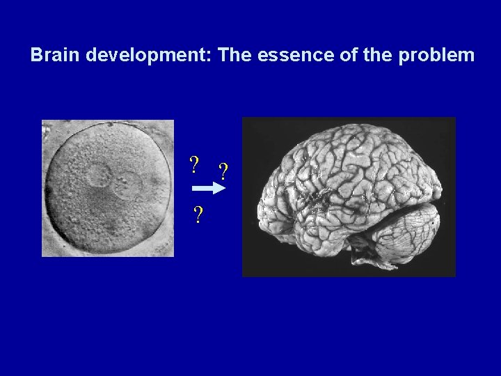
Brain development: The essence of the problem ? ? ?
Outline Part 1 General principles: Where the nervous system comes from. Part 2: Development of the midbrain dopaminergic neurons Part 3: Development of the neocortex
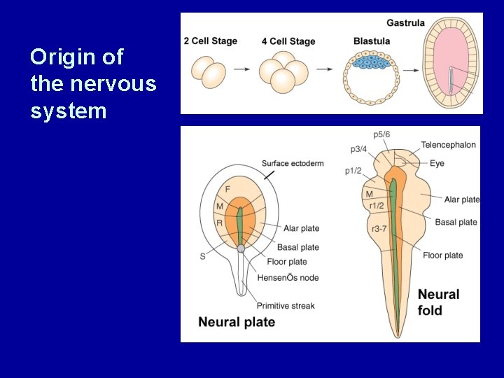
Origin of the nervous system
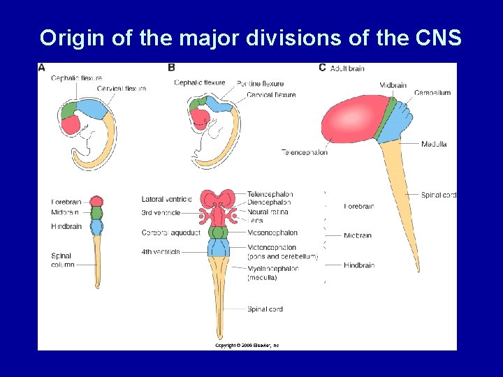
Origin of the major divisions of the CNS
The origin of the peripheral nervous system Trunk neural crest M. Bronner-Fraser Neurogenic placodes
CNS development: the spinal cord Structure: Neural Plate Neural fold Neural tube Spinal cord Event: Gastrulation Notochord formation Floorplate induction Segmental and dorsoventral patterning Neural crest migration Differentiation of specific neuronal types Radial migration; Exit from cell cycle Establishment of neuronal connections
The notochord patterns the dorsoventral axis Forebrain Midbrain Hindbrain Somites Spinal cord The ectopic notochord, n’ repatterns Pax 3 expression in the spinal cord
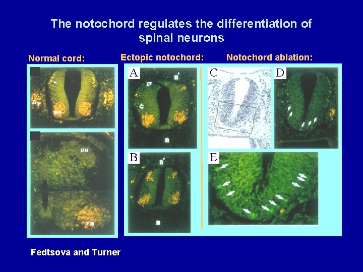
The notochord regulates the differentiation of spinal neurons Normal cord: Ectopic notochord: Fedtsova and Turner Notochord ablation: A C B E D
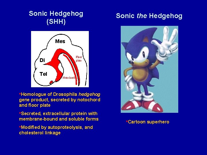
Sonic Hedgehog (SHH) Sonic the Hedgehog Mes Hindbrain Floor plate Di Tel Forebrain Notochord • Homologue of Drosophila hedgehog gene product, secreted by notochord and floor plate • Secreted, extracellular protein with membrane-bound and soluble forms • Modified by autoproteolysis, and cholesterol linkage • Cartoon superhero
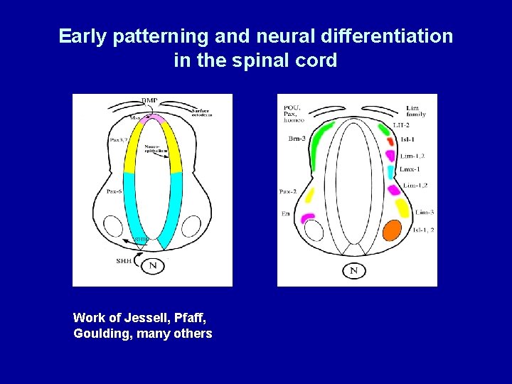
Early patterning and neural differentiation in the spinal cord Work of Jessell, Pfaff, Goulding, many others
Neural patterning in two axes: Development of the dopaminergic neurons of the midbrain Basic developmental principles are conserved between spinal cord and midbrain Dorsoventral patterning --Sonic again Rostrocaudal patterning --Multiple cell-autonomous and diffusible signals
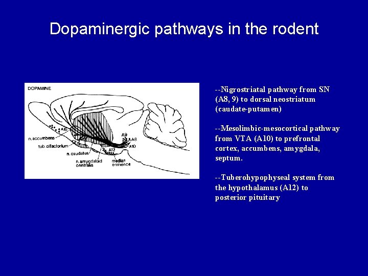
Dopaminergic pathways in the rodent --Nigrostriatal pathway from SN (A 8, 9) to dorsal neostriatum (caudate-putamen) --Mesolimbic-mesocortical pathway from VTA (A 10) to prefrontal cortex, accumbens, amygdala, septum. --Tuberohypophyseal system from the hypothalamus (A 12) to posterior pituitary
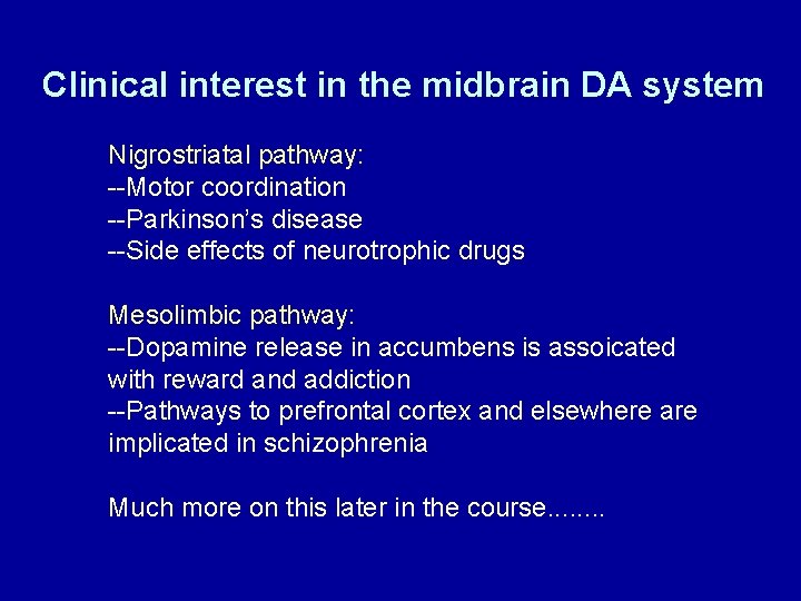
Clinical interest in the midbrain DA system Nigrostriatal pathway: --Motor coordination --Parkinson’s disease --Side effects of neurotrophic drugs Mesolimbic pathway: --Dopamine release in accumbens is assoicated with reward and addiction --Pathways to prefrontal cortex and elsewhere are implicated in schizophrenia Much more on this later in the course. . . .
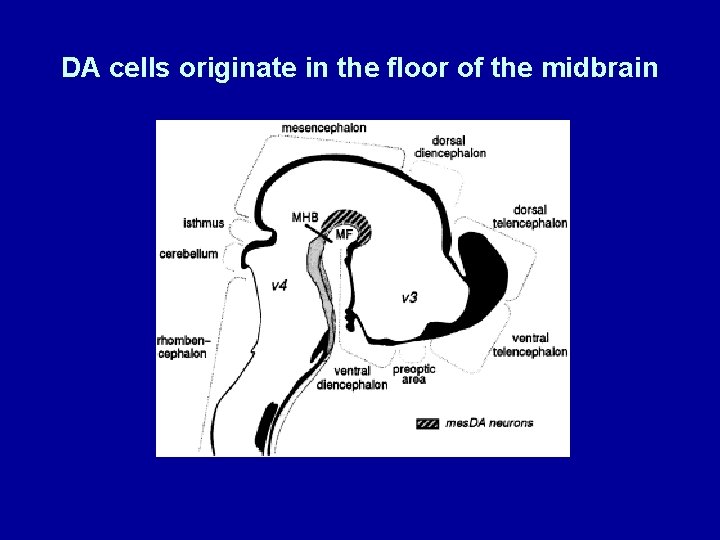
DA cells originate in the floor of the midbrain
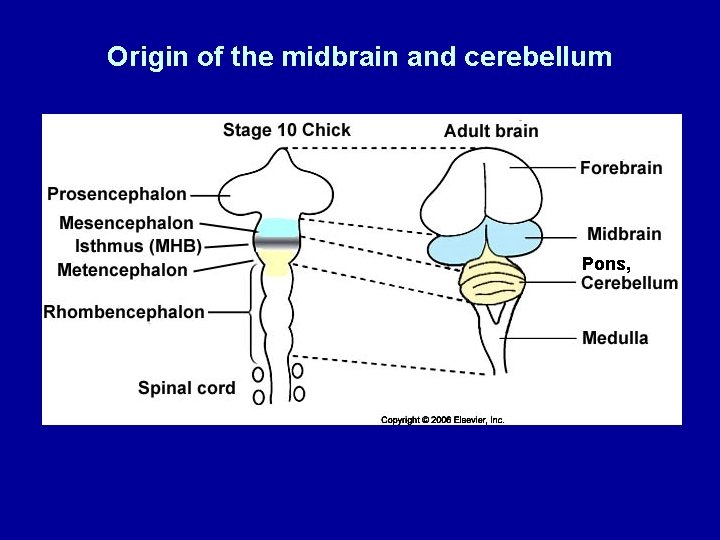
Origin of the midbrain and cerebellum Pons,
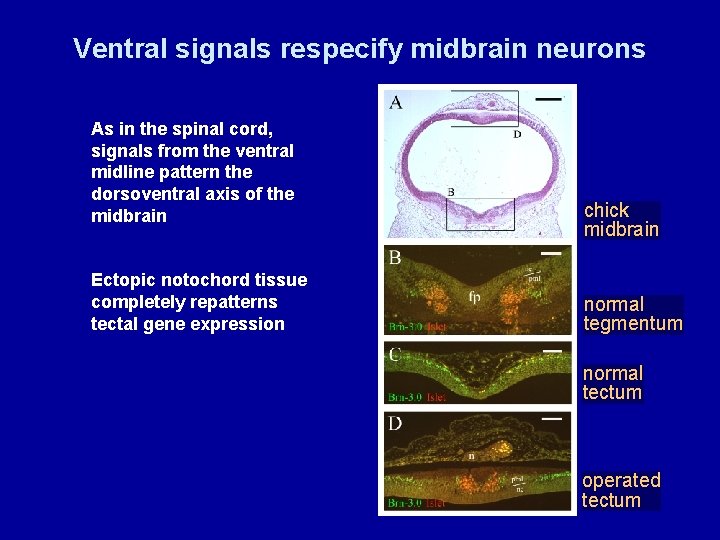
Ventral signals respecify midbrain neurons As in the spinal cord, signals from the ventral midline pattern the dorsoventral axis of the midbrain Ectopic notochord tissue completely repatterns tectal gene expression chick midbrain normal tegmentum normal tectum operated tectum
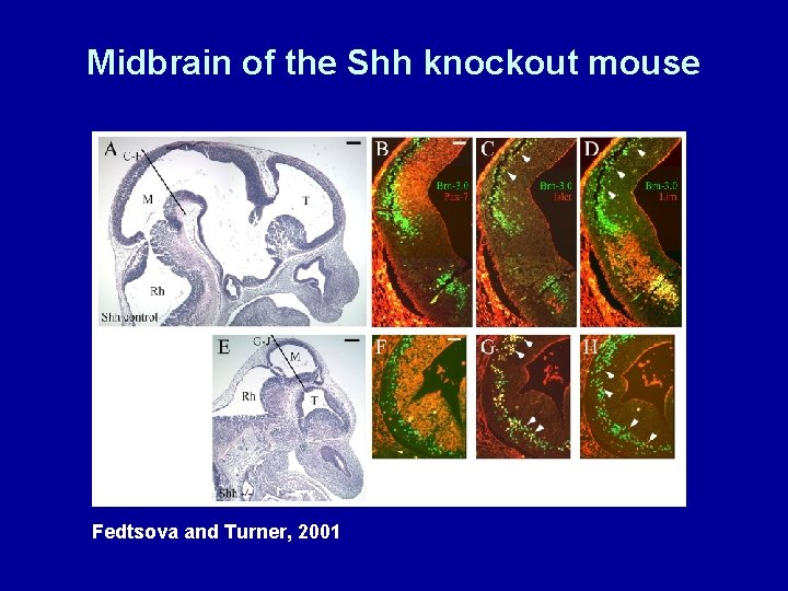
Midbrain of the Shh knockout mouse Fedtsova and Turner, 2001
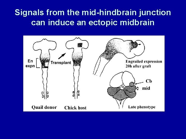
Signals from the mid-hindbrain junction can induce an ectopic midbrain
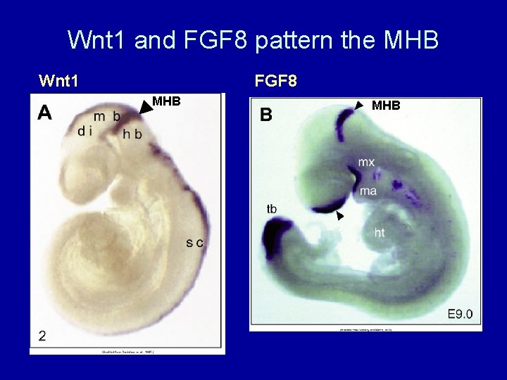
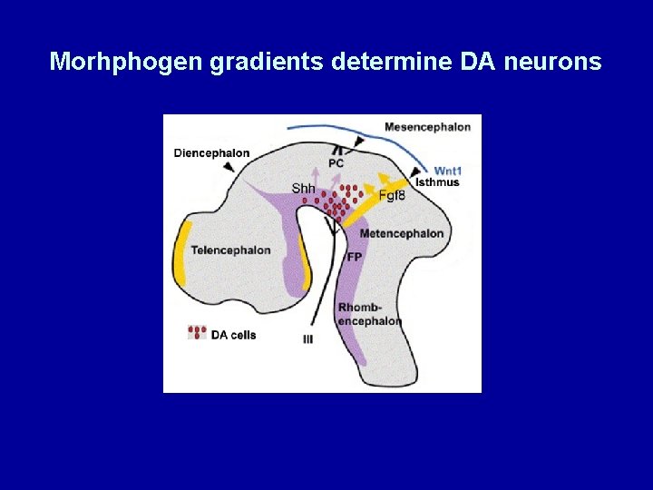
Wnt 1 and FGF 8 pattern the MHB Wnt 1 FGF 8 MHB
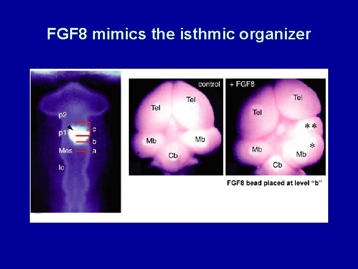
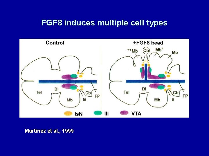
FGF 8 mimics the isthmic organizer
FGF 8 induces multiple cell types Martinez et al. , 1999
Morhphogen gradients determine DA neurons
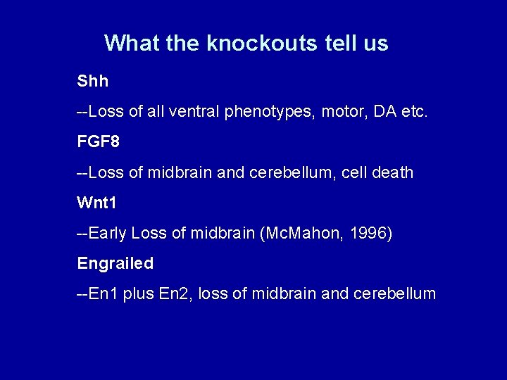
What the knockouts tell us Shh --Loss of all ventral phenotypes, motor, DA etc. FGF 8 --Loss of midbrain and cerebellum, cell death Wnt 1 --Early Loss of midbrain (Mc. Mahon, 1996) Engrailed --En 1 plus En 2, loss of midbrain and cerebellum
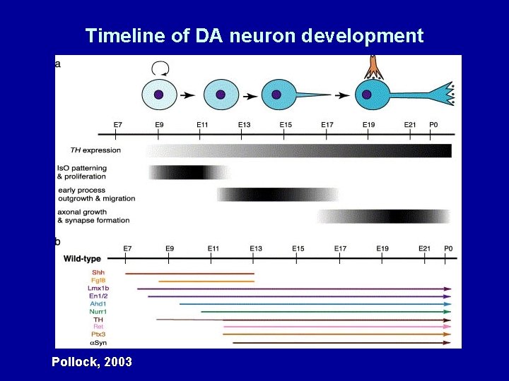
Timeline of DA neuron development Pollock, 2003
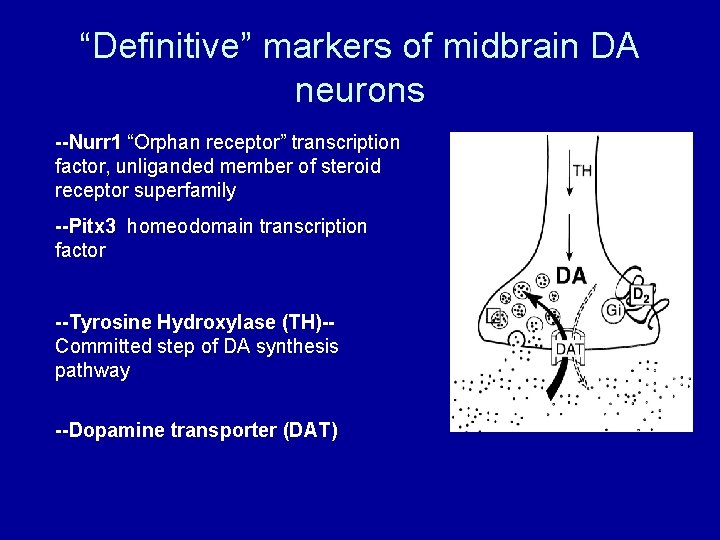
“Definitive” markers of midbrain DA neurons --Nurr 1 “Orphan receptor” transcription factor, unliganded member of steroid receptor superfamily --Pitx 3 homeodomain transcription factor --Tyrosine Hydroxylase (TH)-Committed step of DA synthesis pathway --Dopamine transporter (DAT)
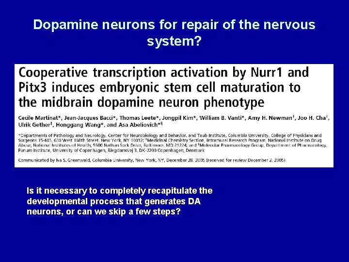
Dopamine neurons for repair of the nervous system? Is it necessary to completely recapitulate the developmental process that generates DA neurons, or can we skip a few steps?
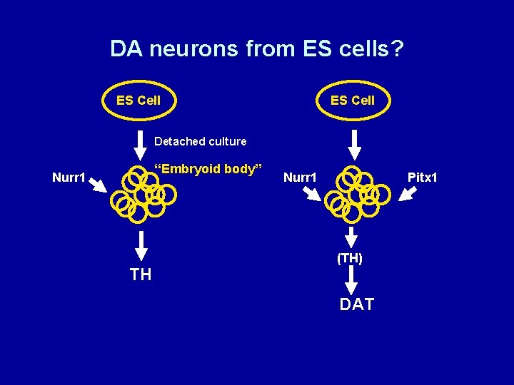
DA neurons from ES cells? ES Cell Detached culture “Embryoid body” Nurr 1 TH Nurr 1 Pitx 1 (TH) DAT
DA neurons from ES cells: summary and critique Biolgogical test: Transplanted Nurr 1/Pitx 1 ES cells rescue apomorphine-induced turning behavior in rats lesioned unilaterally with 6 -OHDA However, the ES cells to not appear to have made appropriate nigro-striatal connections How many genes does it take to execute the entire program of DA neuron development?
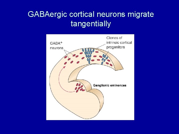
Development of the mammalian neocortex Part 1: Generation of the glutamatergic, or pyramidal cells of the neocortex by radial migration Part 2: Tangential migration of the GABAergic neurons of the neocortex Part 3: Lissencephaly-- a Mendelian disorder of cortical development Part 4: Is schizophrenia a developmental disorder of the neocortex?
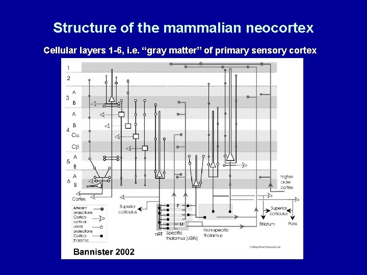
Structure of the mammalian neocortex Cellular layers 1 -6, i. e. “gray matter” of primary sensory cortex
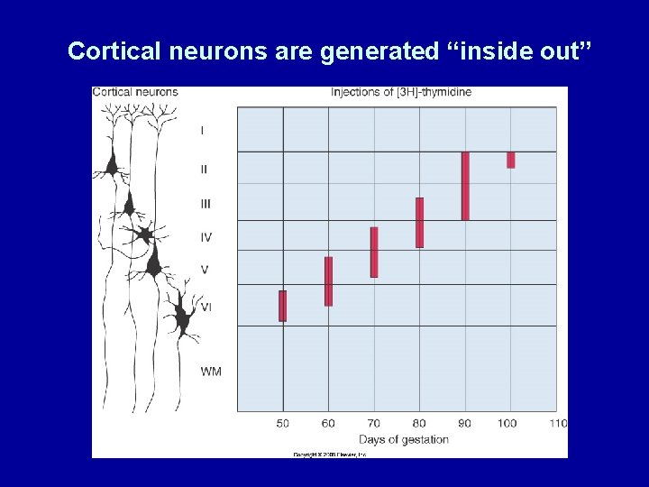
Cortical neurons are generated “inside out”
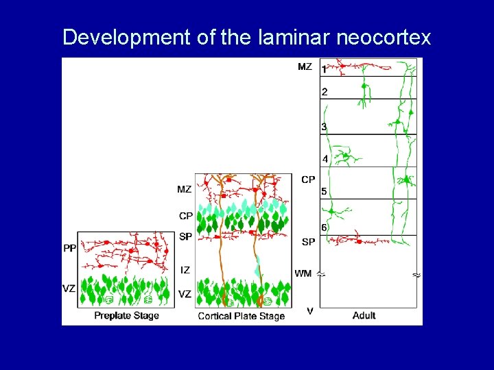
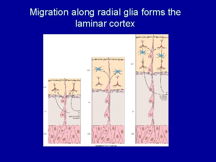
Development of the laminar neocortex
Migration along radial glia forms the laminar cortex
GABAergic cortical neurons migrate tangentially
Lissencephaly--a profound disorder of cortical development Thanks to: Joe Gleeson Male patient, X-linked lissencephaly
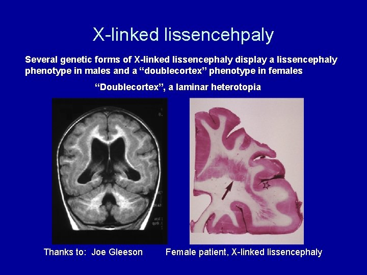
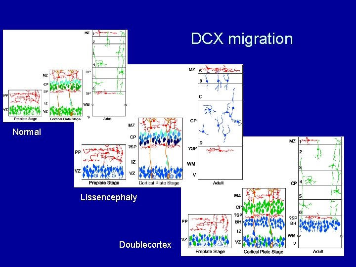
X-linked lissencehpaly Several genetic forms of X-linked lissencephaly display a lissencephaly phenotype in males and a “doublecortex” phenotype in females “Doublecortex”, a laminar heterotopia Thanks to: Joe Gleeson Female patient, X-linked lissencephaly
X-inactivation makes chimeric females Thanks to: Joe Gleeson
DCX migration Normal Lissencephaly Doublecortex
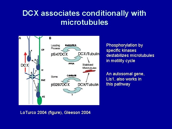
DCX associates conditionally with microtubules Phosphorylation by specific kinases destabilizes microtubules in motility cycle An autosomal gene, Lis 1, also works in this pathway Lo. Turco 2004 (figure), Gleeson 2004
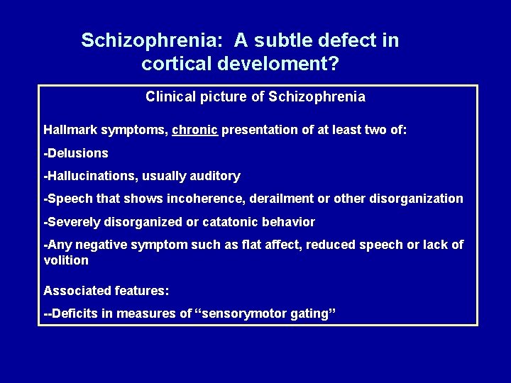
Schizophrenia: A subtle defect in cortical develoment? Clinical picture of Schizophrenia Hallmark symptoms, chronic presentation of at least two of: -Delusions -Hallucinations, usually auditory -Speech that shows incoherence, derailment or other disorganization -Severely disorganized or catatonic behavior -Any negative symptom such as flat affect, reduced speech or lack of volition Associated features: --Deficits in measures of “sensorymotor gating”
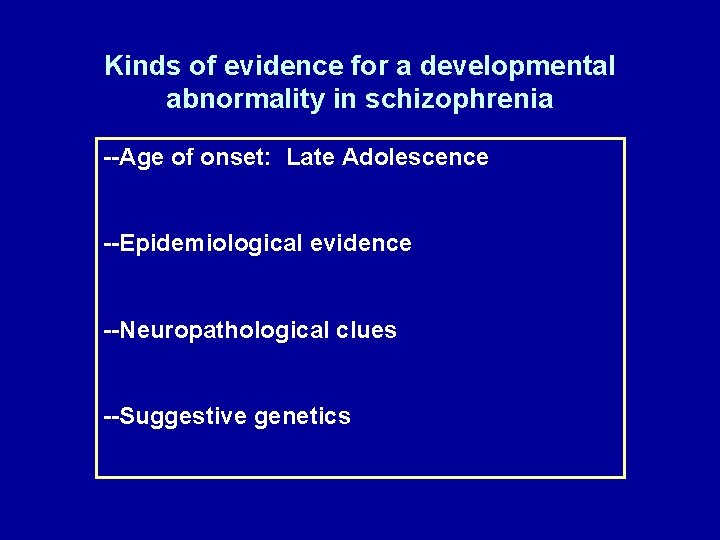
Kinds of evidence for a developmental abnormality in schizophrenia --Age of onset: Late Adolescence --Epidemiological evidence --Neuropathological clues --Suggestive genetics
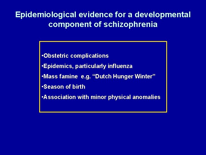
Epidemiological evidence for a developmental component of schizophrenia • Obstetric complications • Epidemics, particularly influenza • Mass famine e. g. “Dutch Hunger Winter” • Season of birth • Association with minor physical anomalies
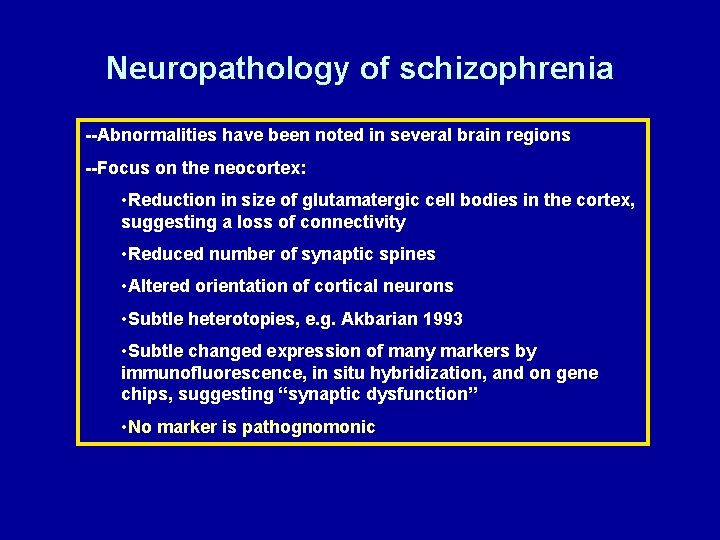
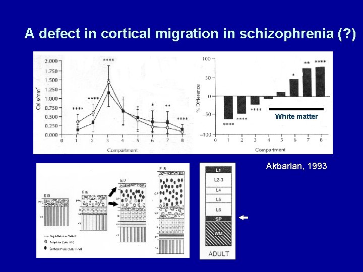
Neuropathology of schizophrenia --Abnormalities have been noted in several brain regions --Focus on the neocortex: • Reduction in size of glutamatergic cell bodies in the cortex, suggesting a loss of connectivity • Reduced number of synaptic spines • Altered orientation of cortical neurons • Subtle heterotopies, e. g. Akbarian 1993 • Subtle changed expression of many markers by immunofluorescence, in situ hybridization, and on gene chips, suggesting “synaptic dysfunction” • No marker is pathognomonic
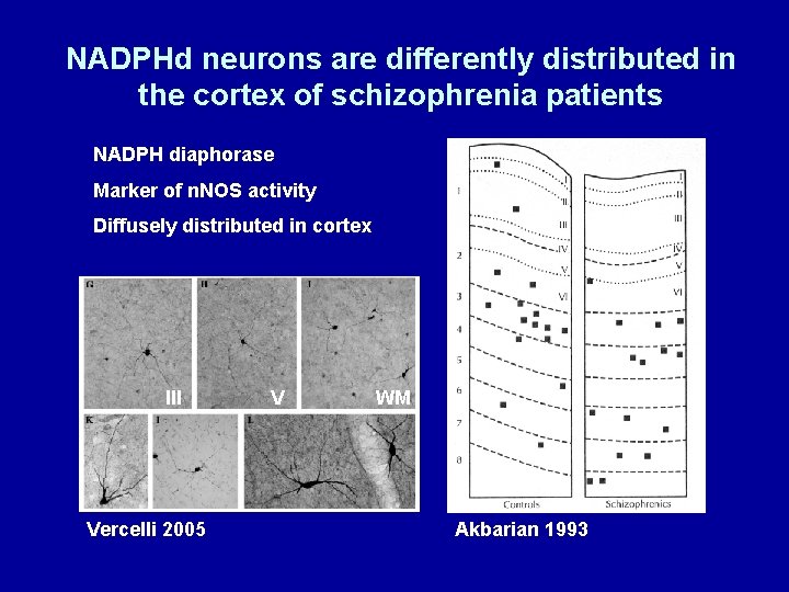
NADPHd neurons are differently distributed in the cortex of schizophrenia patients NADPH diaphorase Marker of n. NOS activity Diffusely distributed in cortex III Vercelli 2005 V WM Akbarian 1993
A defect in cortical migration in schizophrenia (? ) White matter Akbarian, 1993
Genetic leads in schizophrenia No “major effect” genes identified, even within families Linkage or association studies with markers on nearly every chromosome Recent genes of developmental interest: • Dysbindin (DTNBP 1) Interacts with dystrophin, defective in muscular dystrophy. Present in synapses, widely expressed. Duchenne’s muscular dystrophy is accompanies by cortical heterotopias. • Disrupted-in-Schizophrenia 1 (DISC 1) Translocation breakpoint associated with schizophrenia. Large multifunctional protein with possible role in microtubule function • Neuregulin 1 (NRG 1) EGF-family receptor. Hideous number of splice forms. Widely expressed, with multiple developmental roles.
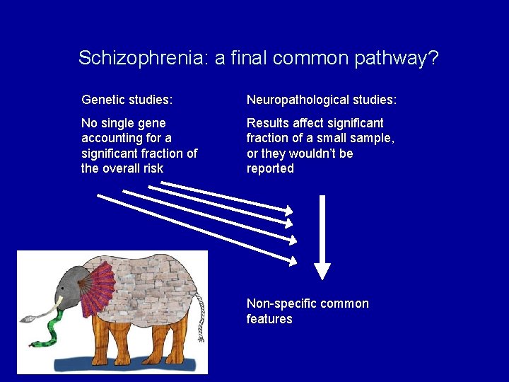
Schizophrenia: a final common pathway? Genetic studies: Neuropathological studies: No single gene accounting for a significant fraction of the overall risk Results affect significant fraction of a small sample, or they wouldn’t be reported Non-specific common features