Alice Thwin Nikon Imaging Center Sample Prep and



















- Slides: 48
Alice Thwin Nikon Imaging Center Sample Prep and Best Practices for Imaging
What is Sample Prep? � Wikipedia definition: the ways in which a sample is treated prior to its analysis
What is Sample Prep? � Wikipedia definition: the ways in which a sample is treated prior to its analysis microscopy.
Order of events: Live imaging ▪ Dyes – Trackers, useful hints ▪ Additives Fixatives Immuno. Histochemistry and Immuno. Cytochemistry ▪ Antibody information for best results Mounting Media ▪ Commercially available Coverslips ▪ The importance of a #1. 5 coverslip Choosing your ▪ Objective ▪ Microscopy technique
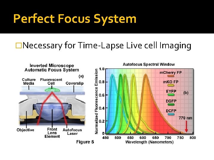
Perfect Focus System �Necessary for Time-Lapse Live cell Imaging
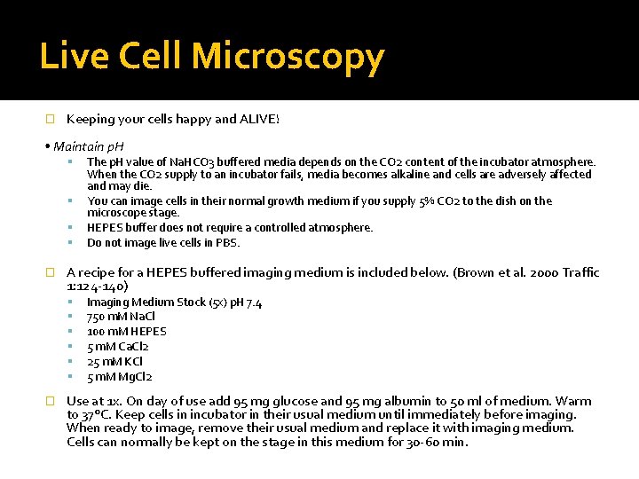
Live Cell Microscopy � Keeping your cells happy and ALIVE! • Maintain p. H � A recipe for a HEPES buffered imaging medium is included below. (Brown et al. 2000 Traffic 1: 124 -140) � The p. H value of Na. HCO 3 buffered media depends on the CO 2 content of the incubator atmosphere. When the CO 2 supply to an incubator fails, media becomes alkaline and cells are adversely affected and may die. You can image cells in their normal growth medium if you supply 5% CO 2 to the dish on the microscope stage. HEPES buffer does not require a controlled atmosphere. Do not image live cells in PBS. Imaging Medium Stock (5 x) p. H 7. 4 750 m. M Na. Cl 100 m. M HEPES 5 m. M Ca. Cl 2 25 m. M KCl 5 m. M Mg. Cl 2 Use at 1 x. On day of use add 95 mg glucose and 95 mg albumin to 50 ml of medium. Warm to 37°C. Keep cells in incubator in their usual medium until immediately before imaging. When ready to image, remove their usual medium and replace it with imaging medium. Cells can normally be kept on the stage in this medium for 30 -60 min.
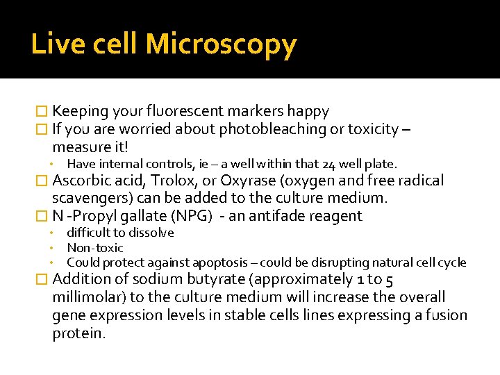
Live cell Microscopy � Keeping your fluorescent markers happy � If you are worried about photobleaching or toxicity – measure it! • Have internal controls, ie – a well within that 24 well plate. � Ascorbic acid, Trolox, or Oxyrase (oxygen and free radical scavengers) can be added to the culture medium. � N -Propyl gallate (NPG) - an antifade reagent • • • difficult to dissolve Non-toxic Could protect against apoptosis – could be disrupting natural cell cycle � Addition of sodium butyrate (approximately 1 to 5 millimolar) to the culture medium will increase the overall gene expression levels in stable cells lines expressing a fusion protein.
Live Cell Microscopy- dyes � Commercially available and good for internal controls for photobleaching and localization studies. ER-Tracker Green dye ▪ green-fluorescent BODIPY® FL dye and glibenclamide. ▪ Glibenclamide (glyburide) binds to the sulphonylurea receptors of ATP-sensitive K+ channels which are prominent on ER Mito-Trackers: per the Invitrogen website: • Mito. Tracker Red or Green FM: mitochondrial stain which appears to localize to mitochondria regardless of mitochondrial membrane potential. • Mito. Tracker Red CMXRos is a red-fluorescent dye that stains mitochondria in live cells and its accumulation is dependent upon membrane potential. • Mito. Tracker Red CM-H 2 XRos is a reduced, nonfluorescent version of Mito. Tracker Red (that fluoresces upon oxidation. This dye also stains mitochondria in live cells and its accumulation is dependent upon membrane potential.
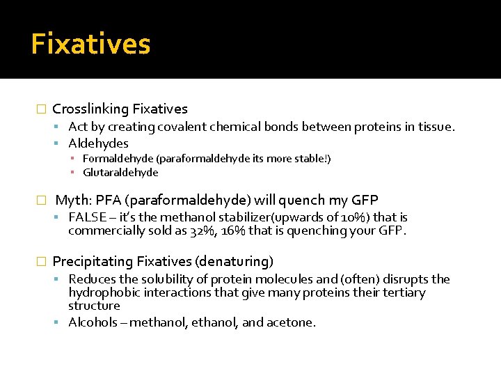
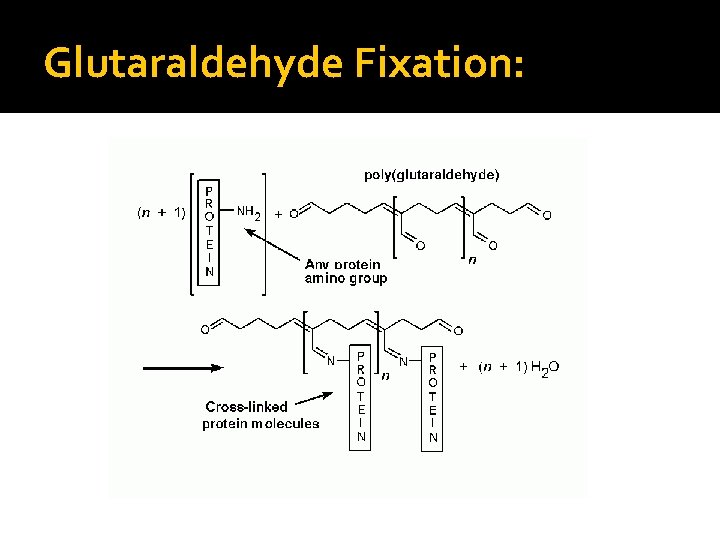
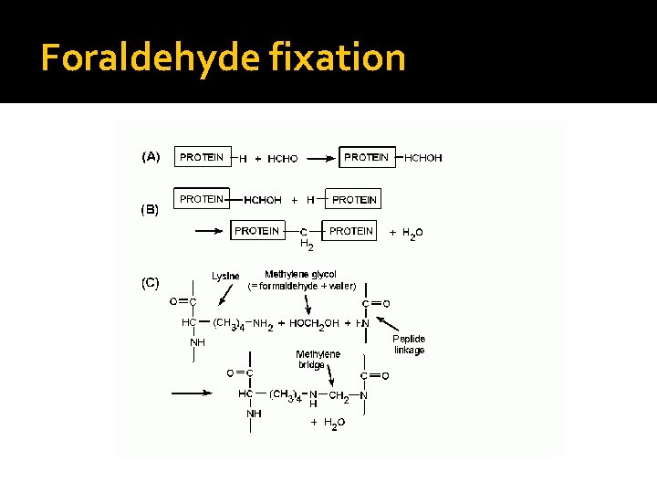
Fixatives � Crosslinking Fixatives Act by creating covalent chemical bonds between proteins in tissue. Aldehydes ▪ Formaldehyde (paraformaldehyde its more stable!) ▪ Glutaraldehyde � Myth: PFA (paraformaldehyde) will quench my GFP FALSE – it’s the methanol stabilizer(upwards of 10%) that is commercially sold as 32%, 16% that is quenching your GFP. � Precipitating Fixatives (denaturing) Reduces the solubility of protein molecules and (often) disrupts the hydrophobic interactions that give many proteins their tertiary structure Alcohols – methanol, and acetone.
Glutaraldehyde Fixation:
Paraformaldehyde
Foraldehyde fixation
Fixation � Per a given antigen rules of thumb (in use since 1973) Antigen Fixative Most proteins, peptides and enzymes of low molecular weight 4% PFA +/- 1%Glutaraldehyde 10% Neutral-Buffered Formalin (NBF) Delicate Tissue Boulin’s Fixative Small molecules 4% PFA + 1% Glutaraldehyde Blood forming organs (liver, spleen, bone marrow); connective tissue Zenker’s solution Helly Solution Nucleic acids Carnoy’s Solution Large Protein antigens (immunoglobulin) Ice cold acetone or Methanol (100%)
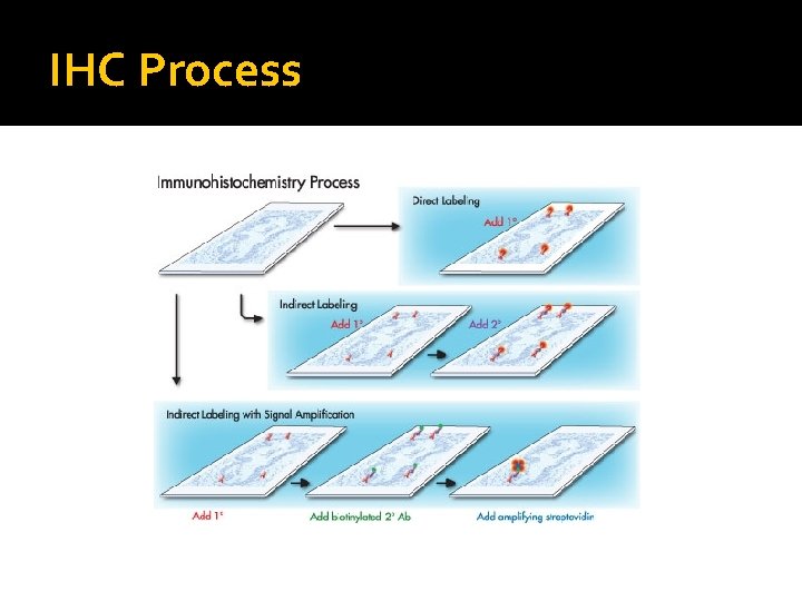
What is the IHC and ICC? � IHC – Immuno. Histochemistry (for tissue) and ICC- Immuno. Cyto. Chemistry (for cell culture) � Combination of immunological and histological techniques Using Antibodies that target epitopes of an antigen of interest to directly or indirectly label ▪ Direct labeling uses a primary antibody that is conjugated. ▪ Indirect labeling is step wise process where a conjugated secondary antibody recognizes the host of the primary antibody.
IHC Process
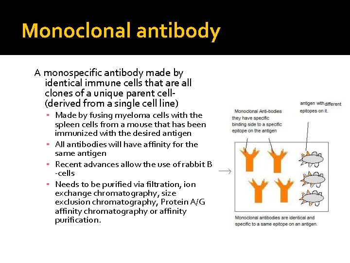
Monoclonal antibody A monospecific antibody made by identical immune cells that are all clones of a unique parent cell(derived from a single cell line) ▪ Made by fusing myeloma cells with the spleen cells from a mouse that has been immunized with the desired antigen ▪ All antibodies will have affinity for the same antigen ▪ Recent advances allow the use of rabbit B -cells ▪ Needs to be purified via filtration, ion exchange chromatography, size exclusion chromatography, Protein A/G affinity chromatography or affinity purification.
Monoclonal Antibody production:
Polyclonal Antibodies � Also known as antisera Immunoglobulin molecules that were secreted-against a specific antigen, each identifying a different epitope. � Produced in the natural immune response � Antibodies obtained are from different B-cell resources An antibody produced recognizes different epitopes on the same antigen and have varying affinities for the each epitope.
Polyclonal antibody production:
Polyclonal Antibody Production
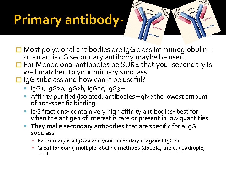
Primary antibody� Most polyclonal antibodies are Ig. G class immunoglobulin – so an anti-Ig. G secondary antibody maybe be used. � For Monoclonal antibodies be SURE that your secondary is well matched to your primary subclass. � Ig. G subclass and how can it be useful? Ig. G 1, Ig. G 2 a, Ig. G 2 b, Ig. G 2 c, Ig. G 3 – Affinity purified (isolated) antibodies – give the lowest amount of non-specific binding. Ig. G fractions- contain very high affinity antibodies- best for when the antigen of interest is rare or present in low quantities. They make secondary antibodies that are specific for a Ig. G subclass ▪ Ex. Primary is a Ig. G 2 a and your secondary is against Ig. G 2 a ▪ Great for doing multiple labeling methods (double, triple, quadruple, etc. )
Primary Antibody
Secondary antibodies � Choosing a secondary The secondary antibody should be against the species that the primary antibody is raised – ex if your primary is raised in mouse, an antimouse secondary antibody should be used. Match the subclass of your secondary to the subclass of your primary also – ex: if your primary is one of mouse Ig. G subclasses( Ig. G 1, Ig. G 2 b, Ig. G 2 c, Ig. G 3) any of the anti-mouse Ig. G can be used. � Labeled or Conjugated Secondary Antibody enzyme labeled – peroxidase, alkaline phosphatase fluorescent labeled – Alexa Fluor 405, 488, 568, 647 biotin conjugated – best amplification of the signal and thus will give greater sensitivity than with enzyme or fluorescent conjugated secondary antibody alone.
Secondary antibodies- a bit more info What if you have a lot of non-specific staining with your secondary? � Structural similarities are sometimes found among immunoglobulins from different species. Consequently, a polyclonal antibody directed to a specific species can also react with Ig. G from other species. This non-specific binding is called cross-reactivity. � To reduce cross-reactivity among immunoglobulins from different species, an additional purification step is applied in which antibodies are pre-adsorbed against potentially cross-reactive species. � Solution- Adsorbed secondary antibody ▪ Adsorbed with an animal or human Ig. G. ▪ Ex: If working with human tissue choose a secondary that is absorbed with human serum or human Ig. G. ▪ *caution - Adsorbed antibodies will reduced epitope recognition.
Take away of an IHC or ICC Indirect method will give you an amplified signal due to its step wise process. � Monoclonal affinity isolated primary antibodies � Target only one epitope on the antigen of interest (could be harder to � � work with) Polyclonal affinity isolated primary antibodies will be batch dependant Target many epitopes on the antigen (easier to work) Choose your secondary antibody accordingly A. B. C. Match the subclass to primary antibody Adsorbed Fluorescently conjugated
Other- more sensitive detection systems – expensive! � If all else fails, and you need to see your protein in vivo. � Polymer detection system Polymerized reporter enzyme staining system based on polymerizing an enzyme and attaching these polymers to an antibody. ▪ ▪ ▪ Envision (Dako Cytomation) Imm. PRESS (Vector Labs) MACH 2 (Biocare Medical) Polink (IHC world) Novolink (Leica Microsystems)
Refraction and the Refractive Index (RI) �Changes in refractive index causes light to deviate from its expected path:
Mounting Media Using the correct mounting medium avoids signal loss and optical aberration and could preserve fluorescence signal with anti-fading properties. � Refractive indexes of interest: � � Water = 1. 33 Immersion Oil = 1. 513 Air = 1 Light is refracted when it crosses the interface between two media of differing refractive indices Mismatching refractive index between the sample’s mounting media and the immersion media is one of the largest components to image degradation in microscopy Mismatched refractive indices can also result in stretching/ compression of the z-axis. ▪ So what you think is a 1 um step size for the objective does not necessarily correspond to a 1 um change in the focal plane.
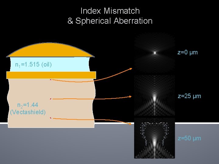
Index Mismatch & Spherical Aberration objective Immersion fluid n 1 Cover glass n 2 Sample Focus at cover slip Focus into sample Spherical aberration unless n 2 = n 1
Index Mismatch & Spherical Aberration z=0 µm n 1=1. 515 (oil) z=25 µm n 2=1. 44 (Vectashield) z=50 µm
Constituents of Mounting Media � A base component- a major factor in the refractive index of the medium Hydrophilic: (do not require dehydration) ▪ Aqueous -(RI ~ 1. 33) ▪ Glyercol (1. 47) Hydrophobic: (require dehydration) � ▪ Natural Oil (1. 53) ▪ Plastic (1. 51) Antifade component - photobleaching is not fully understood – but it seems as though when the fluorescent dye is in its excited triplet state, the dyes react with molecular oxygen and lose the ability to become fluorescent most antifade reagents are reactive oxygen species scavengers P-Phenylenediamine (PPD) seems to be most effective – except cyanine dyes (Cy 2 especiallly)- toxic. N-Propyl gallate (NPG) - difficult to dissolve – is non-toxic and can be used with live cells – could even protect against apoptosis 1, 4 -Diazadicyclo-octane (DABCO) aka – triethylenediamine – less effective then PPD, and also less toxic.
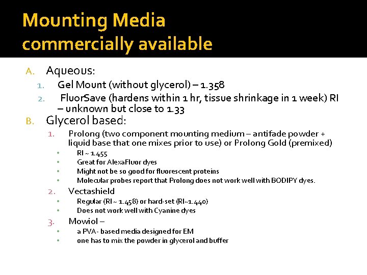
Mounting Media commercially available Aqueous: A. Gel Mount (without glycerol) – 1. 358 Fluor. Save (hardens within 1 hr, tissue shrinkage in 1 week) RI – unknown but close to 1. 33 1. 2. B. Glycerol based: 1. • • 2. 3. • • Prolong (two component mounting medium – antifade powder + liquid base that one mixes prior to use) or Prolong Gold (premixed) RI ~ 1. 455 Great for Alexa. Fluor dyes Might not be so good for fluorescent proteins Molecular probes report that Prolong does not work well with BODIPY dyes. Vectashield Regular (RI ~ 1. 458) or hard-set (RI~1. 440) Does not work well with Cyanine dyes Mowiol – a PVA- based media designed for EM one has to mix the powder in glycerol and buffer
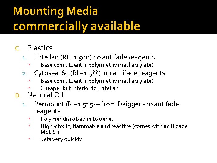
Mounting Media commercially available Plastics C. 1. • 2. • • Entellan (RI ~1. 500) no antifade reagents Base constituent is poly(methylmethacrylate) Cytoseal 60 (RI ~1. 5? ? ) no antifade reagents Base constituent is poly(methylmethacrylate) Cheaper but inferior to Entellan Natural Oil D. 1. • • • Permount (RI~1. 515) – from Daigger -no antifade reagents Polymer dissolved in toluene. Highly toxic, flammable and reactive (comes with an 8 page MSDS!) Sets very quickly
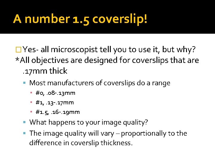
A number 1. 5 coverslip! �Yes- all microscopist tell you to use it, but why? *All objectives are designed for coverslips that are. 17 mm thick Most manufacturers of coverslips do a range ▪ #0, . 08 -. 13 mm ▪ #1, . 13 -. 17 mm ▪ #1. 5, . 16 -. 19 mm What happens to your image quality? The image quality will vary – proportionally to the difference in coverslip thickness.
Use a number. 17 mm thick coverslip � To reduce variability in performance get “high- performance” coverslips that are to be accurate to. 001 mm ( from Zeiss can order a. 17 mm thick coverslip) Numerical Aperture 0. 01 mm Deviation 0. 02 mm Deviation 0. 30 none 0. 45 none 0. 70 2 percent 8 percent 0. 85 19 percent 57 percent 0. 95 55 percent 71 percent
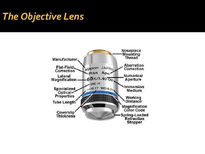
The Objective Lens
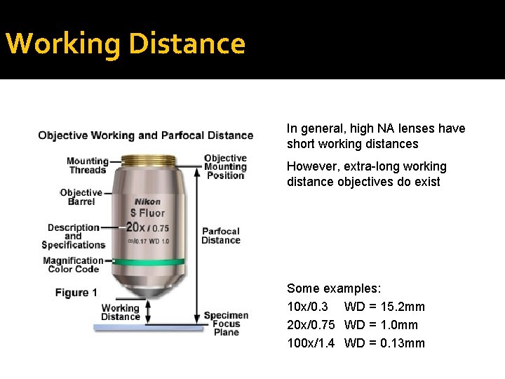
Working Distance In general, high NA lenses have short working distances However, extra-long working distance objectives do exist Some examples: 10 x/0. 3 WD = 15. 2 mm 20 x/0. 75 WD = 1. 0 mm 100 x/1. 4 WD = 0. 13 mm
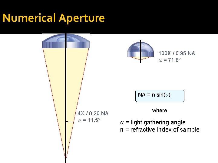
Numerical Aperture 100 X / 0. 95 NA = 71. 8° NA = n sin( ) 4 X / 0. 20 NA = 11. 5° where = light gathering angle n = refractive index of sample
Resolution of the Microscope limited by the point-spread function Resolution: X-Y, 0. 61 l / NA; Z, ln / NA 2 Resolution for some common objectives, in nm: NA 0. 3 0. 75 0. 95 1. 4 X-Y 1017 407 321 218 Z 16830 2690 1680 770
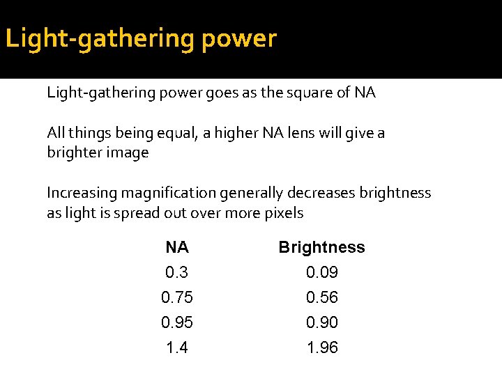
Light-gathering power goes as the square of NA All things being equal, a higher NA lens will give a brighter image Increasing magnification generally decreases brightness as light is spread out over more pixels NA Brightness 0. 3 0. 75 0. 95 1. 4 0. 09 0. 56 0. 90 1. 96
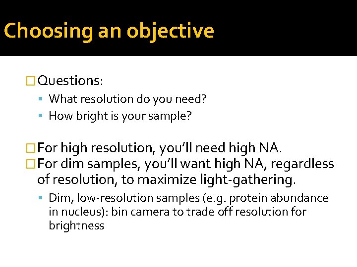
Choosing an objective �Questions: What resolution do you need? How bright is your sample? �For high resolution, you’ll need high NA. �For dim samples, you’ll want high NA, regardless of resolution, to maximize light-gathering. Dim, low-resolution samples (e. g. protein abundance in nucleus): bin camera to trade off resolution for brightness
Choosing an objective �Questions: What resolution do you need? How bright is your sample? �When to use low NA? Bright samples at low resolution / low magnification If you need long working distance If spherical aberration is a concern If you want large depth of field to get whole structures in focus at once (avoid Z-stacks)
Which imaging technique should I use? 1 -5 mm TIRF (for samples at the coverslip) Spinning Disk Confocal Line-scanning confocal >20 mm Point scanning Confocal >50 mm 2 -photon confocal Slow 10 -100 mm Sensitivity Sample Thickness 1 -20 mm Fast Wide-field (+deconvolution)
Microscope choice �Epifluorescence – routine work, low magnification, or thin samples where you don’t need high-resolution 3 D reconstruction �TIRF – samples at the membrane or otherwise at the coverslip surface; very high signal-tonoise; single molecule imaging �Spinning Disk Confocal – Live tissue culture cells, yeast, etc, or thin (<30 mm) tissue sections when you need 3 D reconstructions �Laser-Scanning Confocal – Thick tissues or specimens
Acknowledgements: Hassel, J. and Hand, A. R. (1973) J. Histochem. Cytochem 22 229 -239 Microscopyu. com IHC world. com Confocal listserv Wright Cell Imaging Facility, Toronto Western Research Institute Wikipedia Commercial websites – Cargille, invitrogen, diagger, jackson immuno labs.
Acknowledgements: �Kurt Thorn, Director of the NIC
Acknowledgements: �Kurt Thorn, Director of the NIC
Thanks! �Go out there and do some sample prep so that I can see your samples under the scope – preferably at the NIC! �nic. ucsf. edu