Whats New in DICOM Workshop SPIE MEDICAL IMAGING









- Slides: 46
What’s New in DICOM Workshop @ SPIE MEDICAL IMAGING February 2012 Bas Revet, Philips Healthcare – (Chair WG 6)
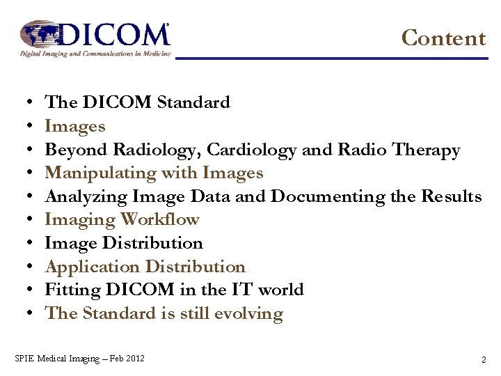
Content • • • The DICOM Standard Images Beyond Radiology, Cardiology and Radio Therapy Manipulating with Images Analyzing Image Data and Documenting the Results Imaging Workflow Image Distribution Application Distribution Fitting DICOM in the IT world The Standard is still evolving SPIE Medical Imaging – Feb 2012 2
What and Who is DICOM The DICOM Standard All almost 30 years going strong … SPIE Medical Imaging – Feb 2012 3
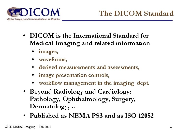
The DICOM Standard • DICOM is the International Standard for Medical Imaging and related information • • • images, waveforms, derived measurements and assessments, image presentation controls, workflow management in the imaging dept. • Beyond Radiology and Cardiology: Pathology, Ophthalmology, Surgery, Dermatology, … • Published as NEMA PS 3 and as ISO 12052 SPIE Medical Imaging – Feb 2012 4
The DICOM Work Process • Continuous process for evolution of the standard – It’s just “DICOM”, not DICOM 3. 0, 3. 1, etc. – Conformance is to SOP Classes, not to a ‘version’ of the Standard • Change Proposals for minor corrections – About 100 / year – Anybody can submit • Supplements for major changes – new object types, services, compression schemes – About 10 / year – Require Work Item approved by DICOM Standards Committee • Consolidated edition published every year – ! w e N – Most recently, August 2011 (2011 version) Available free at DICOM web site SPIE Medical Imaging – Feb 2012 5
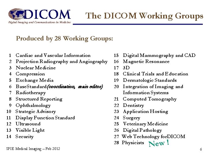
The DICOM Working Groups Produced by 28 Working Groups: 1 2 3 4 5 6 7 8 9 10 11 12 13 14 Cardiac and Vascular Information Projection Radiography and Angiography Nuclear Medicine Compression Exchange Media Base Standard (coordination, main editor) Radiotherapy Structured Reporting Ophthalmology Strategic Advisory Display Function Standard Ultrasound Visible Light Security SPIE Medical Imaging – Feb 2012 15 16 17 18 19 20 21 22 23 24 25 26 27 28 Digital Mammography and CAD Magnetic Resonance 3 D Clinical Trials and Education Dermatologic Standards Integration of Imaging and Information Systems Computed Tomography Dentistry Application Hosting Surgery Veterinary Medicine Digital Pathology Web Technology for DICOM Physicists ew ! N 6
Images Where it all started … SPIE Medical Imaging – Feb 2012 7
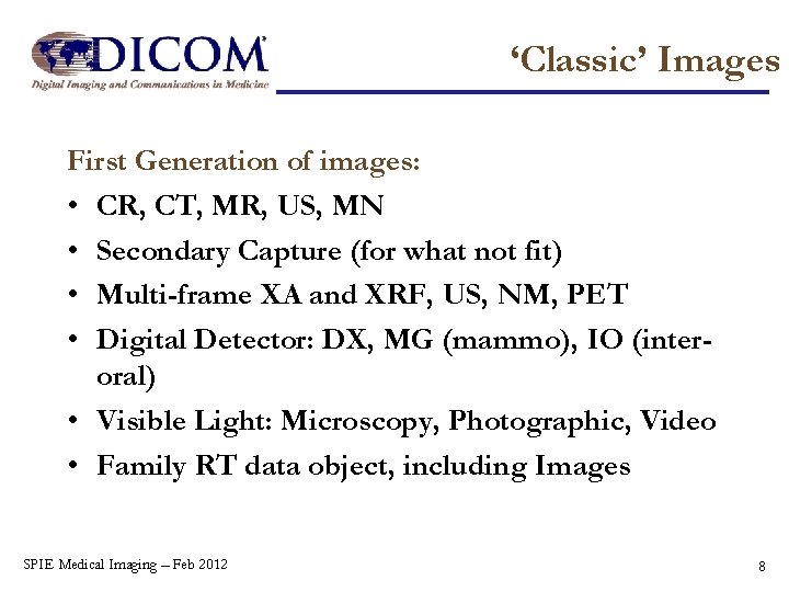
‘Classic’ Images First Generation of images: • CR, CT, MR, US, MN • Secondary Capture (for what not fit) • Multi-frame XA and XRF, US, NM, PET • Digital Detector: DX, MG (mammo), IO (interoral) • Visible Light: Microscopy, Photographic, Video • Family RT data object, including Images SPIE Medical Imaging – Feb 2012 8
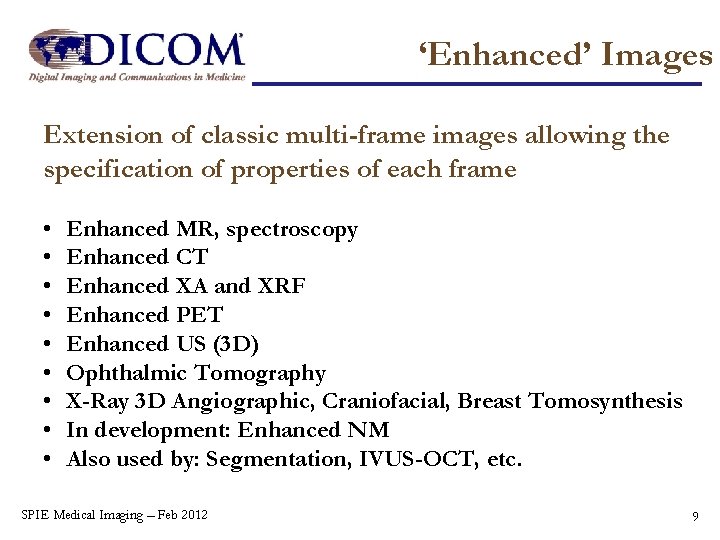
‘Enhanced’ Images Extension of classic multi-frame images allowing the specification of properties of each frame • • • Enhanced MR, spectroscopy Enhanced CT Enhanced XA and XRF Enhanced PET Enhanced US (3 D) Ophthalmic Tomography X-Ray 3 D Angiographic, Craniofacial, Breast Tomosynthesis In development: Enhanced NM Also used by: Segmentation, IVUS-OCT, etc. SPIE Medical Imaging – Feb 2012 9
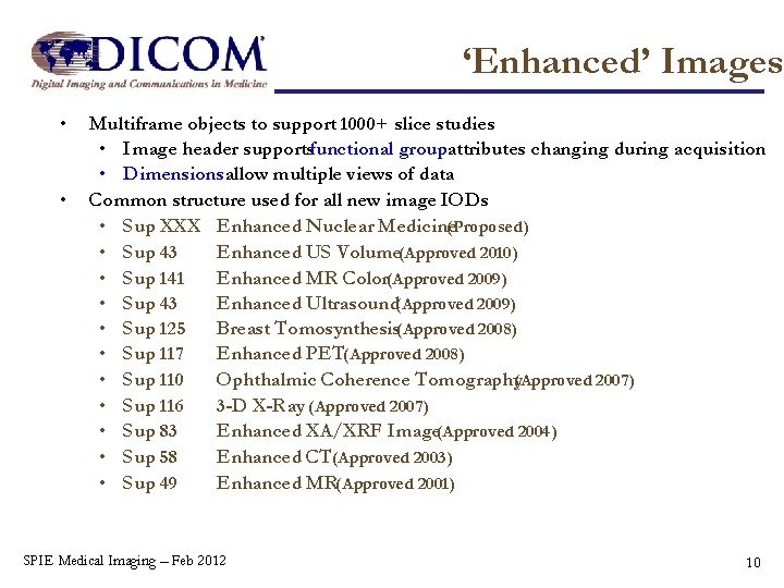
‘Enhanced’ Images • • Multiframe objects to support 1000+ slice studies • Image header supportsfunctional groupattributes changing during acquisition • Dimensionsallow multiple views of data Common structure used for all new image IODs • Sup XXX Enhanced Nuclear Medicine(Proposed) • Sup 43 Enhanced US Volume(Approved 2010) • Sup 141 Enhanced MR Color(Approved 2009) • Sup 43 Enhanced Ultrasound(Approved 2009) • Sup 125 Breast Tomosynthesis(Approved 2008) • Sup 117 Enhanced PET(Approved 2008) • Sup 110 Ophthalmic Coherence Tomography(Approved 2007) • Sup 116 3 -D X-Ray (Approved 2007) • Sup 83 Enhanced XA/XRF Image(Approved 2004) • Sup 58 Enhanced CT(Approved 2003) • Sup 49 Enhanced MR(Approved 2001) SPIE Medical Imaging – Feb 2012 10
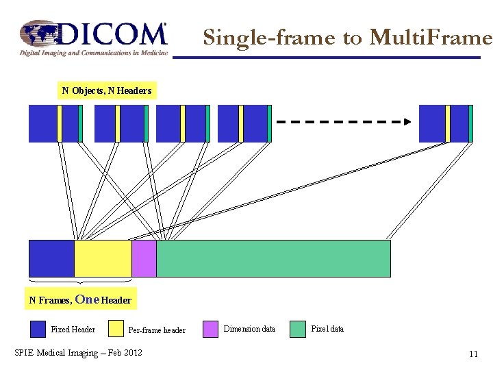
Single-frame to Multi. Frame N Objects, N Headers N Frames, One Header Fixed Header Per-frame header SPIE Medical Imaging – Feb 2012 Dimension data Pixel data 11
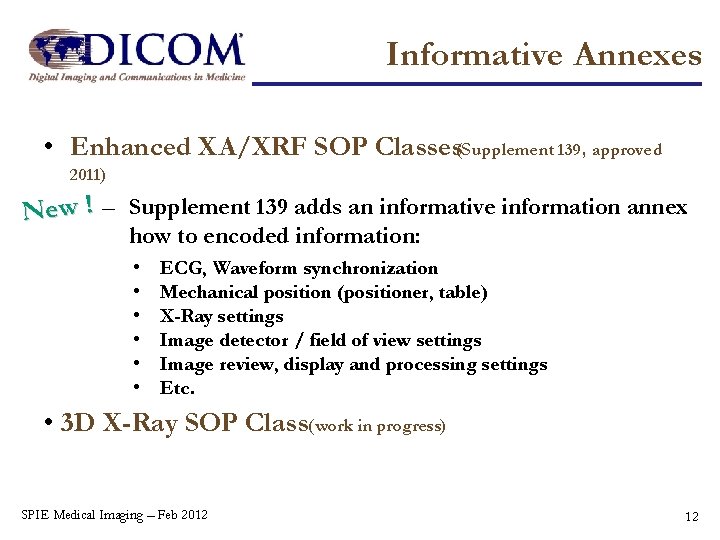
Informative Annexes • Enhanced XA/XRF SOP Classes(Supplement 139, approved 2011) New ! – Supplement 139 adds an informative information annex how to encoded information: • • • ECG, Waveform synchronization Mechanical position (positioner, table) X-Ray settings Image detector / field of view settings Image review, display and processing settings Etc. • 3 D X-Ray SOP Class(work in progress) SPIE Medical Imaging – Feb 2012 12
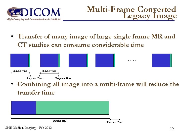
Multi-Frame Converted Legacy Image • Transfer of many image of large single frame MR and CT studies can consume considerable time …. Transfer Time Response Time • Combining all image into a multi-frame will reduce the transfer time Transfer Time SPIE Medical Imaging – Feb 2012 Response Time 13
Multi-Frame Converted Legacy Image • Build analog to ‘Enhanced’ multi-frame definitions for CT, MR or PET • Intended for PACS – PACS transfers or PACS workstation transfers • Receiving side may unpack multi-frame or handle it the same way and enhanced SOP Instance • Standard will specify how to convert the legacy images to multi-frame • Benefit for implementers: the first step to implement the complete SPIE Medical Imaging – Feb 2012 14
New Clinical Domains Beyond Radiology, Cardiology and Radio Therapy Reusing proven concepts … SPIE Medical Imaging – Feb 2012 15
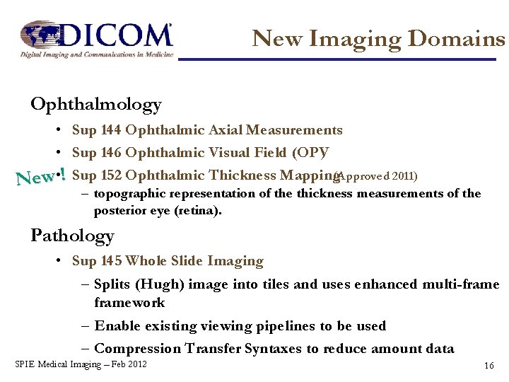
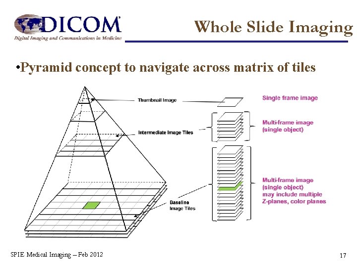
New Imaging Domains Ophthalmology • Sup 144 Ophthalmic Axial Measurements • Sup 146 Ophthalmic Visual Field (OPV) New • ! Sup 152 Ophthalmic Thickness Mapping(Approved 2011) – topographic representation of the thickness measurements of the posterior eye (retina). Pathology • Sup 145 Whole Slide Imaging – Splits (Hugh) image into tiles and uses enhanced multi-framework – Enable existing viewing pipelines to be used – Compression Transfer Syntaxes to reduce amount data SPIE Medical Imaging – Feb 2012 16
Whole Slide Imaging • Pyramid concept to navigate across matrix of tiles SPIE Medical Imaging – Feb 2012 17
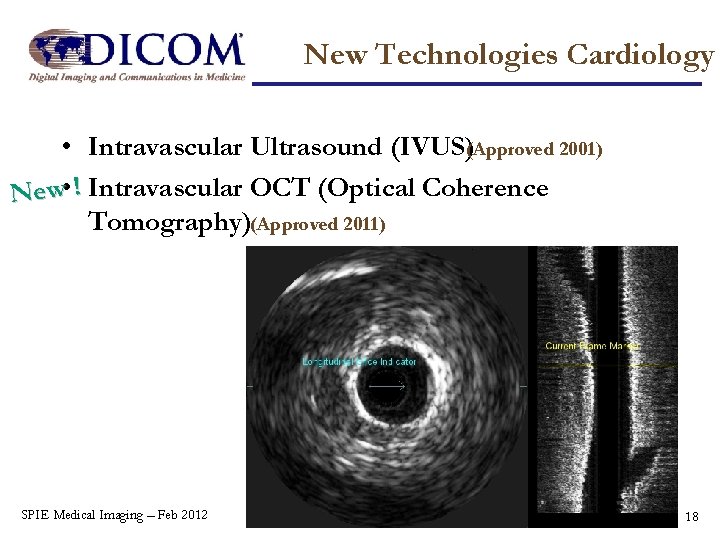
New Technologies Cardiology • Intravascular Ultrasound (IVUS)(Approved 2001) New • ! Intravascular OCT (Optical Coherence Tomography)(Approved 2011) SPIE Medical Imaging – Feb 2012 18
Radiotheraphy RT Second Generation • Radiotherapy Working Group decided to create a new family of SOP Classes • Based on the new Enhanced Multi-frame paradigm and other new DICOM developments • Preparing version for Public Comment (Supplement 147) SPIE Medical Imaging – Feb 2012 19
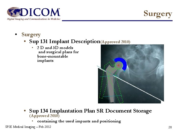
Surgery • Sup 131 Implant Description (Approved 2010) • 2 D and 3 D models and surgical plans for bone-mountable implants • Sup 134 Implantation Plan SR Document Storage (Approved 2010) • containing the used impants and positioning SPIE Medical Imaging – Feb 2012 20
New IT developements New • ! MPEG-4 (Approved 2011) • Higher compression ratio than MPEG-2 • Higher computational requirements than MPEG-2 • These transfer syntaxes will be available for any image modality, although visible light is the expected type. • !Blue Ray Disk in addition to CD and DVD media New (Approved 2011) SPIE Medical Imaging – Feb 2012 21
Image Presentation Manipulating with Images Besides storage there is more … SPIE Medical Imaging – Feb 2012 22
Image Presentation • Displaying and annotating images: • • • Grayscale Softcopy Presentation State Color Softcopy Presentation State Pseudo-Color Softcopy Presentation State Blending Extension with Compound Graphical Objects • Organizing the display of studies • Structured Display (single study) • Hanging Protocols Storage (multi study cases) SPIE Medical Imaging – Feb 2012 23
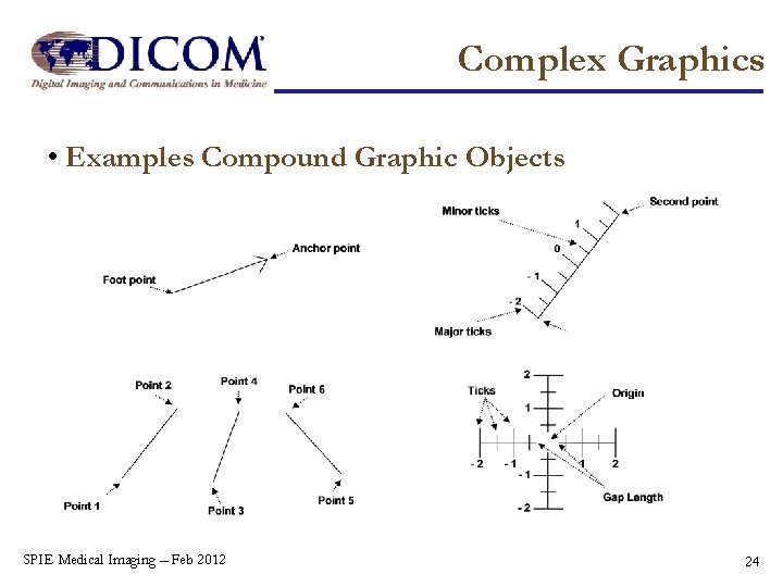
Complex Graphics • Examples Compound Graphic Objects SPIE Medical Imaging – Feb 2012 24
Image Presentation Linking image data: • Spatial Registration (rigid) • Deformable Spatial Registration (flexible) Selecting and annotating parts of images or volumes: • Segmentation • Surface Segmentation (using vertices) SPIE Medical Imaging – Feb 2012 25
Measurements & Reporting Analyzing Image Data and Documenting the Results On a well structured and interoperable way … SPIE Medical Imaging – Feb 2012 26
Report Formats Highly structured: • Diagnostic and Evidence Reports in DICOM Structured Report Format Dedicated SOP Classes: • ECG and other Waveform Documents in a DICOM ‘jacket’: • Encapsulated PDF and HL 7 CDA document streams SPIE Medical Imaging – Feb 2012 27
DICOM Structured Reports • Report is stored in a tree of meaningful leaves (Content Items) • Documented relationship between the Content Items in a Template published in the standard • Robustness by specifying in a fixed nomenclature (Codes from SNOMED, LOINC, UCUM, DICOM, etc) • Encoded as normal DICOM Composite object • More complex than image due to nested structure • Equal format as image for easy storage in PACS, etc. • Templates (in Part 16) for diagnostic reports • evident reports (measurements, logs, etc. ) • X-Ray dose reports • SPIE Medical Imaging – Feb 2012 28
Example of SR in Workflow Cardiology Echocardiography ECG Report Hemo Report IVUS Report LVA/QAA Report Procedure Report Cardiac Information System Cardiac Cath Report Images, Waveforms, Presentations States CT/MR images Cardio/Vascular Analysis Radiology X-ray images CAD Application Mammography Chest X-ray (PACS/RIS) Workstation Radiology OB/GYN (US) Vascular US Procedure (PACS/RIS) US Workstation Radiology Report (basic text, enhanced, comprehensive) Key Object Selection US images, Presentations States SPIE Medical Imaging – Feb 2012 29
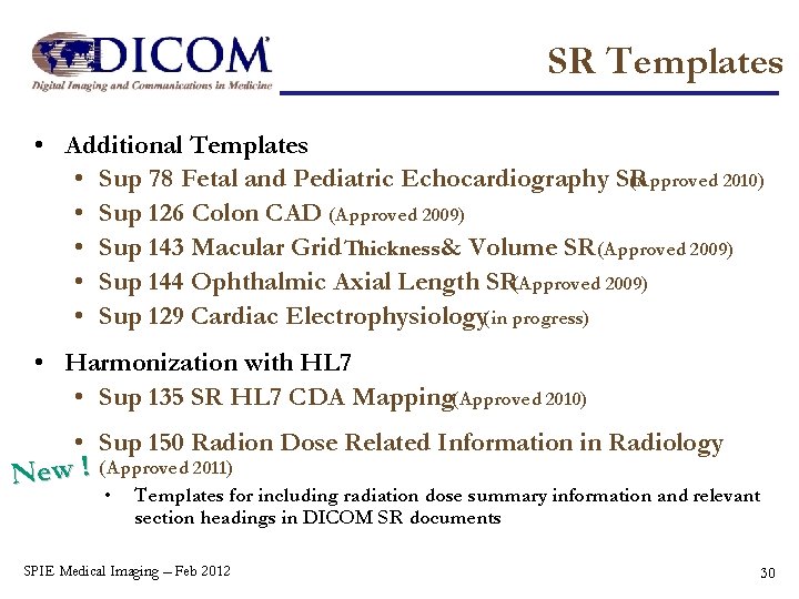
SR Templates • Additional Templates • Sup 78 Fetal and Pediatric Echocardiography SR (Approved 2010) • Sup 126 Colon CAD (Approved 2009) • Sup 143 Macular Grid Thickness& Volume SR (Approved 2009) • Sup 144 Ophthalmic Axial Length SR(Approved 2009) • Sup 129 Cardiac Electrophysiology(in progress) • Harmonization with HL 7 • Sup 135 SR HL 7 CDA Mapping(Approved 2010) • Sup 150 Radion Dose Related Information in Radiology New ! (Approved 2011) • Templates for including radiation dose summary information and relevant section headings in DICOM SR documents SPIE Medical Imaging – Feb 2012 30
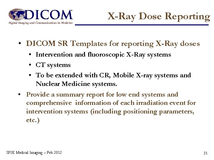
X-Ray Dose Reporting • DICOM SR Templates for reporting X-Ray doses • Intervention and fluoroscopic X-Ray systems • CT systems • To be extended with CR, Mobile X-ray systems and Nuclear Medicine systems. • Provide a summary report for low end systems and comprehensive information of each irradiation event for intervention systems (including positioning parameters, etc. ) SPIE Medical Imaging – Feb 2012 31
Workflow Imaging Workflow To enable a consistent distribution of information … SPIE Medical Imaging – Feb 2012 32
Workflow • In a distributed environment it is crucial that identifying meta data is consistent on all systems • Cooperating system must able to keep track of progress • DICOM Worklist management and Procedure Step management implements dedicated work flow management control: • Pull model for querying a next (set of) activities • Push model to start and control activities • Reports with status and results of activities SPIE Medical Imaging – Feb 2012 33
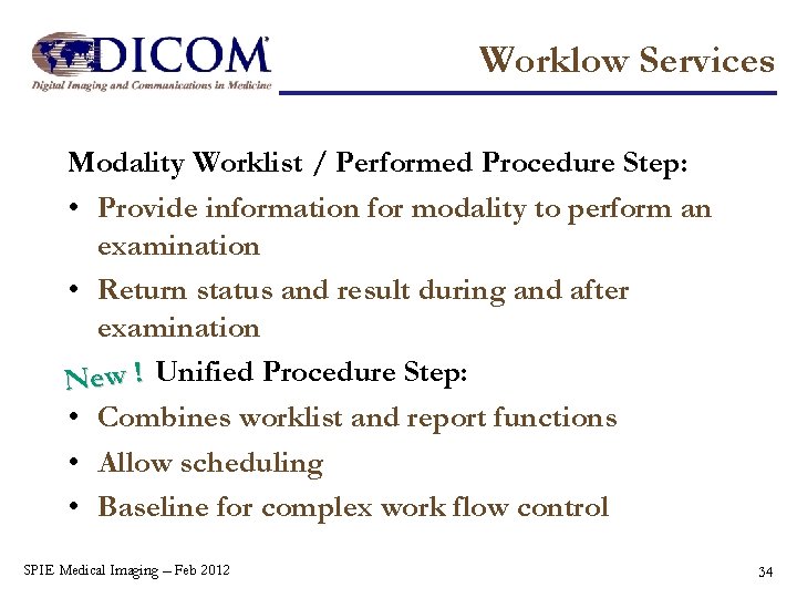
Worklow Services Modality Worklist / Performed Procedure Step: • Provide information for modality to perform an examination • Return status and result during and after examination New ! Unified Procedure Step: • Combines worklist and report functions • Allow scheduling • Baseline for complex work flow control SPIE Medical Imaging – Feb 2012 34
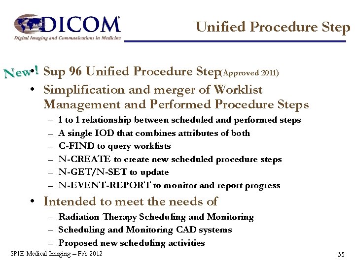
Unified Procedure Step New • ! Sup 96 Unified Procedure Step(Approved 2011) • Simplification and merger of Worklist Management and Performed Procedure Steps – – – 1 to 1 relationship between scheduled and performed steps A single IOD that combines attributes of both C-FIND to query worklists N-CREATE to create new scheduled procedure steps N-GET/N-SET to update N-EVENT-REPORT to monitor and report progress • Intended to meet the needs of – Radiation Therapy Scheduling and Monitoring – Scheduling and Monitoring CAD systems – Proposed new scheduling activities SPIE Medical Imaging – Feb 2012 35
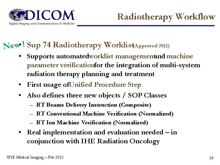
Radiotherapy Workflow New • ! Sup 74 Radiotherapy Worklist(Approved 2011) • Supports automatedworklist managementand machine parameter verificationfor the integration of multi-system radiation therapy planning and treatment • First usage of. Unified Procedure Step • Also defines three new objects / SOP Classes – RT Beams Delivery Instruction (Composite) – RT Conventional Machine Verification (Normalized) – RT Ion Machine Verification (Normalized) • Real implementation and evaluation needed – in conjunction with IHE Radiation Oncology SPIE Medical Imaging – Feb 2012 36
Image Distribution Access to images without DICOM protocols … SPIE Medical Imaging – Feb 2012 37
Other Protocols ‘Traditional’ DICOM protocols require a dedicated client and server not available outside Modality / PACS / Workstation domain DICOM WADO defines access to DICOM formatted data using ‘web’ protocols: • HTTP New ! • Web services (Supplement 148, approved 2011) Enables web based applications (browsers, DICOM web viewers) accessing from (PACS) servers supporting this protocols anywhere on internet or intranet. SPIE Medical Imaging – Feb 2012 38
Application Hosting Application Distribution Moving around applications … SPIE Medical Imaging – Feb 2012 39
Application Hosting Today specialized and dedicated application for multi-platform requires an interface for each platform (expensive, little reuse) DICOM Application Hosting defines a common interface (independent technology) for control, data exchange and with the DICOM data model as basis. It is all in Suppement 118(Approved 2010) SPIE Medical Imaging – Feb 2012 40
IT, Privacy Fitting DICOM in the IT world Standards, IT management, Privacy … SPIE Medical Imaging – Feb 2012 41
IT Standards DICOM is using and following ‘common sense standards’: • TCP/IP for connection between application • ISO-OSI model for application / presentation layers • Web protocols for access non-DICOM application • IT management: DNS, NTP, DHCP, LDAP • Audit trail: SYSLOG – TLS(Supplement 95, approved 2010) SPIE Medical Imaging – Feb 2012 42
Privacy DICOM Meta Data consist of many sensitive data: names, IDs, addresses, phone numbers and other information that allows identification of a patient Depending on the purpose information must removed or replaced: teaching, clinical trials, remote access, remote service, etc. Rules specifying what action should take place are New ! added in Supplement 142(Approved 2011) Several options are available to extend the basic profile as needed SPIE Medical Imaging – Feb 2012 43
DICOM Work in Progress The Standard is still evolving And needs YOUR input and collaboration … SPIE Medical Imaging – Feb 2012 44
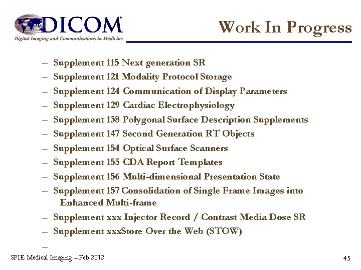
Work In Progress – – – – – Supplement 115 Next generation SR Supplement 121 Modality Protocol Storage Supplement 124 Communication of Display Parameters Supplement 129 Cardiac Electrophysiology Supplement 138 Polygonal Surface Description Supplements Supplement 147 Second Generation RT Objects Supplement 154 Optical Surface Scanners Supplement 155 CDA Report Templates Supplement 156 Multi-dimensional Presentation State Supplement 157 Consolidation of Single Frame Images into Enhanced Multi-frame – Supplement xxx Injector Record / Contrast Media Dose SR – Supplement xxx. Store Over the Web (STOW) – ngle Frame Images into Enhanced Multi-frame SPIE Medical Imaging – Feb 2012 45
Get Involved! • Links to all information at MITA/NEMA – http: //dicom. nema. org/ • Get the DICOM Standard (free!) – http: //medical. nema. org/dicom/2011/ • Ask questions/discuss on the DICOM Newsgroup – http: //groups. google. com/group/comp. protocols. dicom • Respond to Supplements released for Public Comment – http: //www. dclunie. com/dicom-status/status. html • Submit Change Proposals – mailto svastagh@medicalimaging. org • Join a DICOM Working Group – See list in http: //medical. nema. org/dicom/geninfo/Strategy. pdf SPIE Medical Imaging – Feb 2012 46