
DICOM n D Presentation State Toshiba CrossSectional Imaging
DICOM n. D Presentation State Toshiba Cross-Sectional Imaging Use Cases Updated 15 Feb, 2011
Modalities Considered n n n CT MR PET 9/25/2020 n. D Presentation State Use Cases 2
Clinical Specialties Considered n n n Radiology Cardiology Gastroenterology 9/25/2020 n. D Presentation State Use Cases 3
CT Coronary Artery Stenosis Input is a gated, contrast-enhanced CT cardiac exam n n n n n Device/user creating derived views and presentation states CT technologist, radiologist or cardiologist using post-processing application Automated / semi-automated segmentation of: n Heart / myocardium n Coronary arteries Automated / semi-automated coronary artery centreline finding Views saved: n Three MPRs aligned with the axes of the heart intersecting at the point of stenosis n Curved MPR following the centreline of the stenotic vessel with stenosis marked n Cross-curved MPR at the point of stenosis with % stenosis measurement n SVR and / or volumetric MIP, oriented to show stenosis Device/user using presentation state to replicate derived views Radiologist / cardiologist performing primary read after tech workup Radiologist / cardiologist reviewing priors alongside follow-up exams Need to reproduce: n Segmentation n Centrelines n Markers / measurements n Position and orientation of MPRs n Position of cross-curved MPR n Orientation and VOI for SVR / MIP n W/L for MPR and MIP views n Opacity for SVR views – accuracy of colour mapping unimportant 9/25/2020 n. D Presentation State Use Cases 4
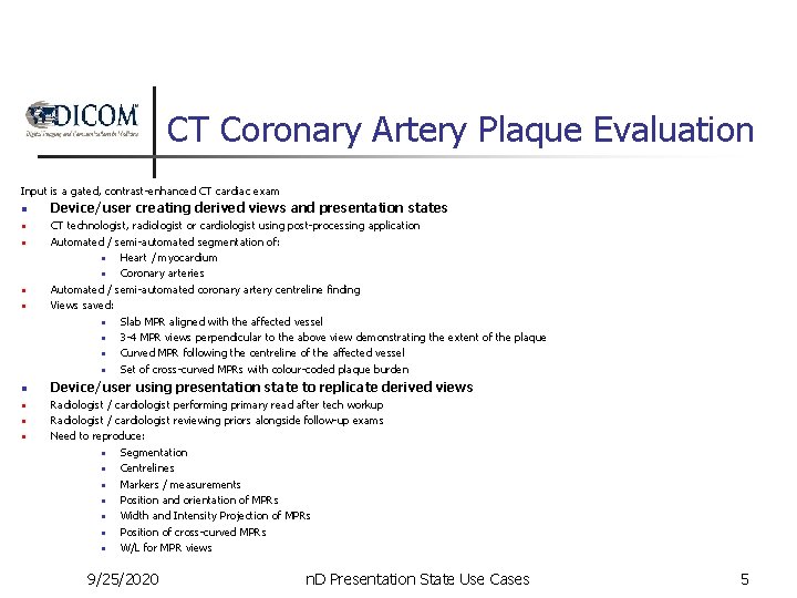
CT Coronary Artery Plaque Evaluation Input is a gated, contrast-enhanced CT cardiac exam n n n n n Device/user creating derived views and presentation states CT technologist, radiologist or cardiologist using post-processing application Automated / semi-automated segmentation of: n Heart / myocardium n Coronary arteries Automated / semi-automated coronary artery centreline finding Views saved: n Slab MPR aligned with the affected vessel n 3 -4 MPR views perpendicular to the above view demonstrating the extent of the plaque n Curved MPR following the centreline of the affected vessel n Set of cross-curved MPRs with colour-coded plaque burden Device/user using presentation state to replicate derived views Radiologist / cardiologist performing primary read after tech workup Radiologist / cardiologist reviewing priors alongside follow-up exams Need to reproduce: n Segmentation n Centrelines n Markers / measurements n Position and orientation of MPRs n Width and Intensity Projection of MPRs n Position of cross-curved MPRs n W/L for MPR views 9/25/2020 n. D Presentation State Use Cases 5
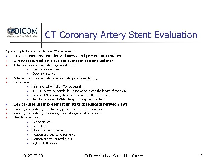
CT Coronary Artery Stent Evaluation Input is a gated, contrast-enhanced CT cardiac exam n n n n n Device/user creating derived views and presentation states CT technologist, radiologist or cardiologist using post-processing application Automated / semi-automated segmentation of: n Heart / myocardium n Coronary arteries Automated / semi-automated coronary artery centreline finding Views saved: n MPR aligned with the affected vessel n 3 -4 MPR views perpendicular to the above along the length of the stent n Curved MPR following the centreline of the affected vessel n Set of cross-curved MPRs along the length of the stent Device/user using presentation state to replicate derived views Radiologist / cardiologist performing primary read after tech workup Radiologist / cardiologist reviewing priors alongside follow-up exams Need to reproduce: n Segmentation n Centrelines n Markers / measurements n Position and orientation of MPRs n Position of cross-curved MPRs n W/L for MPR views 9/25/2020 n. D Presentation State Use Cases 6
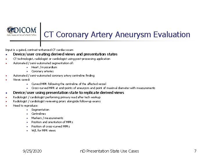
CT Coronary Artery Aneurysm Evaluation Input is a gated, contrast-enhanced CT cardiac exam n n n n n Device/user creating derived views and presentation states CT technologist, radiologist or cardiologist using post-processing application Automated / semi-automated segmentation of: n Heart / myocardium n Coronary arteries Automated / semi-automated coronary artery centreline finding Views saved: n Curved MPR following the centreline of the affected vessel n Cross-curved MPR at end-points of aneurysm and point of maximal diameter with measurements Device/user using presentation state to replicate derived views Radiologist / cardiologist performing primary read after tech workup Radiologist / cardiologist reviewing priors alongside follow-up exams Need to reproduce: n Segmentation n Centrelines n Markers / measurements n Position and orientation of MPRs n Position of cross-curved MPRs n W/L for MPR views 9/25/2020 n. D Presentation State Use Cases 7
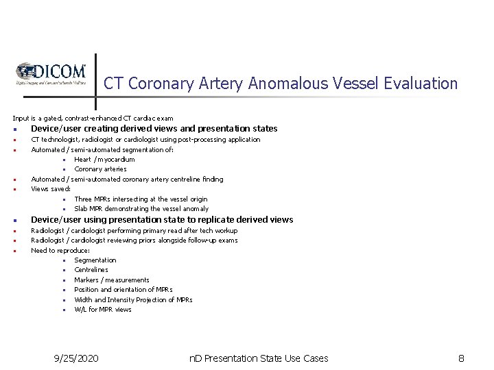
CT Coronary Artery Anomalous Vessel Evaluation Input is a gated, contrast-enhanced CT cardiac exam n n n n n Device/user creating derived views and presentation states CT technologist, radiologist or cardiologist using post-processing application Automated / semi-automated segmentation of: n Heart / myocardium n Coronary arteries Automated / semi-automated coronary artery centreline finding Views saved: n Three MPRs intersecting at the vessel origin n Slab MPR demonstrating the vessel anomaly Device/user using presentation state to replicate derived views Radiologist / cardiologist performing primary read after tech workup Radiologist / cardiologist reviewing priors alongside follow-up exams Need to reproduce: n Segmentation n Centrelines n Markers / measurements n Position and orientation of MPRs n Width and Intensity Projection of MPRs n W/L for MPR views 9/25/2020 n. D Presentation State Use Cases 8
CT Coronary Artery CABG Evaluation Input is a gated, contrast-enhanced CT cardiac exam n n n n n Device/user creating derived views and presentation states CT technologist, radiologist or cardiologist using post-processing application Automated / semi-automated segmentation of: n Heart / myocardium n Coronary arteries Automated / semi-automated coronary artery centreline finding Views saved: n Curved MPR following the centreline of the bypass graft n SVR views oriented to show junction of bypass graft and coronary artery with different VOIs (survey and focused on junction) Device/user using presentation state to replicate derived views Radiologist / cardiologist performing primary read after tech workup Radiologist / cardiologist reviewing priors alongside follow-up exams Need to reproduce: n Segmentation n Centrelines n Markers / measurements n Position and orientation of MPRs n Orientation and VOI for SVR / MIP n W/L for MPR views n Opacity for SVR views – accuracy of colour mapping unimportant 9/25/2020 n. D Presentation State Use Cases 9
CT Cardiac 4 D Valve Evaluation Input is a retrospectively gated, contrast-enhanced CT cardiac exam n Device/user creating derived views and presentation states n CT technologist, radiologist or cardiologist using post-processing application Automated / semi-automated segmentation of: n Left ventricle endocardium n Left ventricle epicardium Views saved: n Three MPRs aligned with the axes of the heart intersecting at the aortic valve cycling through cardiac phases n SVR view oriented to show the aortic valve cycling through cardiac phases n Three MPRs aligned with the axes of the heart intersecting at the mitral valve cycling through cardiac phases n SVR view oriented to show the mitral valve cycling through cardiac phases n Device/user using presentation state to replicate derived views n n n Radiologist / cardiologist performing primary read after tech workup Radiologist / cardiologist reviewing priors alongside follow-up exams Need to reproduce: n Segmentation n Position and orientation of MPRs n Cine details? n Orientation and VOI for SVR n W/L for MPR views n Colour / opacity guideline – accuracy of colour and opacity mapping unimportant 9/25/2020 n. D Presentation State Use Cases 10
CT Cardiac 4 D Myocardial Contractility Input is a retrospectively gated, contrast-enhanced CT cardiac exam n n n n Device/user creating derived views and presentation states CT technologist, radiologist or cardiologist using post-processing application Automated / semi-automated segmentation of: n Left ventricle endocardium n Left ventricle epicardium Views saved: n Three MPRs aligned with the axes of the heart intersecting at the left ventricle cycling through cardiac phases n SVR view oriented to show the left ventricle valve cycling through cardiac phases Device/user using presentation state to replicate derived views Radiologist / cardiologist performing primary read after tech workup Radiologist / cardiologist reviewing priors alongside follow-up exams Need to reproduce: n Segmentation n Position and orientation of MPRs n Orientation and VOI for SVR n W/L for MPR views n Colour / opacity guideline – accuracy of colour and opacity mapping unimportant 9/25/2020 n. D Presentation State Use Cases 11
CT Calcium Scoring Input is a retrospectively gated, non-enhanced CT cardiac phase n n n Device/user creating derived views and presentation states CT technologist, radiologist or cardiologist using post-processing application Automated / semi-automated segmentation of: n Coronary artery calcification Output saved: n Assignment of calcification to coronary arteries Device/user using presentation state to replicate derived views n Radiologist / cardiologist performing primary read after tech workup Radiologist / cardiologist reviewing priors alongside follow-up exams Need to reproduce: n Segmentation n Assignment of calcification to coronary arteries n Use case may be more appropriate to a CAD-like object than to an n. D PS n n 9/25/2020 n. D Presentation State Use Cases 12
CT Colonography Input is a combination of prone, supine and decubitus CT volumes, with or without contrast n n n n Device/user creating derived views and presentation states CT technologist, radiologist or gastroenterologist using post-processing application Automated / semi-automated segmentation of: n Luminal surface of colon n Colonic centerline n Air in small intestine n Polyps/flat lesions n Contrast tagged stool n Residual fluid Views saved: n SVR of air in colon n MPR/slab views to illustrate individual lesions n Endoscopic (SVR/SSD) of colon n Dissected SVR view Device/user using presentation state to replicate derived views Radiologist / gastroenterologist performing primary read after tech workup Radiologist / gastroenterologist reviewing priors alongside follow-up exams Need to reproduce: n Segmentation n Centrelines n Position and orientation of MPRs n Orientation and VOI for SVR n W/L for MPR views n Opacity /threshold for SVR/SSD views – accuracy of colour mapping unimportant 9/25/2020 n. D Presentation State Use Cases 13
CT Angiography Input is a contrast-enhanced CT angiography exam n n n n n Device/user creating derived views and presentation states CT technologist or radiologist using post-processing application Automated / semi-automated segmentation of: n Arteries to be analyzed n Bone adjacent to arteries of interest Automated / semi-automated artery centreline finding Views saved: n Three MPRs aligned with the artery intersecting at the point of stenosis n Curved MPR following the centreline of the stenotic vessel with stenosis marked n Cross-curved MPR at the point of stenosis with % stenosis measurement n SVR and / or boneless volumetric MIP, oriented to show stenosis Device/user using presentation state to replicate derived views Radiologist performing primary read after tech workup Radiologist reviewing priors alongside follow-up exams Need to reproduce: n Segmentation n Centrelines n Markers / measurements n Position and orientation of MPRs n Position of cross-curved MPR n Orientation and VOI for SVR / MIP n W/L for MPR and MIP views n Opacity for SVR views – accuracy of colour mapping unimportant 9/25/2020 n. D Presentation State Use Cases 14
MR Cardiac LV Analysis Input is a gated, contrast-enhanced MR cardiac exam n n n n Device/user creating derived views and presentation states MR technologist, radiologist or cardiologist using post-processing application Automated / semi-automated segmentation of: n Left ventricle endocardium n Left ventricle epicardium Views saved: n Set of LV short axis views with overlaid segmentation Device/user using presentation state to replicate derived views Radiologist / cardiologist performing primary read after tech workup Radiologist / cardiologist reviewing priors alongside follow-up exams Need to reproduce: n Segmentation n W/L for MPR views 9/25/2020 n. D Presentation State Use Cases 15
MR Cardiac Perfusion Input is a contrast-enhanced MR cardiac perfusion exam n n n n Device/user creating derived views and presentation states MR technologist, radiologist or cardiologist using post-processing application Automated / semi-automated segmentation of: n Left ventricle endocardium n Left ventricle epicardium Views saved: n Set of LV short axis views with functional overlays – stress and rest acquisitions Device/user using presentation state to replicate derived views Radiologist / cardiologist performing primary read after tech workup Radiologist / cardiologist reviewing priors alongside follow-up exams Need to reproduce: n Segmentation n W/L for MPR views 9/25/2020 n. D Presentation State Use Cases 16
MR Cardiac Delayed Enhancement Input is a delayed-enhancement MR cardiac exam n n n n n Device/user creating derived views and presentation states MR technologist, radiologist or cardiologist using post-processing application Automated / semi-automated determination of infarct enhancement threshold Automated / semi-automated segmentation of infarct Views saved: n Cross-sectional view highlighting infarct with overlays and measurements Device/user using presentation state to replicate derived views Radiologist / cardiologist performing primary read after tech workup Radiologist / cardiologist reviewing priors alongside follow-up exams Need to reproduce: n Segmentation n Signal intensity threshold used to perform infarct segmentation 9/25/2020 n. D Presentation State Use Cases 17
PET / CT Fusion – Added 15 Feb Input is a set of registered PET /CT exams, typically from a combined PET / CT scanner. A set of prior exams may also be loaded. n Device/user creating derived views and presentation states n Technologist or radiologist using post-processing application If views are not already registered, registration is performed Views saved: n Blended CT / PET MIP views demonstrating areas of interest with measurement overlays (point measurements or ROIs) n 3 D MIP view with an inverted W/L n Device/user using presentation state to replicate derived views n n n Radiologist performing primary read after tech workup Radiologist reviewing priors alongside follow-up exams Need to reproduce: n MPR positions n Blending function n W/L and or color maps n Measurement position and ROI details 9/25/2020 n. D Presentation State Use Cases 18
- Slides: 18