A ThreeArm Randomized Trial of Different Renal Denervation


- Slides: 21

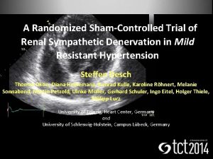
A Three-Arm Randomized Trial of Different Renal Denervation Devices and Techniques in Patients with Resistant Hypertension (RADIOSOUND-HTN) Philipp Lurz, Karl Fengler, Karl-Philipp Rommel, Stephan Blazek, Christian Besler, Philipp Hartung, Maximilian von Roeder, MD; Martin Petzold, Sindy Winkler, Robert Höllriegel, Steffen Desch, Holger Thiele Heart Center Leipzig at University of Leipzig

Disclosure Statement of Financial Interest Speaker’s Name: Philipp Lurz Within the past 12 months, I or my spouse/partner have had a financial interest/arrangement or affiliation with the organization(s) listed below Affiliation/Financial Relationship Company Institutional Grant/Research Support Abbott, Re. Cor, Occlutech Consulting Fees to Institution Abbott, Re. Cor, Rox Medical Personal Fees None Major Stock Shareholder/Equity None Royalty Income None Ownership/Founder None Intellectual Property Rights None

Background • Hypertension represents a major health problem worldwide Ø Prevalence: 1 billion people Ø Up to 1/3 are uncontrolled despite treatment • Renal denervation targets the sympathetic nervous system to lower blood pressure • The SPYRAL-HTN trial program demonstrated efficacy of renal denervation in patients with and without antihypertensive drugs • The RADIANCE-HTN SOLO study provided further evidence for the potential of renal denervation in absence of antihypertensive drugs

Background SPYRAL-HTN ON MED RADIANCE-HTN SOLO Change in 24 h systolic ABPM Change in daytime systolic ABPM Kandzari DE et al. Lancet 2018, 9; 391: 2346 -2355 Azizi M et al. Lancet 2018, 9; 391: 2335 -2345

Radiofrequency Based Renal Denervation • Symplicity spiral catheter • Multi-electrode catheter with quadrantic vessel contact • Simultaneous ablation in 4 electrodes • Ablations delivery to renal main artery and branches

Ultrasound Based Renal Denervation • Paradise endovascular ultrasound ablation catheter • Ring of ablative energy (depth of 1 -6 mm) • Endothelial cooling by water circulating through balloon • 2 -3 ablations delivered to each main renal artery Ultrasonic Heating + Water Cooling Thermal Profile

Objective To compare the effects of renal denervation applying: 1: Radiofrequency main renal artery ablation 2: Radiofrequency main and branch renal artery ablation 3: Ultrasound main renal artery ablation in patients with resistant hypertension.

Study Design – Inclusion/Exclusion Design • Design: Prospective, single-blind, singlecenter, three-arm randomized trial (1: 1: 1) • Population: Patients aged >18 or <75 years with resistant hypertension despite treatment with ≥ 3 drug classes at ≥ 50% maximum dosage including ≥ 1 diuretic. • Primary endpoint: Group difference of change in daytime ambulatory systolic blood pressure at 3 months • Powered to detect a 6 mm. Hg difference between treatment groups with 80% power Screening office BP measurements Antihypertensive medication stable for at least 4 weeks Daytime ABPM systolic ≥ 135 mm. Hg Exclusion of secondary hypertension; lab testing for hyperaldosteronism in all; further diagnostics as appropriate MRA, renal duplex, renal angiography Inclusion: 1 or more renal artery ≥ 5. 5 mm Exclusion: stenosis, unsuitable anatomy, main artery <4 mm Radiofrequency main renal artery Radiofrequency main and branch renal artery Ultrasound main renal artery Primary endpoint @ 3 months between group difference of change in systolic daytime ABPM g

Subject Flow 1884 patients with elevated office blood pressure screened for eligibility 1695 excluded 555 patient or general practitioner denied 1140 had normotensive ABPM 189 patients assessed for final eligibility 120 patients met final inclusion criteria Radiofrequency main renal artery ablation, n=39 Radiofrequency combined main and branch renal artery ablation, n=39 69 excluded 35 were considered non-adherent to medication 14 had renal artery stenosis 13 had renal artery diameter outside inclusion criteria 6 were enrolled in concurrent trial 1 had no femoral access Ultrasound main renal artery ablation, n=42 1 lost to follow-up 1 died from acute aortic dissection after 2 months 2 lost to follow-up 1 unable to attend follow-up 1 withdrew consent 0 lost to follow-up 38 available for primary analysis 37 available for primary analysis 42 available for primary analysis

Baseline Characteristics RF main only (n = 39) RF main+branches (n = 39) Ultrasound (n = 42) p-value Age 63. 8 ± 9. 9 62. 1 ± 10. 2 64. 6 ± 8. 0 0. 48* Body mass index [kg/m²] 30. 6 ± 5. 4 31. 6 ± 5. 9 32. 6 ± 5. 4 0. 27* 13 (33) 15 (38) 10 (24) 0. 36† 79. 3 ± 15. 2 76. 9 ± 18. 0 76. 2 ± 20. 3 0. 72* Smoker, n (%) 17 (44) 20 (51) 18 (43) 0. 75† Diabetes, n (%) 15 (38) 18 (46) 22 (52) 0. 59† Peripheral arterial disease, n (%) 3 (8) 4 (10) 0. 92† Coronary artery disease, n (%) 9 (23) 15 (38) 19 (45) 0. 11† Previous stroke, n (%) 2 (5) 0. 99† Previous myocardial infarction, n (%) 3 (8) 7 (18) 8 (19) 0. 30† 7 (18) 6 (15) 8 (19) 0. 91† Female, n (%) e. GFR [ml/min/1. 73 m²] Atrial fibrillation, n (%) * p-value by ANOVA, † p-value by Pearson’s Chi Square test)

Baseline Medications RF main only (n = 39) RF main+branches (n = 39) Ultrasound (n = 42) p-value 4. 7 ± 1. 4 5. 3 ± 1. 4 5. 0 ± 1. 5 0. 22* Five or more drug classes, n (%) 20 (51) 26 (67) 23 (55) 0. 27† ACE-I, n (%) 12 (31) 13 (33) 13 (31) 0. 96† ARB, n (%) 30 (77) 25 (64) 30 (71) 0. 46† 1 (3) 2 (5) 1 (3) 0. 75† Beta blockers, n (%) 34 (87) 32 (82) 38 (90) 0. 53† Calcium channel blockers, n (%) 29 (74) 31 (79) 31 (74) 0. 72† Diuretics, n (%) 38 (97) 37 (95) 42 (100) 0. 34† Second diuretic, n (%) 4 (10) 10 (26) 10 (24) 0. 18† Mineralocorticoid antagonists, n (%) 6 (15) 12 (31) 10 (24) 0. 27† Vasodilators, n (%) 2 (5) 6 (15) 5 (12) 0. 33† Alpha blockers, n (%) 9 (23) 17 (44) 12 (29) 0. 13† Centrally acting sympathicolytics, n (%) 20 (51) 22 (56) 15 (36) 0. 15† Number of antihypertensive drug classes Renin antagonists, n (%) * p-value by ANOVA, † p-value by Pearson’s Chi Square test)

Baseline Blood Pressures RF main only (n = 39) RF main+branches (n = 39) Ultrasound (n = 42) p-value Systolic Diastolic Daytime average ABMP 147. 4 ± 10. 9 83. 6 ± 10. 4 150. 6 ± 11. 4 83. 5 ± 14. 5 151. 3 ± 13. 0 83. 6 ± 12. 5 0. 31* 0. 99* Systolic 151. 3 ± 12. 3 154. 2 ± 11. 4 153. 9 ± 13. 6 0. 53* Diastolic 86. 7 ± 11. 0 86. 3 ± 15. 3 86. 0 ± 13. 3 0. 98* Systolic 135. 3 ± 13. 9 140. 3 ± 15. 9 143. 9 ± 15. 9 0. 043* Diastolic 73. 9 ± 11. 7 75. 1 ± 15. 2 76. 7 ± 12. 3 0. 62* 20 (51) 22 (56) 20 (48) 0. 73† 24 h average ABMP Nighttime average ABMP Isolated systolic hypertension (%) * p-value by ANOVA, † p-value by Pearson’s Chi Square test

Procedural Details RF main only (n = 39) RF main+branches (n = 39) Ultrasound (n = 42) p-value Right renal artery diameter [mm] 5. 7 ± 0. 8 5. 9 ± 0. 7 5. 9 ± 0. 6 0. 41* Left renal artery diameter [mm] 6. 1 ± 0. 8 5. 9 ± 0. 9 6. 0 ± 0. 7 0. 53* Ablation points right renal artery 9. 1 ± 3. 0 18. 3 ± 6. 1 3. 2 ± 0. 8 <0. 001† Ablation points left renal artery 8. 1 ± 2. 2 16. 8 ± 6. 0 3. 2 ± 0. 9 <0. 001† Right renal arteries treated 1. 1 ± 0. 4 3. 3 ± 0. 9 1. 0 ± 0. 0 <0. 001† Left renal arteries treated 1. 1 ± 0. 2 3. 2 ± 1. 0 ± 0. 2 <0. 001† Contrast agent used [ml] 90. 8 ± 54. 8 143. 1 ± 66. 6 98. 7 ± 52. 9 <0. 001* 8. 9 ± 5. 6 16. 8 ± 8. 0 8. 1 ± 6. 5 <0. 001* Cincefluoroscopy time [min] * p-value by ANOVA, † p-value by Kruskal Wallis)

Primary Endpoint Group Difference of Change in Daytime Ambulatory Systolic BP at 3 Months Systolic Diastolic 0 -3. 2 mm. Hg p<0. 001 -5 -6. 5 mm. Hg p<0. 001 -6. 1 mm. Hg -8. 3 mm. Hg -7. 6 mm. Hg p<0. 001 -10 -13. 2 mm. Hg n. s. -15 n. s. p=0. 002 p=0. 042 -20 Between group difference RF main artery vs. ultrasound -6. 7 mm Hg (98. 3% CI, -13. 2 to -0. 2) Radiofrequency main artery Between Group Difference RF main artery vs. Ultrasound -4. 6 mm Hg (98. 3% CI, -8. 8 to -0. 52) Radiofrequency main artery + branches Ultrasound main artery

Primary Endpoint Group Difference of Change in Daytime Ambulatory Systolic BP at 3 Months Systolic Diastolic 0 -3. 2 mm. Hg p<0. 001 -5 -6. 5 mm. Hg p<0. 001 -6. 1 mm. Hg -8. 3 mm. Hg -7. 6 mm. Hg p<0. 001 -10 -13. 2 mm. Hg n. s. -15 n. s. p=0. 002 p=0. 042 -20 Between group difference RF main artery vs. ultrasound -6. 7 mm Hg (98. 3% CI, -13. 2 to -0. 2) Radiofrequency main artery Between Group Difference RF main artery vs. Ultrasound -4. 6 mm Hg (98. 3% CI, -8. 8 to -0. 52) Radiofrequency main artery + branches Ultrasound main artery

Primary Endpoint Group Difference of Change in Daytime Ambulatory Systolic BP at 3 Months Systolic Diastolic 0 p<0. 001 -3. 2 mm. Hg p<0. 001 -5 -6. 5 mm. Hg p<0. 001 -8. 3 mm. Hg -7. 6 mm. Hg p<0. 001 -10 -13. 2 mm. Hg n. s. -15 p<0. 001 -6. 1 mm. Hg n. s. p=0. 002 p=0. 042 -20 Between group difference RF main artery vs. ultrasound -6. 7 mm Hg (98. 3% CI, -13. 2 to -0. 2) Radiofrequency main artery Between Group Difference RF main artery vs. Ultrasound -4. 4 mm Hg (98. 3% CI, -8. 8 to -0. 52) Radiofrequency main artery + branches Ultrasound main artery

Radiofrequency ablation main artery 30 20 10 0 -10 -20 -30 -40 -50 66% responder Radiofrequency ablation main artery + branches 30 20 10 0 -10 -20 -30 -40 -50 73% responder Change in systolic daytime ambulatory blood pressure (mm. Hg) Individual Patient Response @ 3 Months Ultrasound ablation main artery 30 20 10 0 -10 -20 -30 -40 -50 67% responder Definition of response: % Patients with ≥ 5 mm Hg Decrease

Safety Data @ 3 Months RF main only (n = 39) RF main+branches (n = 39) Ultrasound (n = 42) Death 1 0 0 New myocardial infarction 0 0 0 Major bleeding 0 0 0 New onset end stage renal disease 0 0 0 Serum creatinine elevation >50% 0 0 0 Embolic events 0 0 0 Vascular complications 1 1 1 Hospitalization for hypertensive crisis 0 1 0 New stroke 0 0 0 New renal artery stenosis > 70% 0 0 0

Limitations • Single-center and relatively small number of patients for a three-arm study design • Study endpoint at 3 months, while data from SPYRAL-HTN-ON-MED suggest later response at 6 months • Number of ablations in main and branch artery group lower than in SPYRAL HTN ON/OFF • Inclusion of patients with isolated systolic hypertension • Long-term effects on safety, specifically effects on the renal arteries unknown • No drug testing to inform on drug adherence • Inclusion of patients with larger renal arteries only (based on the assumption that sympathetic fibers are in greater distance from the lumen than in smaller arteries and therefore renal branch artery ablation or higher penetration depth more relevant).

Conclusion • Renal denervation effectively lowered blood pressure in resistant hypertension • All three denervation techniques/technologies exhibited favourable safety profile • Endovascular ultrasound based renal denervation was superior to radiofrequency ablation of the main renal arteries • The differences in magnitude of blood pressure reduction did not translate into better responder rates • Longer follow up and larger, multi-center studies investigating different renal denervation technologies and techniques are needed to determine long term safety as well as efficacy • This pilot study precludes definite recommendations regarding the preferable renal denervation approach

Simultaneous Publication in Circulation
 Mark wholey md
 Barostim
 Ira pré renal renal e pós renal
 Sindrome nefrótica
 Advantage of randomized controlled trial
 Vasa recta vs peritubular capillaries
 Randomized skip list
 Randomized algorithm in daa
 Randomized hill climbing
 Rbd design example
 Probabilistic analysis and randomized algorithms
 Matched pair design vs block design
 Two-way anova table
 R o x o research design
 Blok atau kelompok bias berupa
 Types of randomized algorithms
 Randomized polynomial time
 Randomized polynomial time
 Expected running time
 Duncan's multiple range test
 Layout of completely randomized design
 What is completely randomized design (crd)
