A spectraldomain OCT study of formerly premature children


- Slides: 36
A spectral-domain OCT study of formerly premature children. Prat Itharat MD May 30, 2008 Vanderbilt Eye Institute Preceptor: Dr. Recchia
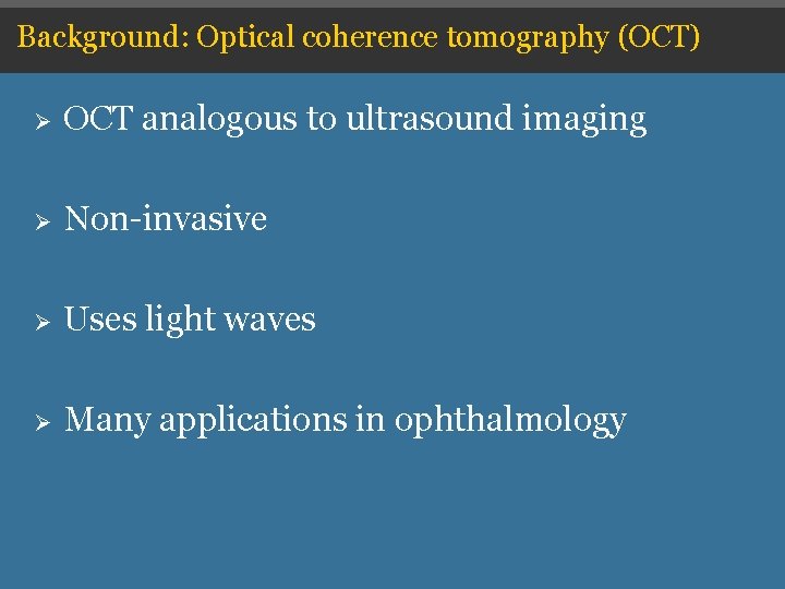
Background: Optical coherence tomography (OCT) Ø OCT analogous to ultrasound imaging Ø Non-invasive Ø Uses light waves Ø Many applications in ophthalmology
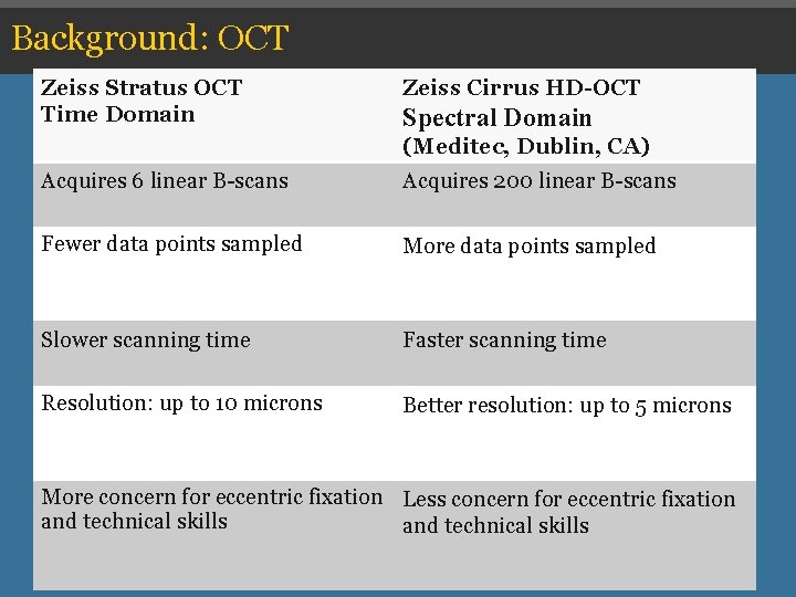
Background: OCT Zeiss Stratus OCT Domain ØTime Zeiss Cirrus HD-OCT Ø Zeiss Stratus OCT Spectral Domain (Meditec, Dublin, CA) Acquires 6 linear B-scans Acquires 200 linear Ø Acquires 6 B-scans linear BØ Acquires 200 linear Bscans Fewer data points sampled More data points sampled Ø Fewer data points Ø More data points sampled ØSlower Faster scanning time Ø Slower time Ø Better resolution: up to 5 Ø Resolution: up to 10 microns Better resolution: up to 5 microns Ø Less concern for eccentric Ø More concern for fixation technical More concernand for eccentric fixation Less concern for eccentric fixation and technical skills
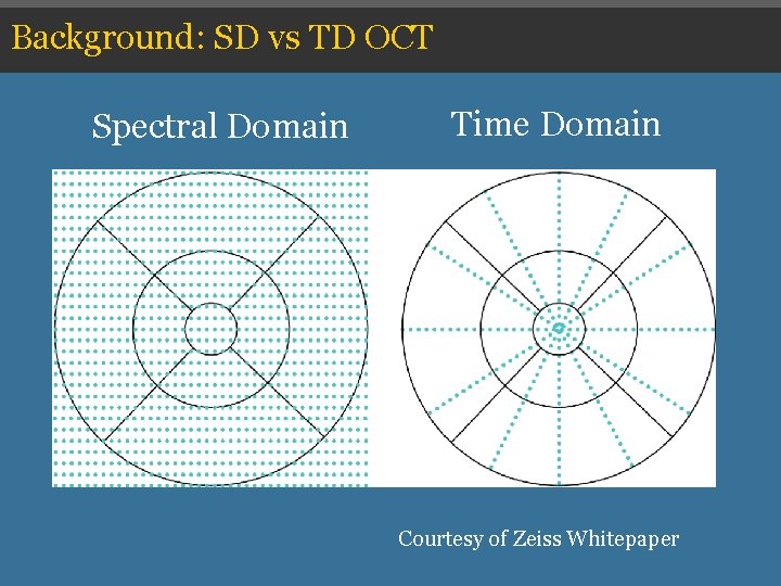
Background: SD vs TD OCT Spectral Domain Time Domain Courtesy of Zeiss Whitepaper
Background: Prematurity
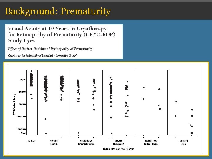
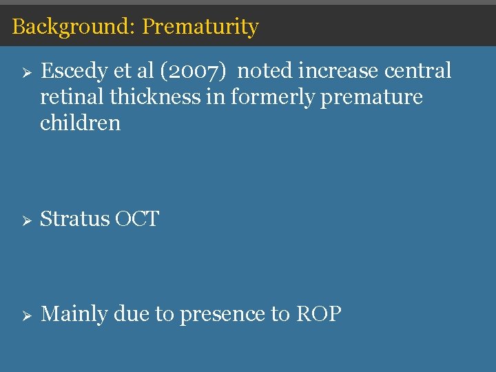
Background: Prematurity Ø Escedy et al (2007) noted increase central retinal thickness in formerly premature children Ø Stratus OCT Ø Mainly due to presence to ROP
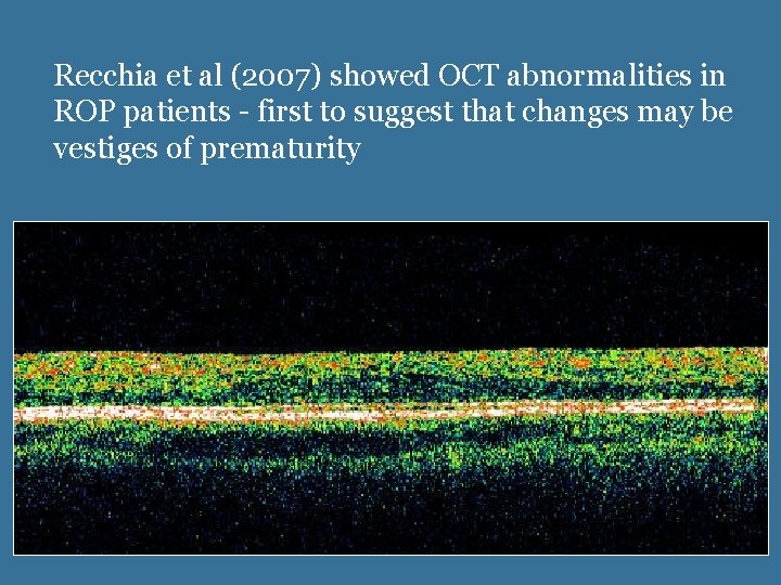
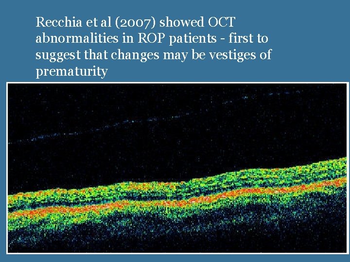
Recchia et al (2007) showed OCT abnormalities in ROP patients - first to suggest that changes may be vestiges of prematurity
Recchia et al (2007) showed OCT abnormalities in ROP patients - first to suggest that changes may be vestiges of prematurity
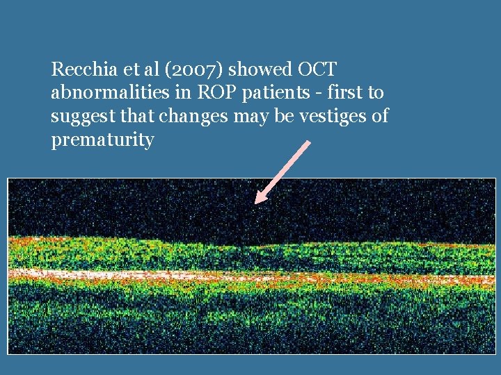
Recchia et al (2007) showed OCT abnormalities in ROP patients - first to suggest that changes may be vestiges of prematurity
Limitations Ø Technologically Ø Methodologically
Hypothesis: OCT abnormalities are more common in formerly premature children than in full term children.
OCT and prematurity Study questions: 1. Is there a difference in macular thickness between premature and full-term children? 2. Is there a difference in foveal contour between these groups? 3. Are the OCT findings correlated to the presence of ROP or another variable?
Methods Ø IRB approval for prospective study Ø Records of formerly premature infants born prior to 1/1/2001 (treated in the Vanderbilt NICU) Ø Control group matched for age and refractive error
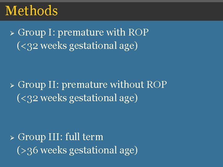
Methods Ø Group I: premature with ROP (<32 weeks gestational age) Ø Group II: premature without ROP (<32 weeks gestational age) Ø Group III: full term (>36 weeks gestational age)
Methods Ø Gestational age, birth weight, ROP status obtained from records Ø Cycloplegic refraction Ø Best corrected visual acuity
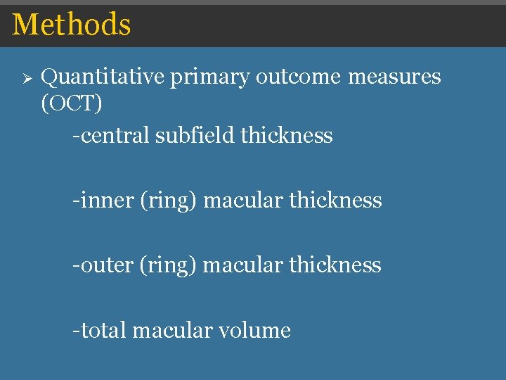
Methods Ø Quantitative primary outcome measures (OCT) -central subfield thickness -inner (ring) macular thickness -outer (ring) macular thickness -total macular volume
Methods Ø Qualitative primary outcome measures (OCT) -presence of foveal depression -preservation of retinal layers
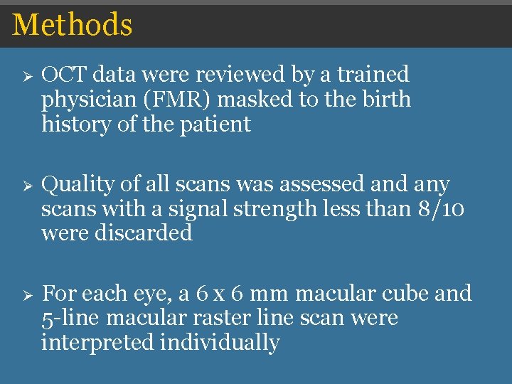
Methods Ø OCT data were reviewed by a trained physician (FMR) masked to the birth history of the patient Ø Quality of all scans was assessed any scans with a signal strength less than 8/10 were discarded Ø For each eye, a 6 x 6 mm macular cube and 5 -line macular raster line scan were interpreted individually
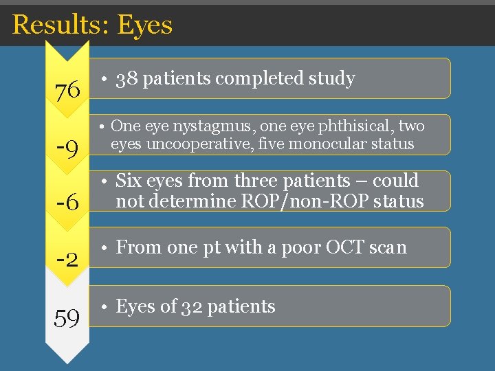
Results: Eyes 76 • 38 patients completed study -9 • One eye nystagmus, one eye phthisical, two eyes uncooperative, five monocular status -6 • Six eyes from three patients – could not determine ROP/non-ROP status -2 • From one pt with a poor OCT scan 59 • Eyes of 32 patients
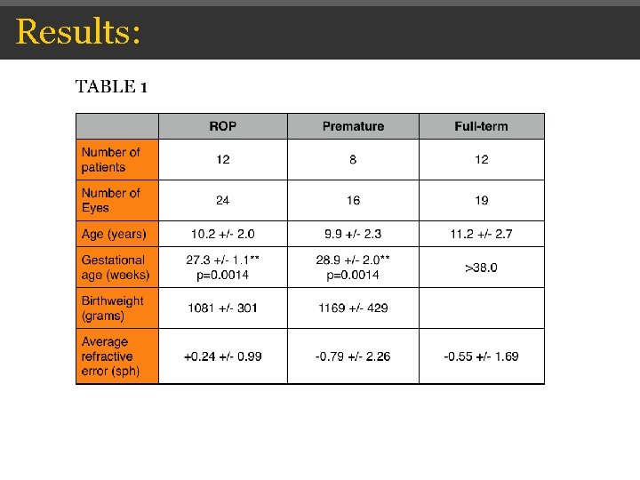
Results:
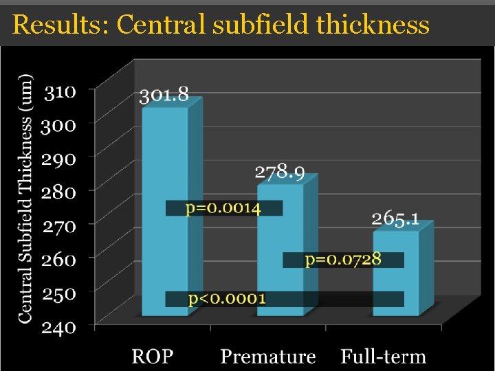
Results: Central subfield thickness
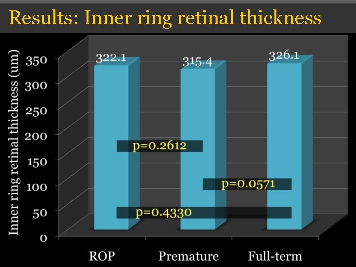
Results: Inner ring retinal thickness
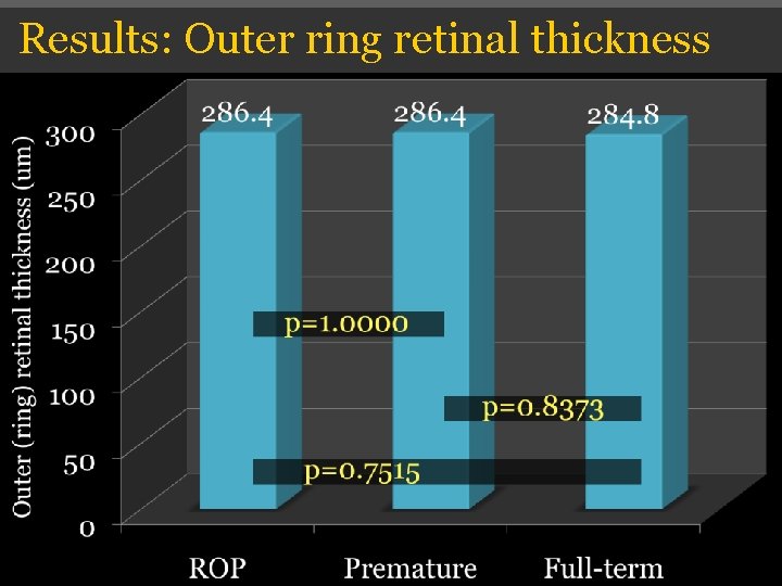
Results: Outer ring retinal thickness
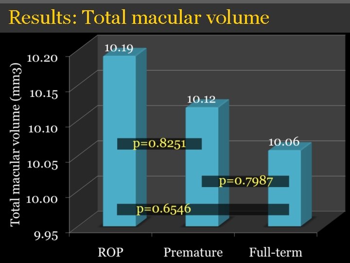
Results: Total macular volume
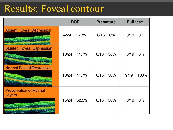
Results: Foveal contour
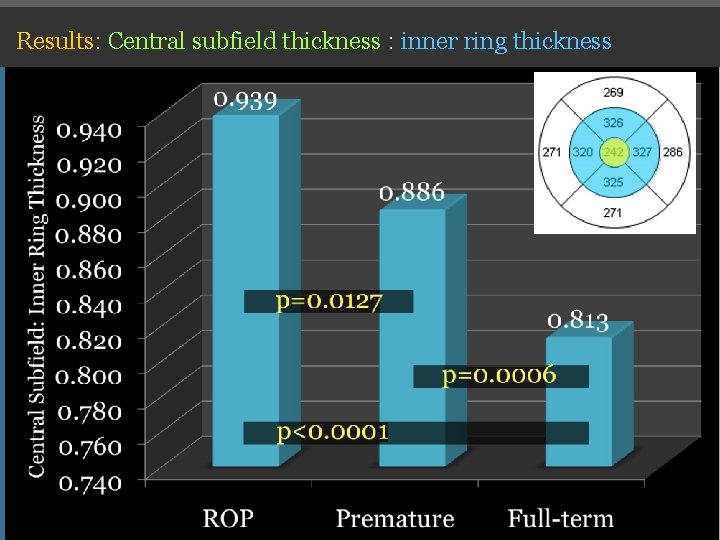
Results: Central subfield thickness : inner ring thickness
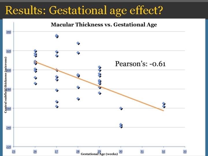
Results: Gestational age effect?
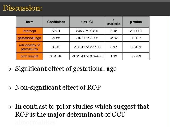
Discussion: Ø Significant effect of gestational age Ø Non-significant effect of ROP Ø In contrast to prior studies which suggest that ROP is the major determinant of OCT abnormalities
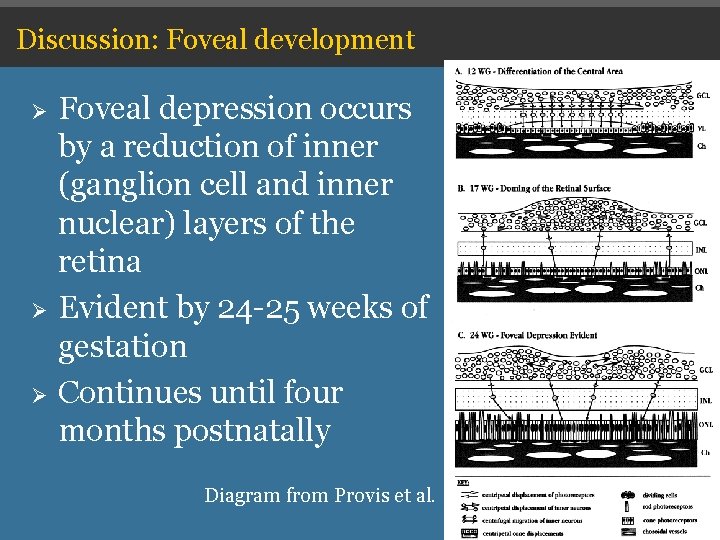
Discussion: Foveal development Ø Ø Ø Foveal depression occurs by a reduction of inner (ganglion cell and inner nuclear) layers of the retina Evident by 24 -25 weeks of gestation Continues until four months postnatally Diagram from Provis et al.
Discussion: Foveal development Ø Mintz-Hittner et al described smaller foveal avascular zone (FAZ) in formerly premature children Ø Provis et al showed that the formation of the FAZ (vascular border) precedes foveal depression Ø These two processes may be interrelated
Discussion: Foveal development Ø Prematurity may change retinal oxygenation Ø Disruption of FAZ and foveal formation -Blunted/absent foveal depression -Thickened central macula -Preservation of retinal layers Ø Interestingly, most of our patients had excellent visual acuity
Limitations Ø Selection bias (less severe ROP, less severe neurological disease) Ø Non-matched gestational age Ø Most ROP staging obtained indirectly through NICU discharge summaries Ø Inter-rater variability of ROP
Conclusions Ø OCT findings such as increased central macular thickness and foveal depression blunting may represent hallmarks of prematurity Ø These findings can be associated with normal visual acuity
Clinical relevance Ø Greater use of OCT in evaluation of patients with decreased visual acuity Ø OCT abnormalities may not represent ocular disease Ø Research studies in which numerical cutoffs are made for inclusion or evaluation of therapeutic response
Acknowledgements Ø Dr. Franco Recchia Ø Tony Adkins Ø Dr. Jeffrey Kammer Ø Rocky Munn Ø Dr. Lori. Ann Kehler Ø Dr. Robert Estes Ø Dr. Cynthia Recchia Ø Dr. Ryan Tarantola Ø Dr. Chun Li Ø Christine Franklin Ø Dr. Jeffrey Sonsino Ø Ø Sandy Owings Family and children volunteers
References • • • Ecsedy M, Szamosi A, Karko C, Zubovics L, Varsanyl B, Nemeth J, Recsan Z. A comparison of Macular Structure Imaged by Optical Coherence Tomography in Preterm and Full-Term Children. Investigative Ophthalmology & Visual Science. November 2007; 48(11), 5207 -5211. Fleck BW, Mc. Intosh N. Pathogenesis of retinopathy of prematurity and possible preventive strategies. Early Human Development 2008; 84: 83 -88. Fulton AB, Hansen RM, Moskowitz A, Barnaby Am. Multifocal ERG in subjects with a history of retinopathy of prematurity. Doc. Ophthal. 2005; 111: 7 -13. Hammer DX, Iftimia N, Ferguson RD, Bigelow CE, Ustun TE, Barnaby AM, Fulton AB. Foveal Fine Structure in Retinopathy of Prematurity: An Adaptive Optics Fourier Domain Optical Coherence Tomography Study. IOVS. May 2008; 49(5), 2061 -2070. Hendrickson AE and Yuodelis C. The morphological development of the huma fovea. Ophthalmology 1984; 91, 603 -612. Leung CKS, Cheung CYL, Weinreb RN, Lee G, Lin D, Pang CP, Lam DSC. Comparison of macular thickness measurements between time domain and spectral domain optical coherence tomography. IOVS 2008 O’Conno AR, Wilson CM, Fielder AR. Ophthalmological problems associated with preterm birth. Eye 2007; 21, 1254 -1260. Provis JM, Diaz CM, Dreher B. Ontogeny of the primate fovea: a central issue in retinal development. Progress in Neurobiology 1998; 54, 549 -581. Provis JM, Sandercoe T, Hendrickson A. Astrocytes and blood vessels define the foveal rim during primate retinal development. IOVS 2000; 41(10), 2827 -2836. Provis, JM. Development of the Primate Retinal Vasculature. Progress in Retinal and Eye Research 20(6), 799 -821. Recchia FM, Recchia CC. Foveal Dysplasia Evident by Optical Coherence Tomography in Patients with a History of Retinopathy of Prematurity. Retina 2007; 27 -1221 -1226. Stout AU, South JM. Retinopathy of Prematuirty. Ped Clin N Am 2003; 50, 77 -87.