
Normal and Abnormal Sexual Differentiation Rukset Attar MD
Normal and Abnormal Sexual Differentiation Rukset Attar, MD, Ph. D Assoc. Prof. of Obs & Gyn Yeditepe University Hospital
Normal Sexual Differentiation n The gender identity of a person (whether an individual identifi es as a male or a female) is determined by their genetic, gonadal, and phenotypic sex and also is influenced by their environment.
Normal Sexual Differentiation n Genetic or chromosomal sex - sex chromosomes, typically XX or XY. Gonadal sex - gonadal differentiation, into ovaries or testes. Phenotypic sex - appearance of the external genitalia and the secondary sexual characteristics that develop at puberty.
Normal Sexual Differentiation n n Gender identity includes all behavior having any sexual connotation, such as body gestures and mannerisms, habits of speech, recreational preferences, and content of dreams Sexual expression, both homo-sexual and heterosexual, reflects the sum of all sexual influences on the individual, both prenatal and postnatal (the role assigned by society in accordance with the individual’s phenotype and behavior).
Normal Sex Differentiation n n Four major steps which constitute normal sexual differentiation: Fertilization and determination of genetic sex Formation of organs common to both sexes Gonadal differentiation Differentiation of the internal ducts and external genitalia
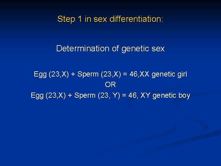
Step 1 in sex differentiation: Determination of genetic sex Egg (23, X) + Sperm (23, X) = 46, XX genetic girl OR Egg (23, X) + Sperm (23, Y) = 46, XY genetic boy
Step 2: Formation of Organs Common to Both Sexes n Both 46, XX and 46, XY fetuses have similar sex organs, specifically the gonadal ridges (recognized by 4 -5 weeks of gestation and n they include the undifferentiated germ cells which will later develop into either eggs or sperm the internal ducts (by 6 -7 weeks of fetal life, fetuses of both sexes have two sets of internal ducts, the Mullerian (female) ducts and the Wolffian (male) ducts) n the external genitalia (at 6 -7 weeks gestation appear female and include a genital tubercle, the genital folds, urethral folds and a urogenital opening)

Step 3: Gonadal Differentiation n The gonadal ridge become either an ovary or a testis In males, the gonadal ridge develops into testes as a result of a product from a gene located on the Y chromosome. This product has been termed the "testis determining factor" or "sex determining region of the Y chromosome" (SRY). In females, the absence of SRY, due to the absence of a Y chromosome, permits the expression of other genes which will trigger the gonadal ridge to develop into ovaries
Step 3 in Sex Differentiation Determination of Gonadal Sex XX fetus = ovary (with no SRY) OR XY fetus = testes (with SRY located on the Y chromosome)
Step 4: Differentiation of the Internal Ducts and External Genitalia n The next step in sex differentiation depends upon the formation of two important hormones: n n n the secretion of Mullerian (female) Inhibiting Substance (MIS) and the secretion of androgens If testes are developing normally, then n n Sertoli cells of the developing testes produce MIS which inhibits the growth of the female Mullerian ducts (the uterus and fallopian tubes) and the Leydig cells of the testes start secreting androgens (produce growth effects on the male Wolffian ducts (the epididymis, vas deferens, seminal vesicles)
Step 4: Differentiation of the Internal Ducts and External Genitalia n If ovaries are developing normally n n n Two products of the developing testes are needed for normal male development. n n n ovaries do not produce androgens as a result, the Wolffian ducts fail to grow and consequently disappear in fetuses with ovarian development ovaries do not produce MIS at the appropriate time, and as a consequence, the Mullerian (female) ducts can develop MIS must be secreted to inhibit female duct growth androgens must be secreted to enhance male duct growth. In contrast, a female fetus will produce neither MIS nor androgens, and hence female ducts will develop and male ducts will disappear
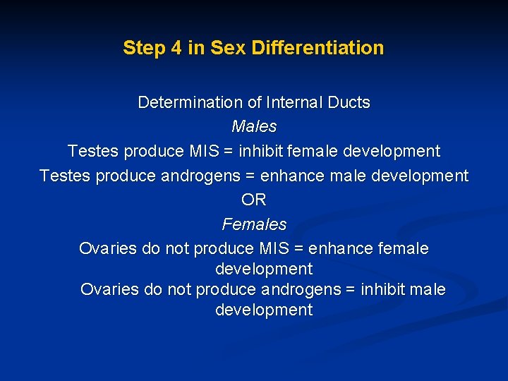
Step 4 in Sex Differentiation Determination of Internal Ducts Males Testes produce MIS = inhibit female development Testes produce androgens = enhance male development OR Females Ovaries do not produce MIS = enhance female development Ovaries do not produce androgens = inhibit male development
External Genitalia n In the female, absence of androgens permits the external genitalia to remain feminine: n the genital tubercle becomes the clitoris, n the genital swellings become the labia majora and n the genital folds become the labia minora
Sexual Differentiation of the Central Nervous System n n Studies in rodents and nonhuman primates suggests strongly that the fetal hormonal environment directs sexual differentiation of not only the genitalia, but also the central nervous system (CNS). Treatment with testosterone during early development increases reproductive and other behaviors more common in males and decreases behaviors more common in females. These observations suggest that testosterone and its metabolites play a role in brain development and neuronal organization. In addition, gender role is influenced heavily by the sex of rearing and by social interactions based upon genital appearance and secondary sexual characteristics.
Disorders of Sex Differentiation n n Sex differentiation is a complex physiological process comprised of many steps. Problems associated with sex differentiation, or syndromes of intersexuality, occur when errors in development take place at any of these steps
Disorders of Sex Differentiation n n Disorders of sexual development (DSD) are congenital conditions characterized by atypical development of chromosomal, gonadal, or phenotypic sex. A true hermaphrodite has both ovarian and testicular tissue. A male pseudohermaphrodite has testes, but a female genital phenotype A female pseudohermaphrodite has ovaries, but masculine genital characteristics
Genetic Sex n Problems can arise at fertilization when chromosomal sex is established n For example; n girls with Turner Syndrome have a 45, XO karyotype boys with Klinefelter Syndrome have a 47, XXY karyotype. some women have a 46, XY or 47, XXY karyotype n some men a 46, XX or 47, XYY karyotype. n n
Gonadal Sex n n n Disorders of sex differentiation can occur when a bipotential gonad is incapable of developing into a testis or an ovary. The inability to develop testes may occur if a gene such as SRY is absent or deficient. Additionally, 46, XY fetuses may begin to develop testes, but this development can be thwarted, and subsequently MIS androgen production may be absent or diminished.
Gonadal Sex n n n SRY mut Sox 9 mut- is required for testis differentiation, but also that Sry activation of Sox 9 may be all that is necessary to activate other genes important to testis development, such as Fgf 9 (fi broblast growth factor 9), and to repress genes that induce ovary development, such as Wnt 4 (a member of the wingless family of genes), Rspo 1 (Rspondin 1), Dax 1 (dosage-sen-sitive sex reversal, adrenal hypoplasia critical region, on chromosome X, gene 1), and Foxl 2 (forkhead box L 2). In mice, inactivating mutations in genes such as Wnt 4, 39, 41 Rspo 1, 42– 44 and Foxl 245– 47 result in partial or complete XX male sex reversal, and activating mutations in b-catenin or Dax 1 result in XY female sex reversal
Mullerian and Wolffian Duct Development n n MIS secretion accompanied by the absence of androgens or the inability to respond to androgens can result in a fetus lacking both male and female internal duct structures. The absence of MIS accompanied by androgen secretion can result in a fetus possessing both male and female internal duct structures to varying degrees.
External Genitalia Babies born with sex differentiation syndromes possess external genitalia that can usually be classified as either: n n normal female ambiguous n normal male but with a very small penis (micropenis) n n Normal female external genitalia develop among 46, XY intersex patients when the genital tubercle, genital swellings, and genital folds either completely lack exposure to, or are totally incapable of responding to, male hormones.
External Genitalia n Ambiguous external genitalia develop n n n in female patients when the external genital structures are exposed to greater-than-normal amounts of male hormones (masculinized females) or in male patients when less-than-normal amounts of male hormones (under-masculinized males) occurs. Thus, in these patients, external genitalia develop in a manner that is neither female nor male, but rather is somewhere in between the two.
Abnormal Sexual Differentiation n Female pseudohermaphrodites n n n Male pseudohermaphrodites n Syndromes of androgen insensitivity Abnormal androgen synthesis n Abnormal Mullerian inhibiting factor (MIF) n n Congenital virilizing adrenal hyperplasia (CAH) Masculinization due to elevated androgens in maternal circulation Ambiguous genitalia
Abnormal Sexual Differentiation n Abnormal gonadogenesis n True agonadism Anorchia True hermaphrodites Gonadal dysgenesis Mixed gonadal dysgenesis n Noonan's syndrome n n
Specific Syndromes of Sex Differentiation n n n n Gonadal Dysgenesis αlfa- 5 -Reductase Deficiency Testosterone Biosynthetic Defects Micropenis Timing Defect Congenital Adrenal Hyperplasia (CAH) in 46, XX Individals Klinefelter Syndrome(47, XXY karyotype) Turner Syndrome (45, XO karyotype ) 45, XO/46, XY Mosaicism
Diagnostic Evaluation n n history, complete physical examination, abdominal/pelvic ultrasonography, a karyotype and FISH for SRY, and endocrine studies of adrenal and gonadal steroid secretion n n 17 OHP—rapidly excludes CAH due to 21 -hydroxylase Electrolytes—should be measured immediately and monitored at least daily until salt wasting can be excluded. , n n ACTH, cortisol, DHEA, 17 a-hydroxypregnenolone, and 11 -desoxycortisol—CAH caused by enzyme deficiencies other than 21 -hydroxylase
Normal and Abnormal Puberty Rukset Attar, MD, Ph. D Assoc. Prof. of Obs & Gyn Yeditepe University Hospital
Normal Puberty n n Puberty is the period that fertility is acquired. The most visible changes during puberty are n growth in stature and n development of secondary sexual characteristics n changes in body composition; n the achievement of fertility; n and changes in most body systems, such as the neuroendocrine axis, bone size, and mineralization; and the cardiovascular system (normal cardiovascular changes, including greater aerobic power reserve, electrocardiographic changes, and blood pressure changes, occur during puberty).
Normal Puberty n n n Data indicate that a substantial proportion of American girls begin pubertal development 6– 12 months earlier than previously observed. On average, black American girls begin puberty between ages 8 and 9, and white American girls by age 10. However, thelarche and/or pubarche can occur normally in black girls as early as age 6 and in white girls as early as age 7.
Normal Puberty n n The cascade of events initiated by the release of pulsatile Gn. RH from prepubertal feedback and central negative inhibition results in increased levels of gonadotropins and steroids with appearance of secondary sexual characteristics and eventually adult function (menarche and ovulation) As puberty approaches 3 critical changes occur; n Adrenarche n Decreasing repression of the gonadostat n gradual amlification of the Gn. RH-gonadotropin and gonadotropin-ovarian steroid interactions leading to gonadarche
Adrenarche n n n is the growth of pubic and axillary hair due to an increased production of adrenal androgens at puberty Adrenarche describes the activation of adrenal androgen secretion that begins before puberty and ultimately stimulates pubarche, the appearance of pubic hair Adrenarche is the term used to describe the increase in adrenal androgen production that begins at approximately 6 years of age in both boys and girls
Adrenarche n n DHEA, DHEA-SO 4, A levels rise due to increased 17 alfa- OH, 17, 20 -Lyase activity and increase in size and differentiation of the inner zone ( zona reticularis ) of the adrenal cortex There is a dissociation of the control mechanism that initiate adrenarche and those governing gonarche( eg premature adrenarche is not associated with abnormal advancement of gonadarche; in gonadal disgenesis adrenarche still occurs)
Adrenarche n n n A pituitary adrenal androgen stimulating factor formed by clevage of a high molecular weight precursor proopiomelanocortin ( POMC) which also contains ACTH and beta lipotropin acting on an ACTH prepared and maintained adrenal has been suggested as the agent stimulating adrenarche (? ) A large glycoprotein has been identified that also displayed adrenal androgen activity (? ) ACTH (? ) Melatonin (? ) Local steroid inhibition of key enzymes
Decreasing Repression of the Gonadostat n n n For approx. 8 years from early infancy to the prepubertal period LH and FSH are suppressed to very low levels The mechanisms for this restraint on gonadotropin secretion are a highly sensitive negative feedback of low gonadal esrogen on hypothalamic and pituitary sites and an intrinsic central inhibitory influence on Gn. Rh that reduces basal gonadotropin concentrations even in agonadal children Central inhibiting factor ? Melatonin? POMC peptides?
Gonadarche n n describes the activation of the hypothalamic-pituitarygonadal axis, which facilitates the pubertal growth spurt, stimulates thelarche, the appearance of breast tissue, and finally menarche, the onset of menses the growth spurt begins before the onset of other signs of puberty. The peak growth velocity occurs at an average of 11 to 12 years, usually 1 year before menarche
Menarche n n n In menarche, vaginal bleeding occurs in response to hormonal changes, specifically production of estrogen by the ovary, for the first time. The average of the first menses is 12 to 13 years. For the first 2 years following menarche, menses are often irregular because of anovulation or sporadic ovulation.
Menarche n n n n Genetic Environment General health and nutrition Sun light Body weight and fat composition Physocologic factors Geografic location
Thelarche n n The onset of breast development usually begins between 9 and 11 years of age. It is a sign of ovarian estrogen production and is completed over approximately 3 years. Tanner stages describe the normal changes in the transition from the prepubertal to the mature breast contour
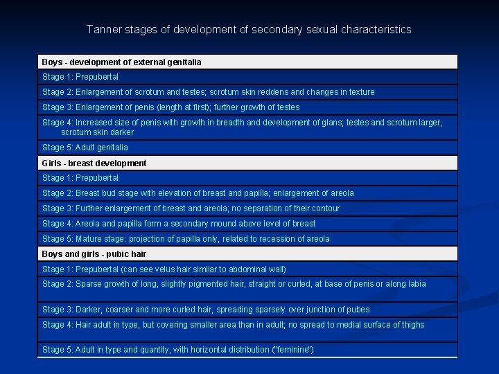
Tanner stages of development of secondary sexual characteristics Boys - development of external genitalia Stage 1: Prepubertal Stage 2: Enlargement of scrotum and testes; scrotum skin reddens and changes in texture Stage 3: Enlargement of penis (length at first); further growth of testes Stage 4: Increased size of penis with growth in breadth and development of glans; testes and scrotum larger, scrotum skin darker Stage 5: Adult genitalia Girls - breast development Stage 1: Prepubertal Stage 2: Breast bud stage with elevation of breast and papilla; enlargement of areola Stage 3: Further enlargement of breast and areola; no separation of their contour Stage 4: Areola and papilla form a secondary mound above level of breast Stage 5: Mature stage: projection of papilla only, related to recession of areola Boys and girls - pubic hair Stage 1: Prepubertal (can see velus hair similar to abdominal wall) Stage 2: Sparse growth of long, slightly pigmented hair, straight or curled, at base of penis or along labia Stage 3: Darker, coarser and more curled hair, spreading sparsely over junction of pubes Stage 4: Hair adult in type, but covering smaller area than in adult; no spread to medial surface of thighs Stage 5: Adult in type and quantity, with horizontal distribution ("feminine")
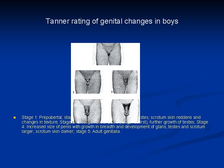
Tanner rating of genital changes in boys n Stage 1: Prepubertal; stage 2: Enlargement of scrotum and testes; scrotum skin reddens and changes in texture; Stage 3: Enlargement of penis (length at first); further growth of testes; Stage 4: Increased size of penis with growth in breadth and development of glans; testes and scrotum larger, scrotum skin darker; stage 5: Adult genitalia.
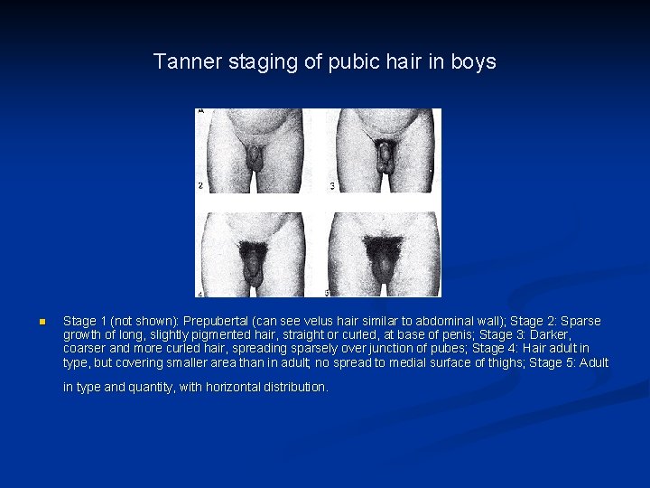
Tanner staging of pubic hair in boys n Stage 1 (not shown): Prepubertal (can see velus hair similar to abdominal wall); Stage 2: Sparse growth of long, slightly pigmented hair, straight or curled, at base of penis; Stage 3: Darker, coarser and more curled hair, spreading sparsely over junction of pubes; Stage 4: Hair adult in type, but covering smaller area than in adult; no spread to medial surface of thighs; Stage 5: Adult in type and quantity, with horizontal distribution.
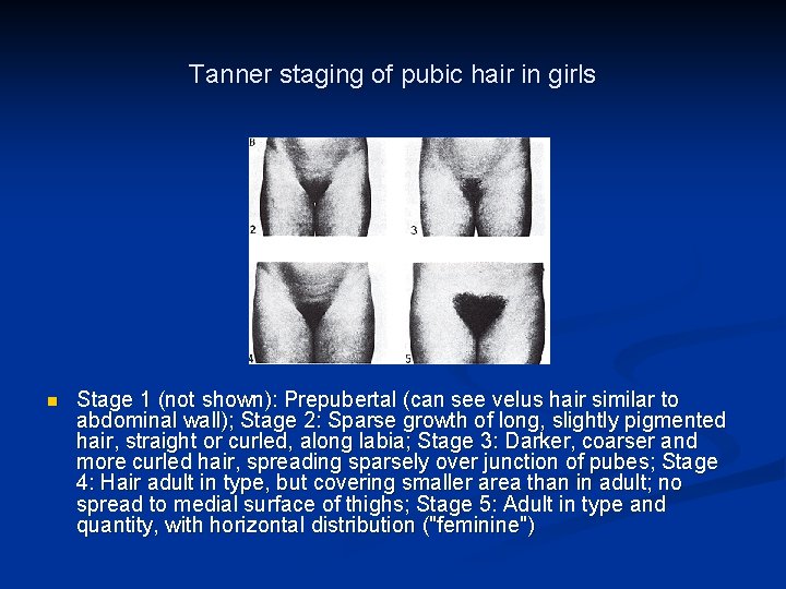
Tanner staging of pubic hair in girls n Stage 1 (not shown): Prepubertal (can see velus hair similar to abdominal wall); Stage 2: Sparse growth of long, slightly pigmented hair, straight or curled, along labia; Stage 3: Darker, coarser and more curled hair, spreading sparsely over junction of pubes; Stage 4: Hair adult in type, but covering smaller area than in adult; no spread to medial surface of thighs; Stage 5: Adult in type and quantity, with horizontal distribution ("feminine")
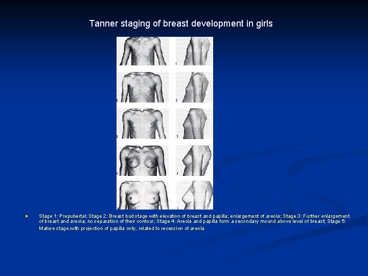
Tanner staging of breast development in girls n Stage 1: Prepubertal; Stage 2: Breast bud stage with elevation of breast and papilla; enlargement of areola; Stage 3: Further enlargement of breast and areola; no separation of their contour; Stage 4: Areola and papilla form a secondary mound above level of breast; Stage 5: Mature stage with projection of papilla only, related to recession of areola
Normal Puberty n Pubertal sequence of n n n growth spurt, Telarche (age 7 -12), Pubarche or Adrenarche (age 10) (age 7 -12), height peek ( age 11. 5) , Menarche ( age 12. 7) (age 11 -14) requires a period of 4. 5 years ( range 1. 5 to 6 years ).
Abnormal Puberty n n Abnormal female development n Precocious puberty n Delayed puberty Growth problems in normal adolescents n Short stature n Tall stature
Precocious Puberty n n Pubertal development begin at an earlier age than is expected based upon established normal standards (before 8 years of age in Caucasian girls and before 7 years of age in African-American girls) The cause of precocious puberty may range from a variant of normal development (eg, premature adrenarche) to pathologic conditions with significant risk of morbidity and even death (eg, malignant germ-cell tumor and astrocytoma). Usually the first sign is an increase in height Sometimes menarche is the first sign
Precocious Puberty n Types of Precocious Puberty n Isosexual ( early pubertal growth) n n n n Idiopathic %80 Ovarian or adrenal estrogen secreting tumors CNS disease Gonadotropin releasing tumors Hypothyroidism Iatrogenic Heterosexual ( with virilisation, acne, hirsutismus) n n n Ovarian ( cyst or tumor) Adrenal tumors Mc. Cune-Albright Syndrome Congenital Adrenal Hyperplasia Ectopic gonadotropin production Cushing syndrome
Types of Precocious Puberty n Central precocious puberty - is caused by early activation of the hypothalamic-pituitary-gonadal axis, leading to the onset of hormonal secretion from the ovaries. n The most common cause is idiopathic (74%). n Other causes are rare and include central nervous system lesions such as infection, craniopharyngioma, astrocytoma, neurofibroma, hemangioma of the hypothalamus, hydrocephalus, and neoplasm of the floor of the third ventricle
Types of Precocious Puberty n Peripheral precocious puberty- caused by secretion of sex steroids from the ovary or exogenous hormone ingestion n hormone-producing ovarian or adrenal tumors, n Mc. Cune-Albright syndrome, n ectopic gonadotropin production, and n primary hypothyroidism
Diagnosis n n Physical and radiologic signs are the basis of diagnosis. Assessment of bone age is critical, usually with a plain film of the wrist of the nondominant hand. An endocrine profile including gonadotropin levels and thyroid function tests must be evaluated. In addition, imaging of the brain with computed tomography (CT) or MRI is essential to rule out an intracranial mass.
Management n n Therapy is aimed at slowing down accelerated growth; reducing pituitary, ovarian, and adrenal function; and inducing regression of secondary sex characteristics. For idiopathic precocious puberty - gonadotropinreleasing hormone (Gn. RH) agonists, which suppress the pituitary and halt the progression through puberty. n n The treatment is continued until an appropriate age has been reached for resumption of pubertal development. In cases of known etiology, recommended therapy is with surgery, chemotherapy, or radiation therapy as indicated
Precocıous Pseudopuberty n n n Occasionally, for reasons that remain unclear, only one sign of pubertal development is present (breast development, pubic hair, or menstruation). It possibly results from transient elevations of the levels of circulating steroid hormones, or possibly from extreme sensitivity of the end organ (eg, breast tissue) to the low, prepubertal levels of sex hormones. Such isolated development, however, may represent the initial sign of precocious puberty, and these patients should be reevaluated at regular intervals.
Premature Thelarche n n n Premature thelarche is the isolated development of breast tissue prior to age 8, most commonly occurring between 1 and 3 years of age. It may affect one or both breasts On examination, the somatic growth pattern is not accelerated, bone age is not advanced, and smear of vaginal secretions fails to show estrogen effect. The diagnosis is made by exclusion of other disorders. Surgical biopsy of the breast is not indicated, as extensive excision of tissue may cause permanent damage to the breast.
Premature Pubarche n n Premature pubarche is the isolated development of pubic or axillary hair prior to age 8 years without other signs of precocious puberty Such hair growth may be idiopathic and of no clinical significance. these children tended to be slightly taller, had marginally advanced bone age, and had slightly elevated serum dehydroepiandrosterone (DHEA) levels. Early pubarche may be a sign of excess androgen production due to enzyme deficiency (congenital adrenal hyperplasia) or tumor (Leydig cell tumor).

Premature Pubarche n The diagnosis of idiopathic premature pubarche is made only after such an evaluation fails to detect an abnormality in Thorough evaluation of adrenal and gonadal function and assessment of androgen production.
Premature Menarche n n n Premature menarche denotes the appearance of cyclic vaginal bleeding in children in the absence of other signs of secondary sexual development. The cause is unknown but may be related to increased end organ sensitivity of the endometrium to low prepubertal levels o estrogens. Alternatively, bleeding may be related to transient elevation of estrogens due to premature follicular development
Premature Menarche n n These patients have estradiol levels in the prepubertal range, and cytologic smears of vaginal secretions indicate lack of estrogenic stimulation. When patients are given Gn. RH, the response of the pituitary gland is similar to that seen in prepubertal children. The diagnosis is by exclusion following investigation of other causes of vaginal bleeding and is confirmed when the cyclic nature of the bleeding becomes apparent. The prognosis is excellent. Adult height is uncompromised; the menstrual pattern is normal; and fertility potential remains unimpaired.
Delayed puberty n n Pubertal development begin at a later age than is expected based upon established normal standards ( after 13 years of age for girls, 14 years of age for boys ) Delayed puberty is characterized by the absence of breast development by age 13 years or the absence of menses by age 16 years
Delayed puberty n Types of Delayed Puberty n n Constitutional %10 -20 Delayed Puberty with sexual infantism n n Hypogonadotropic hypogonadism ( CNS diseases, Kallman Syndrome, multipl hypophisial hormon deficiency, functional hypophisial hormon deficiency-chronic systemic diseases, hypothroidism, Cushing, DM, Hyperprolactinemia, hypothalamic suppression by stress, severe disease, or malnutrition, anorexia nervosa, and tumor invasion of the pituitary (prolactinoma or craniopharyngioma)) Hypergonadotropic hypogonadism - almost 50% - ( Klinifelter S, Turner S, idiopathic premature ovarian failure, autoimmune ovarian failure, gonadal dysgenesis, and ovarian failure secondary to radiation therapy or chemotherapy)
n The initial evaluation of delayed puberty begins in the same way as that for precocious puberty, with a careful history, physical examination, and a measurement of bone age
n The laboratory evaluation of girls with delayed puberty is aimed first at differentiating primary (hypergonadotropic) from secondary (hypogonadotropic) hypogonadism, which typically can be accomplished by measuring the serum FSH, LH, and estradiol concentrations.
n n n TSH and f. T 4 - primary or secondary hypothyroidism, particularly if growth velocity has slowed and the bone age is grossly delayed. DHEA-S concentration may be helpful for distinguishing constitutional delay of puberty from congenital Gn. RH deficiency. Patients with congenital Gn. RH deficiency are more likely to have a normal adrenarche than those with constitutional delay, although values in the two groups frequently overlap. Other laboratory tests to identify occult chronic illness, such as chronic infl ammatory bowel disease, liver disease, or anorexia nervosa, and should include a complete blood count, erythrocyte sedimentation rate, and liver function tests.
n n pelvic ultrasonography can be used to determine the presence or absence of a uterus in virginal girls, it must be interpreted cautiously because results can be misleading when the reproductive organs are immature and very small, and generally is unnecessary Cranial MRI should be obtained in patients with hypogondotropic hypogonadism and those with neurologic signs or symptoms to detect mass lesions, and the presence or absence of the olfactory bulbs and tracts (absent in Kallmann syndromes).
n A karyotype should be obtained in all girls with hypergonadotropic hypogonadism to detect chromosomal abnormalities, except when a history of previous chemotherapy or gonadal radiation provides an obvious explanation.
Management n n n Treatment is based on the etiology of the delay. In cases of gonadal dysgenesis when a Y chromosome or fragment of a Y chromosome is present, the gonads should be removed at diagnosis because of the risk of neoplastic degeneration. Otherwise, hormone replacement with estrogen, and subsequently both estrogen and progesterone, is required to promote sexual development and menarche.
n n In general, sex hormone therapy should be limited to girls over 12 years of age having few or no signs of sexual maturation causing significant distress or anxiety. The goals of short-term hormone therapy are to foster age-appropriate secondary sexual development and to induce a growth spurt and a normal adolescent increase in bone density without causing premature epiphyseal closure, which requires that bone age be monitored at 6 monthly intervals during treatment.
n In those who prove ultimately to have an isolated Gn. RH deficiency, the longer-term goals are to maintain sex hormone levels in the normal physiologic range and to induce ovulation with exogenous gonadotropin therapy when fertility becomes a priority.
n Oral or transdermal estrogen therapy can be used, beginning at doses well below those used for adults (e. g. , 0. 25– 0. 5 mg oral micronized estradiol or its equivalent), increasing gradually at intervals of 3– 6 months according to response (Tanner stage, bone age), with the goal of completing sexual maturation over a period of 2– 3 years.
n n A progestin should not be added to the treatment regimen until there is substantial breast development and full contour breast growth has plateaued, because premature progestin treatment can adversely affect breast growth or contours. In general, progestin therapy can safely begin once menses have begun, or after 12– 24 months of estrogen treatment.
n n Once breast development has been accomplished and menses are established, hormone therapy can be discontinued for 1– 3 months, at intervals, to observe whether spontaneous menses will begin, as can be expected in girls with constitutional delay of puberty. Persistent hypogonadism beyond 18 years of age clearly suggests congenital Gn. RH deficiency.
Growth Problems in Normal Adolescents n n n a child’s height potential can be estimated by calculating the midparental height; for girls, midparental height is calculated as follows: (father’s height– 13 cm + mother’s height) / 2 Target height represents a range of heights encompassing the 3 rd to 97 th percentiles for expected adult height, equating with the midparental height ± 8. 5 cm. The basic and essential laboratory test in the evaluation of perceived abnormal growth is a left hand/wrist x-ray for bone age. The Bayley-Pinneau tables can be used to determine a predicted adult height, based on current height and bone age, in reference to the Greulich-Pyle Atlas.
Short Stature n n n is defined as height 2 or more standard deviations below the mean height for children of the same sex and chronologic age, as determined by plotting height on an appropriate growth chart. Accurate serial measurements of height and growth velocity are perhaps the most useful tool in the evaluation of children with growth failure. The pattern of growth is more important than any single point measurement. Slowing growth that increasingly deviates from a previously defined pattern (percentile) is the key finding. Children should grow at a rate of at least 5 cm/year from age 4 year to the onset of puberty.
Causes n n n The most common causes of short stature are familial (genetic) and constitutional delay of growth; Chromosomal (Down syndrome, Turner syndrome) Genetic disorders (Noonan syndrome, Russell-Silver syndrome), and Commonly is associated with intrauterine growth restriction or infections and Maternal exposures during pregnancy such as smoking and alcohol.
Causes n n n Endocrine disorders such as Cushing syndrome, GH deficiency, and hypothyroidism usually are overweight for height. In contrast, those with malnutrition due to an eating disorder, excessive exercise, malabsorption or other systemic illness typically are underweight for height and merit evaluation for gastrointestinal, heart, pulmonary, and renal disease. Idiopathic short stature describes children whose height is more than 2 standard deviations below the mean for age with no identifiable cause
Evaluation n n n n Bone age A complete blood count and sedimentation Electrolytes, creatinine, bicarbonate, calcium, phosphate, ALP Albumin TSH, free T 4, IGF-I (and IGFBP-3 in children under 3 years of age) Anti-endomysial antibodies (a serologic screen for celiac disease) Karyotype (to exclude Turner synd. or other X ch. Abnor. ) A head MRI is not required, but should be considered in children with known GH deficiency and those with signs or symptoms of hypothalamic-pituitary dysfunction.
Treatment n GH therapy for idiopathic short stature n Most children with idiopathic short stature, particularly those with constitutional delay of growth, exhibit catch -up growth during puberty without treatment n Moreover, the average increase in height is only approximately 4– 6 cm after more than 5 years of treatment. n The optimal age for starting treatment is between age 5 years and early puberty.
Treatment n n An alternative approach to treatment involves the use of a long-acting Gn. RH agonist to delay pubertal development and epiphyseal fusion. However, the modest impact of such treatment (ranging up to only 4 cm in increased height) comes at the cost of a substantial decrease in bone mineral density accretion. Although treatment with an aromatase inhibitor might also seem logical, such therapy actually slows growth in girls via profound inhibition of estrogen production.
Tall Stature n n is defined as height 2 or more standard deviations above the mean height for children of the same sex and chronological age. Most children with tall stature, like those with short stature, represent the extremes of a normal distribution of heights and only a few have a specific growth abnormality.
- Slides: 78