A 35 YearOld Woman with Thyrotoxicosis with fatigue

")


")


 was positive in 34 out")
, 239 hypothyroid (Hypo) with initial TSH")


 Unsubtracted levothyroxine")


 elevated free")


- Slides: 70
A 35 -Year-Old Woman with Thyrotoxicosis (with fatigue & frequent syncope)
Patient History The patient is a 35 -year-old woman who presented to a physician with complaints associated with hyperthyroidism 5 years ago, for first time and has been treated initially with Methimazole and then with radioactive iodine with a diagnosis of Graves' disease. Despite a high RAIU (always > 40%), Radioiodine therapy was repeated 3 times due to RAI resistance and disease recurrence. After the third round of radioiodine therapy, severe Graves ophthalmopathy has occurred that has been controlled with multiple injections of intravenous glucocorticoid (Methyl Prednisolone). Finally, thyroidectomy was performed 4 years ago due to resistant hyperthyroidism and thereafter, replacement therapy with LT 4 (700 µg/week) was started.
Patient History Six months after starting treatment with LT 4, the patient returned complaining of severe headache and 5 kg weight gain. Physical Exam Lab. Results Mild puffiness TSH : 40 m. IU/L Nonpalpable thyroid T 4 : 2. 5 µg/d. L LT 4 was restarted and the importance of taking medication regularly was emphasized to the patient.
Patient History After 2 months, the patient referred with complaints of hyperthyroidism symptoms. Lab. Results TSH : <0. 01 m. IU/L f. T 4 : 3 ng/d. L Despite stopping LT 4, TSH was suppressed and f. T 4 was elevated. Lab. Results TRAb : 3. 93 IU/L (NL. <1. 75 )
Patient History Based on a diagnosis of recurrence of Graves’ Disease, the patient was treated with Methimazole 10 mg/day and this treatment was continued for 3 years. Lab. Results (Average) f. T 4 : 2. 16 ng/d. L (Average) T 3 : 210 ng/d. L What is your plan/recommendation ?
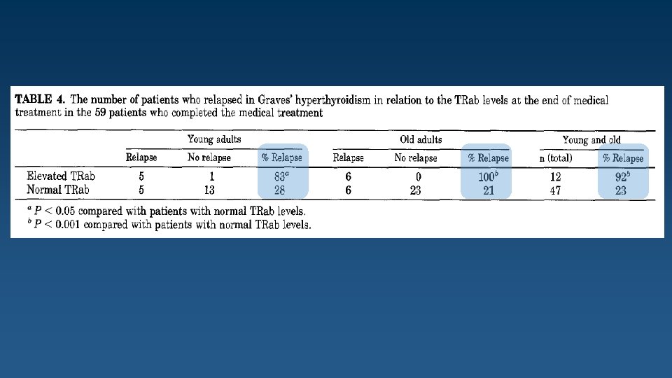
179 patients between 1983 and 1990 with hyperthyroidism caused by Graves’ disease. Young adult : 20 -34 Yr. Old adult : 35 -55 Yr.
Clinical outcome in the five treatment groups
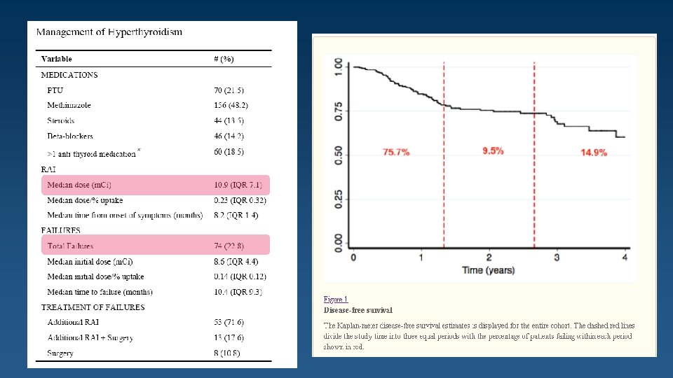
A retrospective review of patients treated with RAI from 2007– 2010. Patient Characteristics (n = 325)
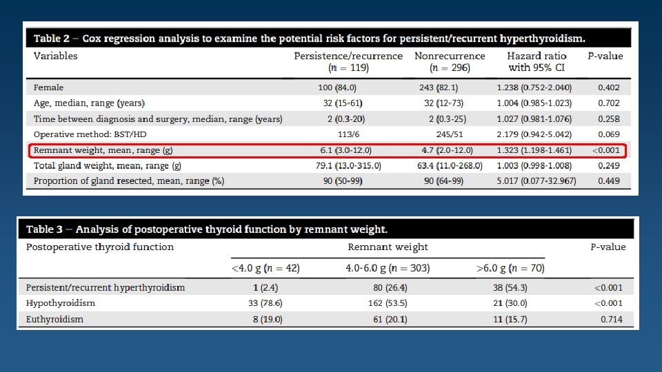
Final Multivariate Model for failure Conclusions Laboratory values at presentation can predict which patients with hyperthyroidism are at risk for failing RAI treatment. Higher doses of RAI or surgical referral may prevent the need for repeat RAI in selected patients.
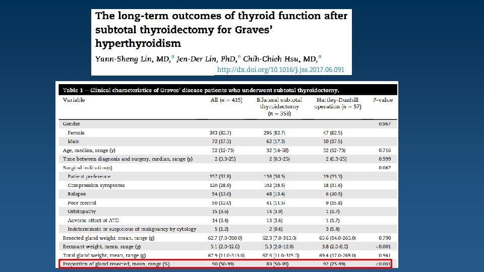
Color echo-doppler of the thyroid lodge in 102 patients who had undergone TT for benign thyroid pathologies: Significant thyroid tissue remnants after TT in 34 cases of 102 (33%). Only 67% were really total thyroidectomy. 12% had near total thyroidectomy( leaving tissue remnants < 1 cm ) 21% had subtotal thyroidectomy( with tissue remnants ≥ 1 cm)
Methods: Patients with hypothyroidism were selected in a cross-sectional study, followed prospectively for 12 months, and classified as subclinical or overt hypothyroidism. The patients were divided into two groups: with and without HAH. 34%
Involving areas Fronto-orbital : 49% Temporal : 37% Posterior part of the head : 15% Conclusion Patients with HAH may present with unilateral, pulsatile, episodic pattern, and nausea/vomiting, which is at odds with the criteria for HAH established by ICHD 3 beta.
Patient History Six months ago, the patient had symptoms compatible with hyperthyroidism and the decision was taken to have another round of treatment with RAI. Lab. Results TSH : <0. 1 m. IU/L f. T 4 : 2. 1 ng/d. L 24 H-RAIU : <5% One month after discontinuation of Methimazole 24 H-RAIU : <5% Whole body scan with RAI : No absorption was found in any point. Neck, Chest and Abdomen CT Scan: Normal findings. The thyroid gland was not seen.
What is your plan/recommendation ?
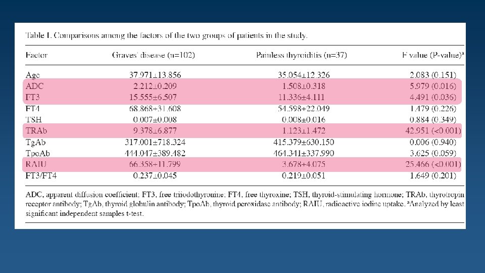
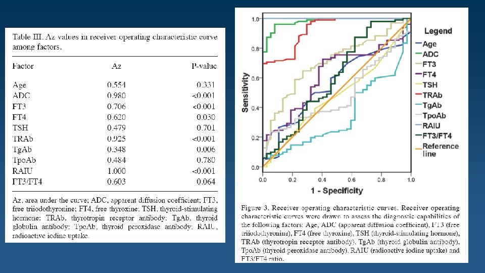
The aim of the present study was to assess the apparent diffusion coefficient (ADC) in diffusion-weighted imaging (DWI), thyroid radioactive iodine uptake (RAIU), thyroid scintigraphy and thyrotropin receptor antibody (TRAb) levels in the differential diagnosis between Graves' disease (GD) and painless thyroiditis (PT). A total of 102 patients with GD and 37 patients with PT were enrolled in the study.
The present study showed that RAIU, ADC and TRAb are all of diagnostic value for reliably differentiating between GD and PT.
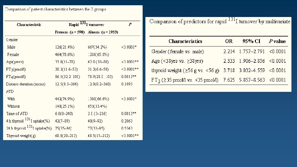
The clinical data on 2543 patients were retrospectively reviewed. Patients were divided into 2 groups depending on present or absent with rapid 131 I turnover defined as a 4 hour to 24 -hour 131 I uptake ratio of ≥ 1. Overall, 590 cases (23. 2%) had a rapid 131 I turnover.
Patient History Lab. Results TSH : <0. 01 m. IU/L f. T 4 : 2. 3 ng/d. L T 3 : 225 ng/d. L Thyroglobulin : 0. 7 ng/ml (NL. 4 -70) × 3 Times Although repeatedly asked, the patient has denied taking thyroid hormones. ?
40 untreated patients with Graves' disease Anti-Tg antibody (Tg-Ab) was positive in 34 out of 40 (85%) untreated cases and its level decreased after treatment. Anti-thyroid peroxidase antibody (TPO-Ab) was positive in 32 of the 40 (80%) untreated Graves' patients and its level significantly decreased after treatment.
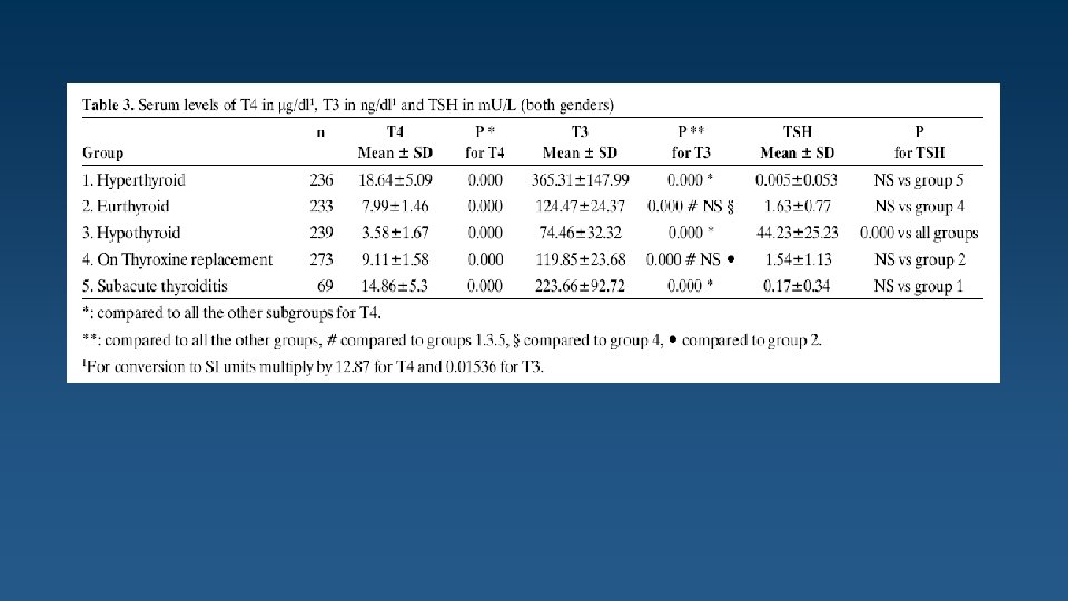
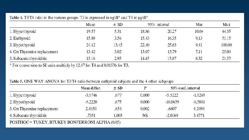
We studied 1050 subjects: 233 were euthyroid (Eu), 239 hypothyroid (Hypo) with initial TSH levels >15 m. U/L, 273 hypothyroid on substitution therapy with L-thyroxine alone (hypo. Rx), 236 hyperthyroid (hyper) 69 in the acute phase of sub acute thyroiditis De Quervain’s (DQ).
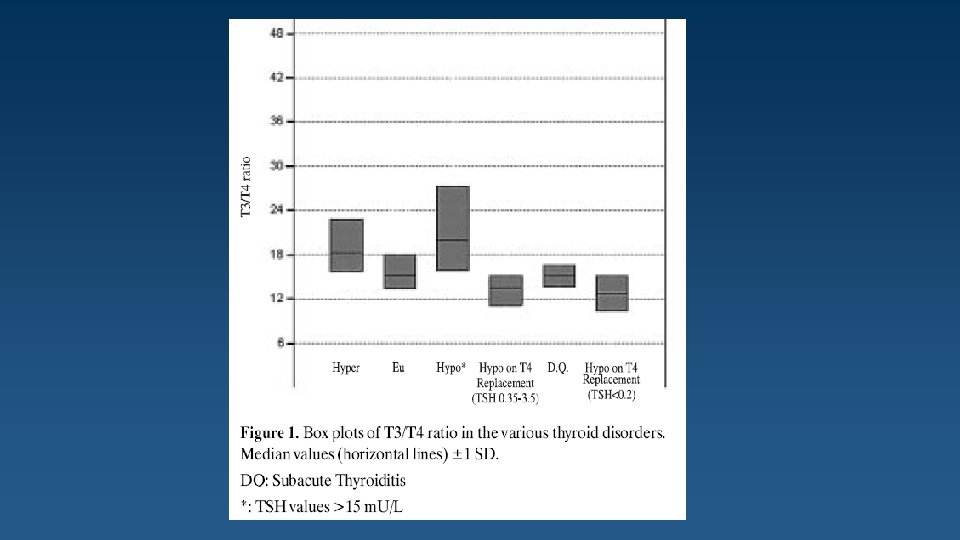
This finding means that in order to achieve normal T 3 levels in hypothyroid patients, we have to give higher doses of L-Thyroxine as replacement therapy. Conclusions In Thyrotoxicosis, a ratio of total T 3/T 4 >18. 9 suggests Graves’ Disease or toxic multinodular goiter whereas T 3/T 4 <16 suggests thyroiditis (sub acute or silent).
A retrospective pathological analysis of 1390 surgically removed ovarian tumors at 2 major academic centers.
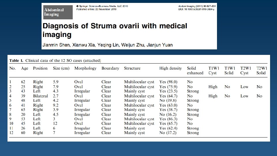
This study was conducted from 1994 to 2007, and included 25 patients with histologically confirmed struma ovarii whose medical records were retrospectively reviewed.
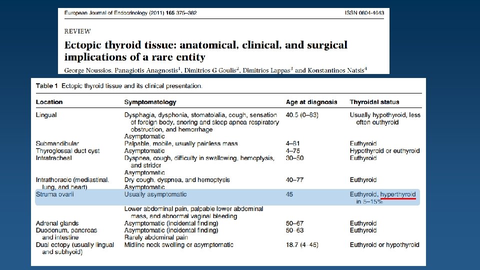
From January 1990 to January 2012, a total of 68 patients were diagnosed struma ovarii. RESULTS: The median age of patients was 42 years old (17 -81 years). [Struma ovarii is most common between the ages of 40 and 60 years. ] Uptodate 94% were diagnosed benign struma ovarii and 6% were malignant struma ovarii. 47% patients were identified with pelvic mass by ultrasonic test, 41% had clinical abdominal pain, 12% touched a mass from abdominal wall. Ascites was present in 6% patients. 75% were mulitcystic lesions, 4% solid lesions and 21% solid-cystic tumors. No patient had hyperthyroidism presentation. [Approximately 5 to 15 percent of women with struma ovarii have hyperthyroidism due to a thyroid adenoma in the struma]Uptodate
Scintigraphy performed with either iodine-123 or iodine-131 is useful for diagnosing a hyperfunctioning struma ovarii on the basis of higher uptake of the radionuclide by the ovarian mass compared with the thyroid gland. a Whole-body I-123 imaging shows significantly increased I-123 uptake within the left hemi-pelvis in a 61 -year-old woman with a history of thyrotoxicosis treated with radioiodine treatment twice over the last 30 years, who ultimately had a thyroidectomy which showed Graves disease but who remained thyrotoxic post surgery.
struma ovarii
Patient History The patient was treated with Cholestyramine 4 gr. QID for 2 months. Lab. Results TSH : 0. 8 m. IU/L f. T 4 : 1. 2 ng/d. L T 3 : 110 ng/d. L ?
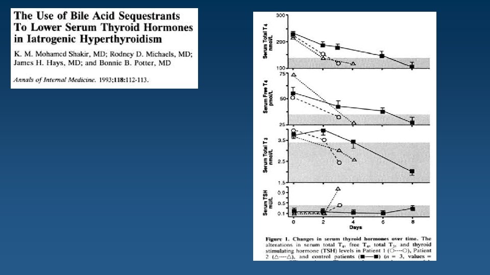
In order to evaluate the effect of Cholestyramine administration on the absorption of thyroxine, absorption studies with thyroxine I 131 were performed on both patients 1 and 2 during Cholestyramine administration and during control periods without Cholestyramine. Each period was of seven days' duration.
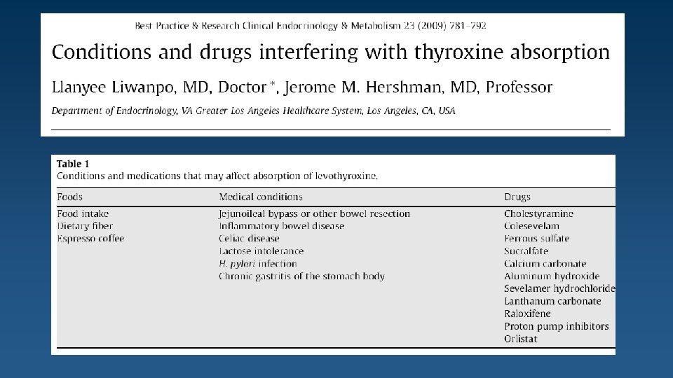
Malabsorption of thyroxine can be avoided to a large extent by allowing a time interval of four to five hours to elapse between the ingestion of the two drugs.
Mean plasma concentration-time profiles for levothyroxine administered alone or with colesevelam. (A) Unsubtracted levothyroxine plasma concentration-time curve. (B) Baseline-subtracted levothyroxine plasma concentration-time curve.
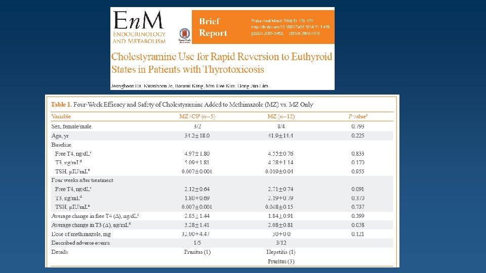
In conclusion, CS could be safely applicable adjunctive therapy for 4 weeks when first-line antithyroid medications are not enough to adequately control severe thyrotoxicosis or side effects of antithyroid drug would be of great concern at the initial stage of treatment.
In two patients who were on thyroid hormone replacement therapy, absorption tests were conducted by administering I 131 - thyroxine with and without cholestyramine. A scanning-bed whole-body counter was used to determine the amount of 131 I-thyroxine retained within the body. Urine and stool samples were collected analysed for 131 Ithyroxine. Treatment with cholestyraminewas associated with a twofold increase in total stool radioactivity and decrease in urinary radioactivity, representing decreased intestinal absorption and subsequent loss of I 131 -thyroxine.
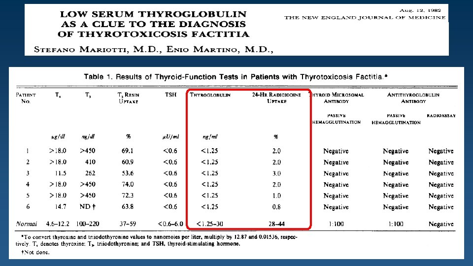
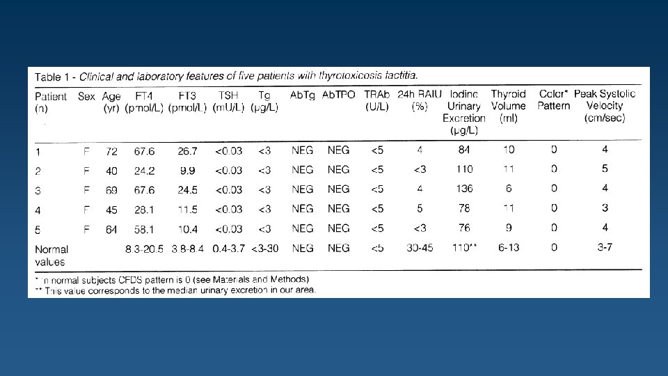
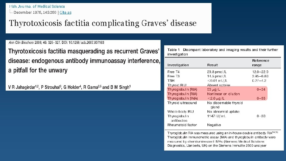
Diagnosis was supported by the finding of elevated free thyroxine (FT 4) elevated free triiodothyronine (FT 3) Undetectable TSH absent anti-thyroid antibodies undetectable serum thyroglobulin very low/suppressed thyroidal radioiodine uptake normal/low urinary iodine excretion. Five women with thyrotoxicosis factitia were evaluated by CFDS (age range 40 -72 yr. , mean 58 yr. ).
CFDS showing an absent intrathyroidal hypervascularity in spite of elevated circulating free thyroid hormones. CFDS can be used as an additional, rapid and noninvasive aid to confirm the diagnosis.
Two patients with prolonged thyrotoxicosis are presented with low radioiodine uptake, low-normal serum thyroglobulin concentrations, and low iodide content of the neck on x-ray fluorescence studies. The surreptitious intake of thyroid hormone was directly proven by a high fecal concentration of T 4 (12. 38 and 23. 99 nmol/g) compared with low concentrations in healthy subjects (1. 03 ± 0. 64 nmol/g; mean ± SD, = 6) and in patients with hyperthyroidism due to Graves' disease (1. 93 ± 1. 86; = 8).
The measurement of fecal T 4 is technically simple and reliable and could help in clarifying the origin of difficult cases of thyrotoxicosis.
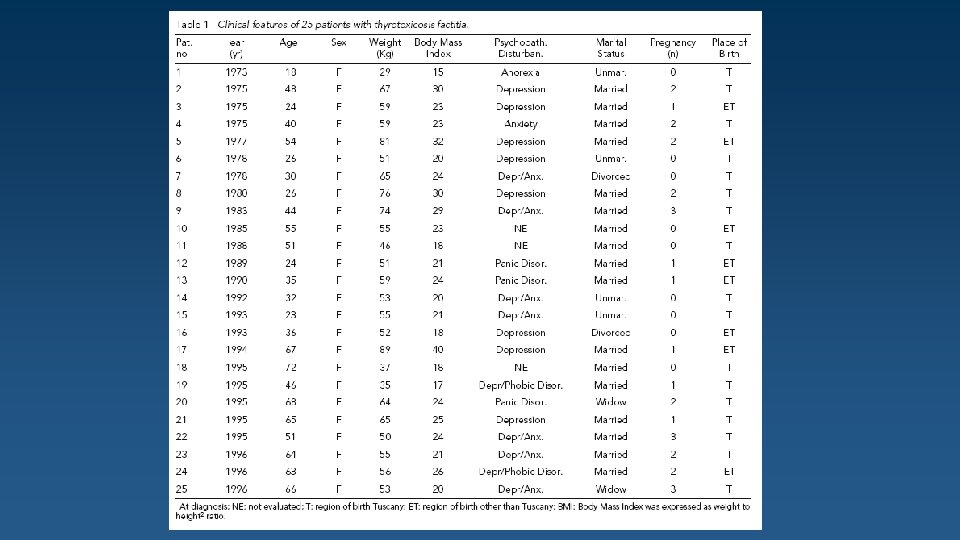
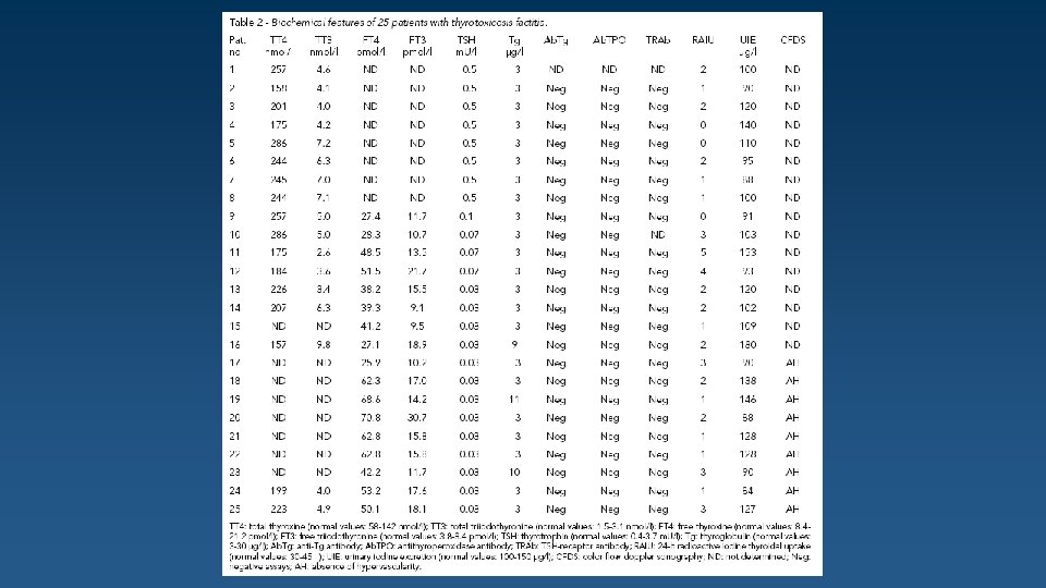
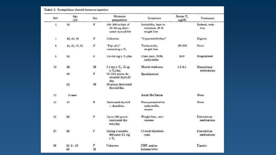
All 25 patients were women. Analysis was restricted to 17 patients who were born and lived in Tuscany (our region). Diagnosis of thyrotoxicosis factitia was based on the following parameters: Elevated serum total and/or free thyroid hormone levels Undetectable serum thyrotropin levels Low/undetectable serum thyroglobulin concentration Normal urinary iodine excretion Low/suppressed thyroidal RAIU Absence of goiter Absence of circulating anti-thyroid antibodies. Age at diagnosis was >50 yr. in 7/17 patients (41%). Patients older than 60 yr. were 5/17 (29%).
Advanced age is not a sufficient reason to rule out thyrotoxicosis factitia.
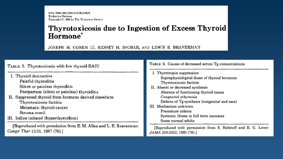
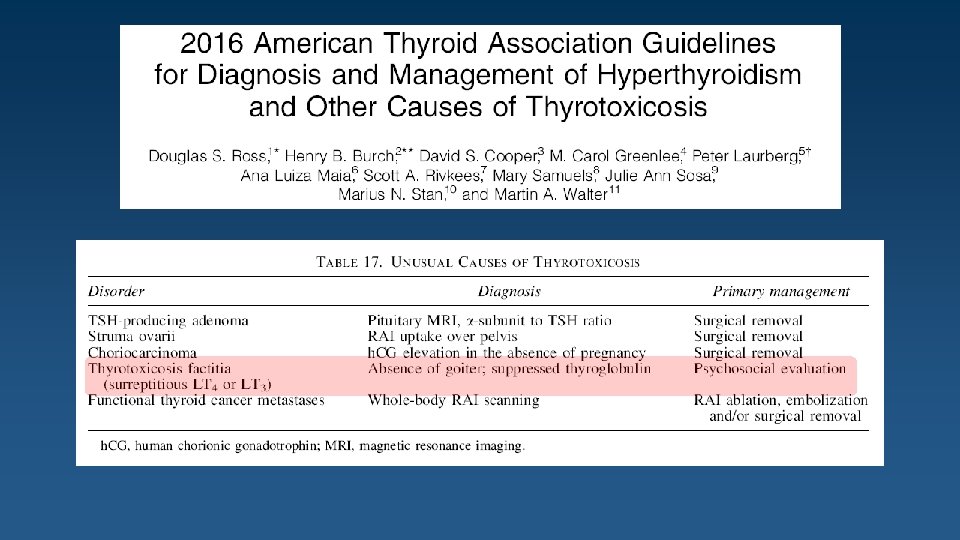
Thyrotoxicosis factitia Clues to this diagnosis : Absence of goiter Suppressed serum TG Decreased RAIU Distinction between painless thyroiditis and factitious thyrotoxicosis in Anti TG Positive : Elevated levels of T 4 A high T 4/T 3 ratio (with exogenous levothyroxine) A small thyroid A low thyroidal RAIU Thyroid ultrasound may be helpful painless thyroiditis: The thyroid has a heterogeneous echo Normal sized or slightly enlarged Ingesting thyroid hormone : Small with a normal echo Fecal levothyroxine A disproportionately elevated T 3 level suggests that the patient may be ingesting Liothyronine or a combination T 4/T 3 preparation.