Invasive Aspergillosis 34 yearold woman Presents with 2


- Slides: 23
Invasive Aspergillosis � 34 -year-old woman • Presents with 2 -day history of weakness, dizziness, left calf pain, and black tarry stools. • Denies chest pain, cough, or shortness of breath �Medical history: • Diabetes leading to renal failure and renal transplant • 3 weeks before presentation, acute graft rejection developed • Began an immunosuppressive regimen Guha, et al. Infect Med 24 (Suppl 8): 8 -11, 2007
Invasive Aspergillosis �On admission • Tachycardic, hypotensive and febrile �Initial chest x-ray was normal �Lab results: • • Anemia WBC = 4800/µl, 80% neutrophils Blood cultures were positive for E. coli Antibiotic therapy initiated Guha, et al. Infect Med 24 (Suppl 8): 8 -11, 2007
Invasive Aspergillosis �Day 6: • Vesicular rash developed on buttocks and left calf • Cultures positive for HSV, antiviral therapy initiated �Day 8: • Renal function continued to decline • Intermittent hemodialysis started �Day 12: • Decreased responsiveness • Intubated for respiratory distress Guha, et al. Infect Med 24 (Suppl 8): 8 -11, 2007
Invasive Aspergillosis �Chest x-ray: • Diffuse bilateral lung nodules �Culture of BAL: • Positive for Aspergillus spp. • Immunesuppression decreased • Liposomal Amphotericin B started �Condition deteriorates: • Acute MI, comatose • Mulitple acute infarcts in frontal lobe and cerebellum by MRI • Multiple skin nodules form on arms and trunk Guha, et al. Infect Med 24 (Suppl 8): 8 -11, 2007
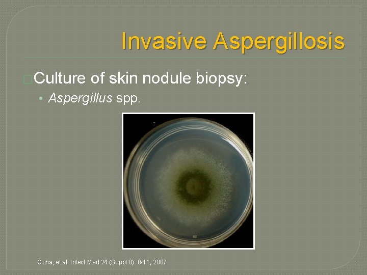
Invasive Aspergillosis �Culture of skin nodule biopsy: • Aspergillus spp. Guha, et al. Infect Med 24 (Suppl 8): 8 -11, 2007
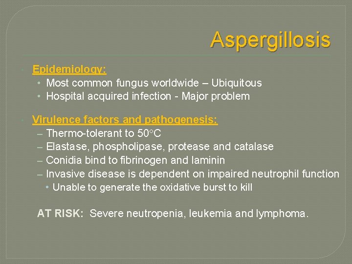
Aspergillosis • Epidemiology: • Most common fungus worldwide – Ubiquitous • Hospital acquired infection - Major problem • Virulence factors and pathogenesis: – Thermo-tolerant to 50 C – Elastase, phospholipase, protease and catalase – Conidia bind to fibrinogen and laminin – Invasive disease is dependent on impaired neutrophil function • Unable to generate the oxidative burst to kill AT RISK: Severe neutropenia, leukemia and lymphoma.
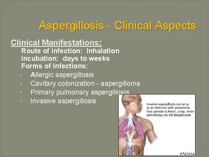
Aspergillosis - Clinical Aspects Clinical Manifestations: � � � Route of infection: Inhalation Incubation: days to weeks Forms of infections: • Allergic aspergillosis • Cavitary colonization - aspergilloma • Primary pulmonary aspergillosis • Invasive aspergillosis
Types of Aspergilloses
ABPA – Allergic broncopulmonary aspergillosis (ABPA) �Asthma �Pulmonary infiltrates �Peripheral eosinophelia �Elevated serum Ig. E �Hypersensitivity to Aspergillus antigen • Skin test
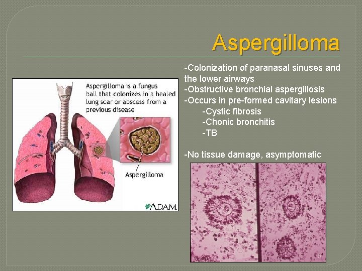
Aspergilloma -Colonization of paranasal sinuses and the lower airways -Obstructive bronchial aspergillosis -Occurs in pre-formed cavitary lesions -Cystic fibrosis -Chonic bronchitis -TB -No tissue damage, asymptomatic
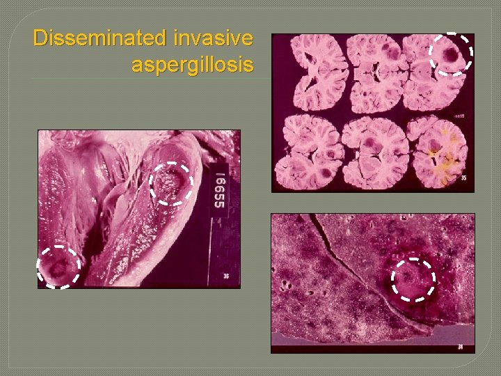
Disseminated invasive aspergillosis
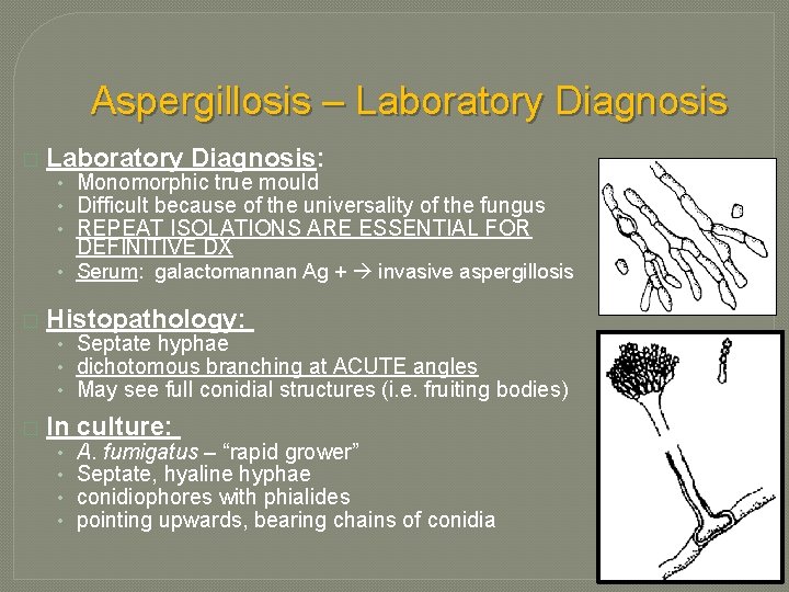
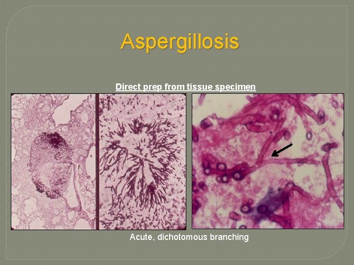
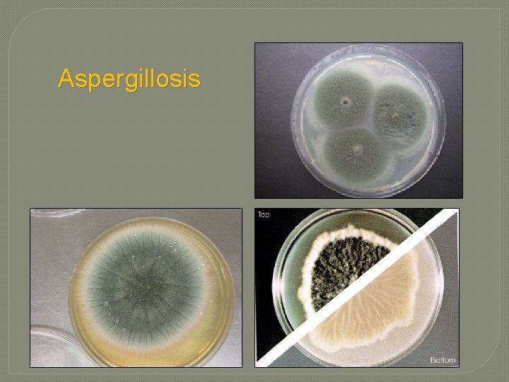
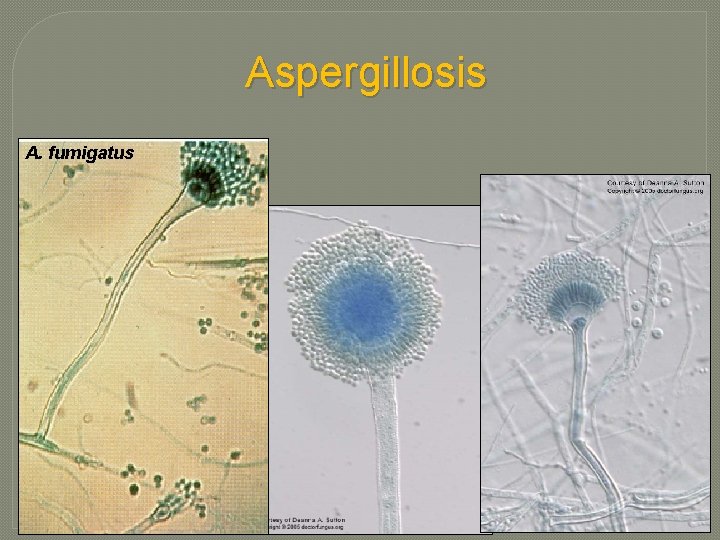
Aspergillosis – Laboratory Diagnosis � Laboratory Diagnosis: • Monomorphic true mould • Difficult because of the universality of the fungus • REPEAT ISOLATIONS ARE ESSENTIAL FOR DEFINITIVE DX • Serum: galactomannan Ag + invasive aspergillosis � Histopathology: • Septate hyphae • dichotomous branching at ACUTE angles • May see full conidial structures (i. e. fruiting bodies) � In culture: • • A. fumigatus – “rapid grower” Septate, hyaline hyphae conidiophores with phialides pointing upwards, bearing chains of conidia
Aspergillosis Direct prep from tissue specimen Acute, dichotomous branching
Aspergillosis Septate hyphae Aspergilloma Conidiophore “fruiting body”
Aspergillosis
Aspergillosis A. fumigatus
Aspergillosis - Treatment � Treatment: • Invasive disease is difficult to treat • Amphotericin B, caspofungin (echinocandins), voriconazole • Decrease immunosuppression or reconstitute immune defenses • Surgical debridement, if possible � Prevention in high-risk patients: • Neutropenic: Filtered air to minimize exposure!
Invasive Aspergillosis �Our patient: • Expired on hospital day 23 • At autopsy, A. flavus was detected in multiple organs: �Heart, lungs, adrenal galnd, thyroid, kidney, and liver �Extreme example of disseminated aspergillosis in an immunocompromised host Guha, et al. Infect Med 24 (Suppl 8): 8 -11, 2007
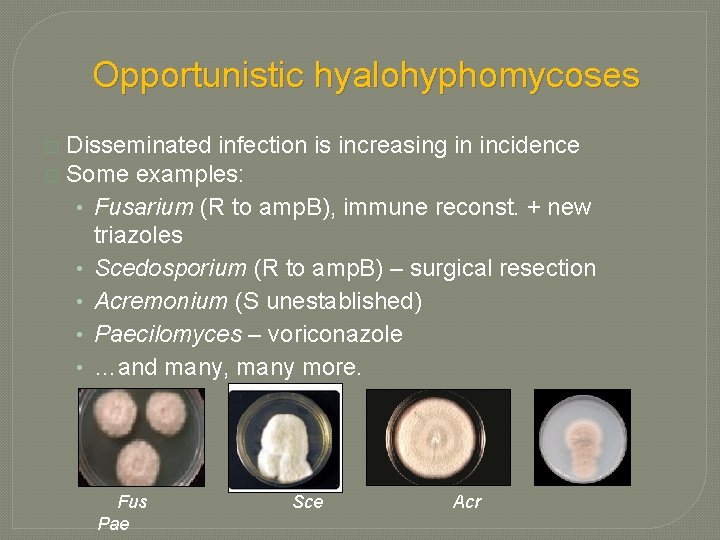
Opportunistic hyalohyphomycoses Diverse agents � Many are ubiquitous – inhaled conidia � Many are resistant to antifungal agents � In tissue, they appear indistinguishable from Aspergillus! (i. e. branching, septate hyphae) • Repeated isolation from multiple sites/multiple times is best criteria to determine clinical significance. � BOTTOM LINE: CULTURE IS CRITICAL FOR DX & TREATMENT
Opportunistic hyalohyphomycoses Disseminated infection is increasing in incidence � Some examples: • Fusarium (R to amp. B), immune reconst. + new triazoles • Scedosporium (R to amp. B) – surgical resection • Acremonium (S unestablished) • Paecilomyces – voriconazole • …and many, many more. � Fus Pae Sce Acr
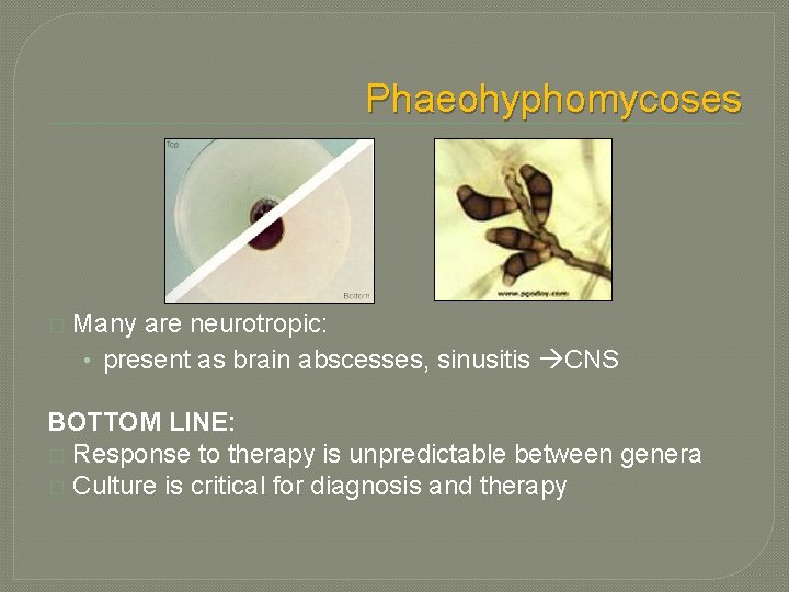
Phaeohyphomycoses � Many are neurotropic: • present as brain abscesses, sinusitis CNS BOTTOM LINE: � Response to therapy is unpredictable between genera � Culture is critical for diagnosis and therapy
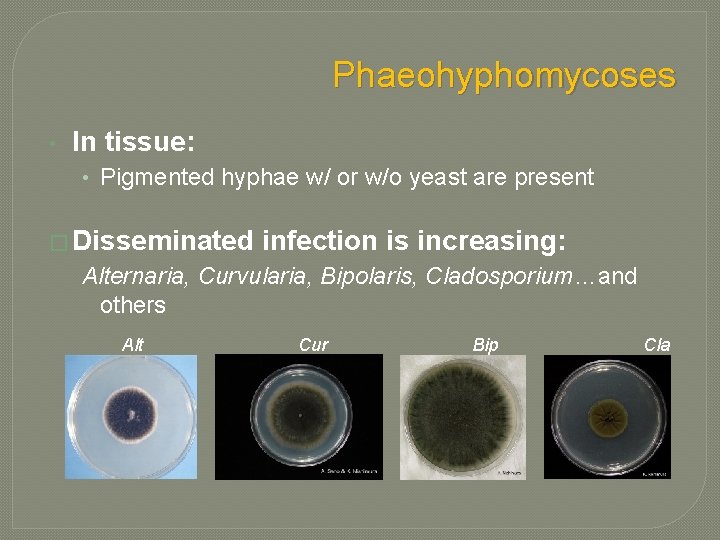
Phaeohyphomycoses • In tissue: • Pigmented hyphae w/ or w/o yeast are present � Disseminated infection is increasing: Alternaria, Curvularia, Bipolaris, Cladosporium…and others Alt Cur Bip Cla
Pneumocystosis �Etiology: Pneumocystis jirovecii �Most common opportunistic infection among individuals with AIDS • Incidence has decreased significantly with HAART • Reservoir in nature unknown • Pneumonia is clearly the most common presentation �Interstitial pneumonitis, mononuclear infiltrate �Onset insidious �Diagnosis based on microscopic examination of BAL