THYROTOXICOSIS Thyrotoxicosis is a hypermetabolic state caused by

THYROTOXICOSIS: � Thyrotoxicosis is a hypermetabolic state caused by elevated circulating levels of free T 3 and T 4. � Causes of thyrotoxicosis: I. Associated with hyperthyroidism (Thyroid hyperfunction): 1. Primary a. Graves disease. b. Hyperfunctioning (toxic ) adenoma. 2. Secondary TSH-secreting pituitary adenoma (rare) II. Not associated with hyperthyroidism: - Such as in thyroiditis early stage � Struma ovarii (ovarian teratoma with thyroid) � Factitious thyrotoxicosis (exogenous thyroxine intake)

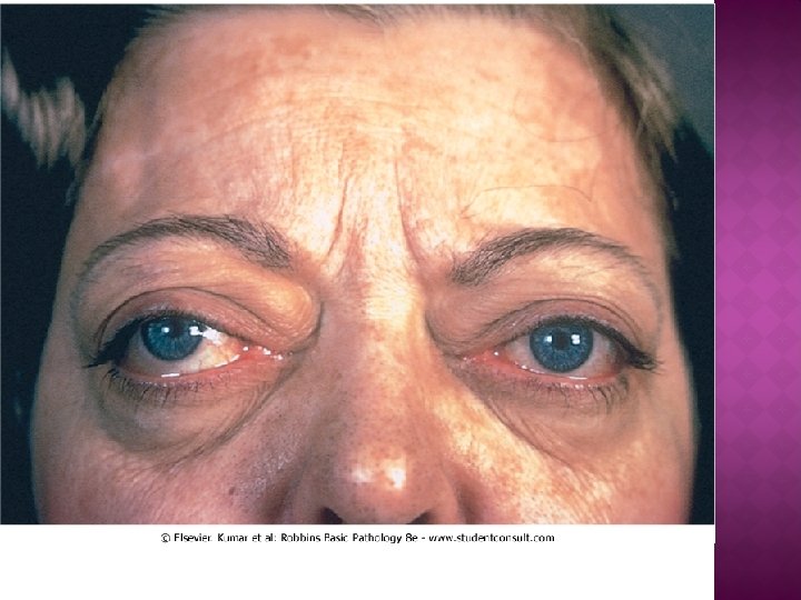
CLINICAL MANIFESTATIONS � - Constitutional symptoms: � soft, warm, flushed skin; � heat intolerance � excessive sweating � Weight loss & increased appetite. � Gastrointestinal: � hypermotility, malabsorption, diarrhea. � Cardiac: � Palpitations and tachycardia. � Neuromuscular: � nervousness, tremor, irritability and muscle weakness (thyroid myopathy). � Ocular manifestations: � a wide, staring gaze and lid lag. However, true thyroid ophthalmopathy associated with proptosis is a feature seen only in Graves disease.

HYPOTHYROIDISM: CAUSES 1. Worldwide, the most common cause is dietary deficiency of iodine. 2. In developed countries – Autoimmune Hashimoto thyroiditis. 3. Genetic defects such as thyroid dysgenesis or dyshormogentic goiter are rare

A. - Cretinism : Refers to hypothyroidism developing in infancy or early childhood. Results in impaired development of the skeletal system and central nervous system, with severe mental retardation, short stature, coarse facial features, a protruding tongue, and umbilical hernia. - Endemic cretinism: caused by dietary iodine deficiency. - Sporadic cretinism: caused by inborn errors in metabolism.

B. Myxedema : Ø occurs in older children and adults Ø manifestations: �Generalized apathy and mental sluggishness, may mimic depression. �Cold intolerant. �Obesity. �Mucopolysaccharide-rich edematous fluid accumulates in skin, subcutaneous tissue, and a number of visceral sites, with resultant broadening and coarsening of facial features, enlargement of the tongue, and deepening of the voice. �Bowel motility is decreased, resulting in constipation.
 thyroiditis : � Most common cause of hypothyroidism")
1. THYROIDITIS A. Chronic lymphocytic (Hashimoto) thyroiditis : � Most common cause of hypothyroidism in areas where iodine is sufficient. � Gradual thyroid failure secondary to autoimmune destruction of the thyroid gland. � Women>men � Diffuse symmetric painless enlargement of the thyroid

Figure 15 -13 Hashimoto thyroiditis, microscopic lymphoid infiltrates, including a lymphoid follicle. The remaining thyroid follicles become atrophic, and the epithelial cells undergo Hürthle cell change, with abundant pink cytoplasm.

B. Subacute granulomatous thyroiditis Self-limited caused by viral infection - patients have history of upper respiratory tract infection. - Acute onset with pain in the neck, fever, and variable enlargement of the thyroid. - Transient hyperthyroidism may occur. -

Figure 15 -15 Granulomatous thyroiditis, microscopic. begins with diffuse painful thyroid enlargement. there is marked acute inflammation along with lymphocytes, macrophages, and prominent giant cells. There is destruction of thyroid follicles. This condition typically follows a viral infection that activates cytotoxic T lymphocytes.

2. GRAVES DISEASE: Most common cause of endogenous hyperthyroidism. Triad of manifestations: • Thyrotoxicosis, caused by a diffusely enlarged, hyperfunctional thyroid, in all cases. • An infiltrative ophthalmopathy with exophthalmos (40%). • A localized, infiltrative dermopathy (pretibial myxedema) seen in a minority of cases. Mostly involves the skin overlying the shins. � � 7: 1 female to male ratio. Elevated serum free T 4 and T 3 and depressed serum TSH

Figure 15 -16 Graves disease, microscopic. thyroid hyperplasia is characterized by many papillary infoldings within follicles. The entire thyroid gland becomes diffusely enlarged to double or triple normal size.

3 -MULTINODULAR GOITER: Ø Ø Goiter: thyroid enlargement most common manifestation of thyroid disease mostly caused by dietary iodine deficiency Impaired thyroid hormone synthesis >> compensatory rise in the serum TSH>>> gross enlargement of the thyroid gland

Figure 15 -20 Thyroid, multinodular goiter, gross and scintigraphic scan often asymmetric. Most patients remain euthyroid, bothered only by the mass effect. In 10%, a hyperfunctioning “toxic” nodule producing T 4 and causing hyperthyroidism. Such a “hot” nodule with increased activity on r(right panel).

Figure 15 -21 Thyroid, goiter, microscopic. enlarged thyroid follicles lined with inactive, flattened epithelial cells and filled with abundant stored colloid. The process starts as a simple, diffuse, nontoxic goiter. Over time, there can be irregular nodular enlargement with fibrosis, hemorrhage, and calcification in areas of cystic change.

1. Endemic goiter: Iodine deficient areas 2. Sporadic goiter: less common, more in females, peak in puberty or young adult life.

- Slides: 17