1 1 a Skeletal and muscular systems Learning








- Slides: 77
1. 1. a – Skeletal and muscular systems Learning objectives To understand the different types of joint, articulating bones and main agonists and antagonists in the body. To be able to explain the different types of muscular contractions. To understand the planes of the body. To describe the characteristics of slow twitch and fast twitch muscle fibres. To be able to explain motor unit recruitment in muscle contraction and the ‘all or none law’.
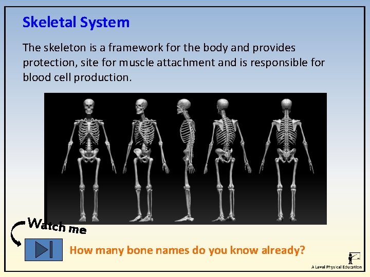
Skeletal System The skeleton is a framework for the body and provides protection, site for muscle attachment and is responsible for blood cell production. Watch me How many bone names do you know already?
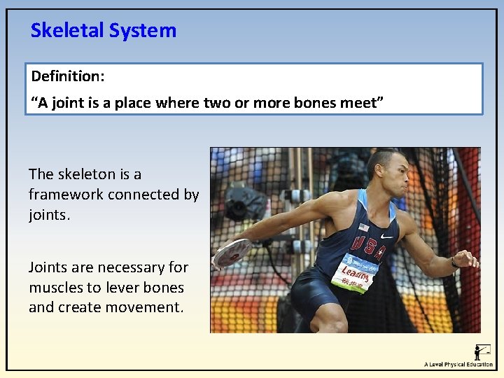
Skeletal System Definition: “A joint is a place where two or more bones meet” The skeleton is a framework connected by joints. Joints are necessary for muscles to lever bones and create movement.
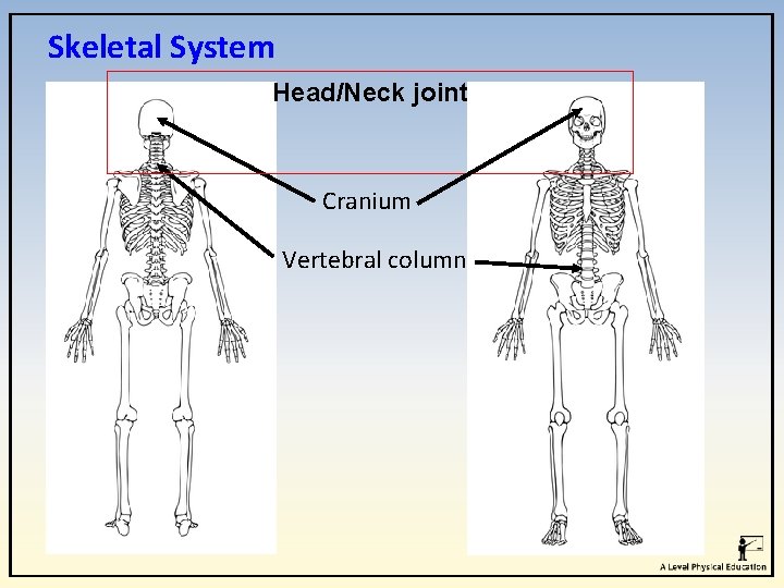
Skeletal System Head/Neck joint Cranium Vertebral column
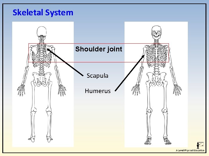
Skeletal System Shoulder joint Scapula Humerus
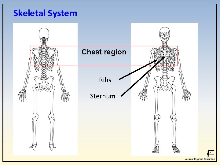
Skeletal System Chest region Ribs Sternum
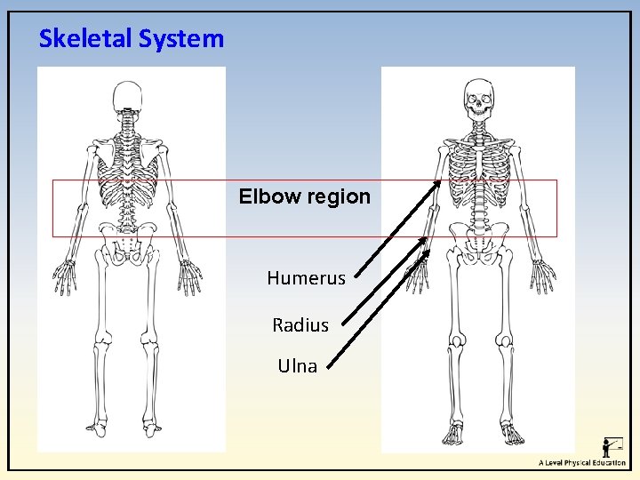
Skeletal System Elbow region Humerus Radius Ulna
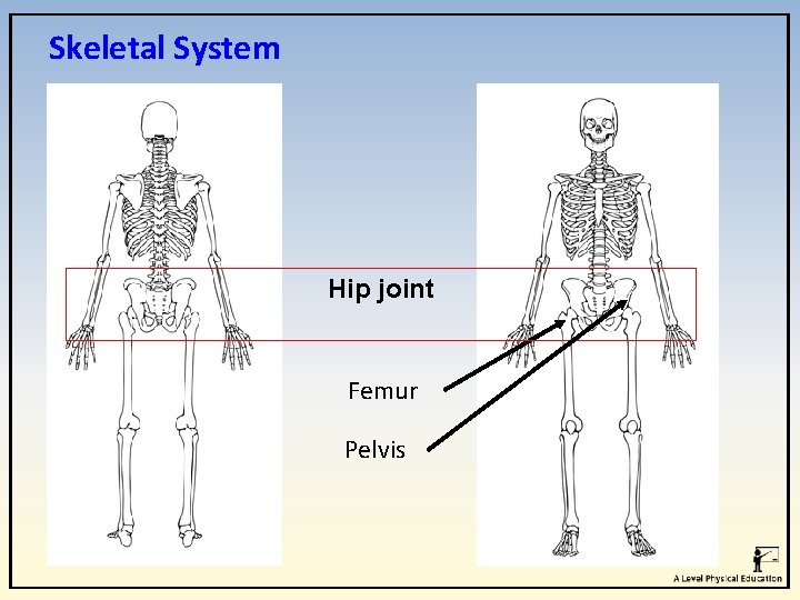
Skeletal System Hip joint Femur Pelvis
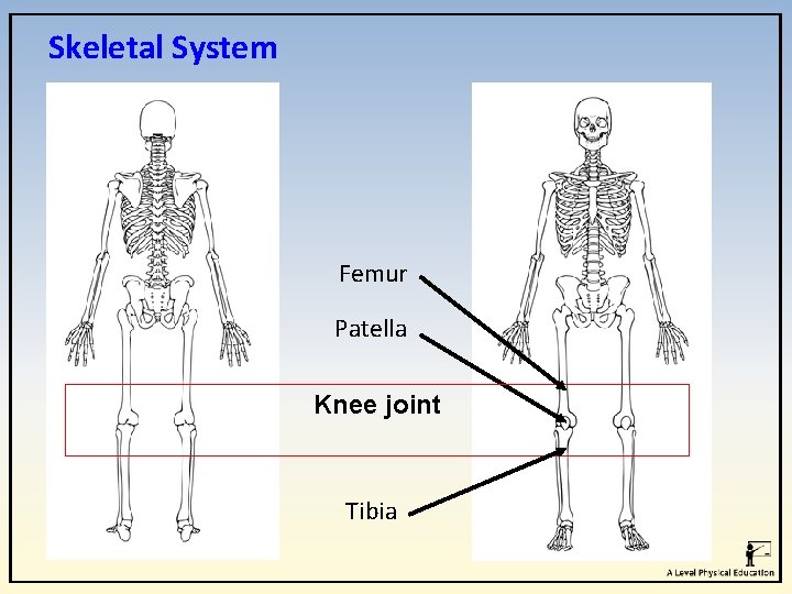
Skeletal System Femur Patella Knee joint Tibia
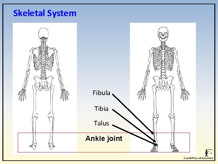
Skeletal System Fibula Tibia Talus Ankle joint
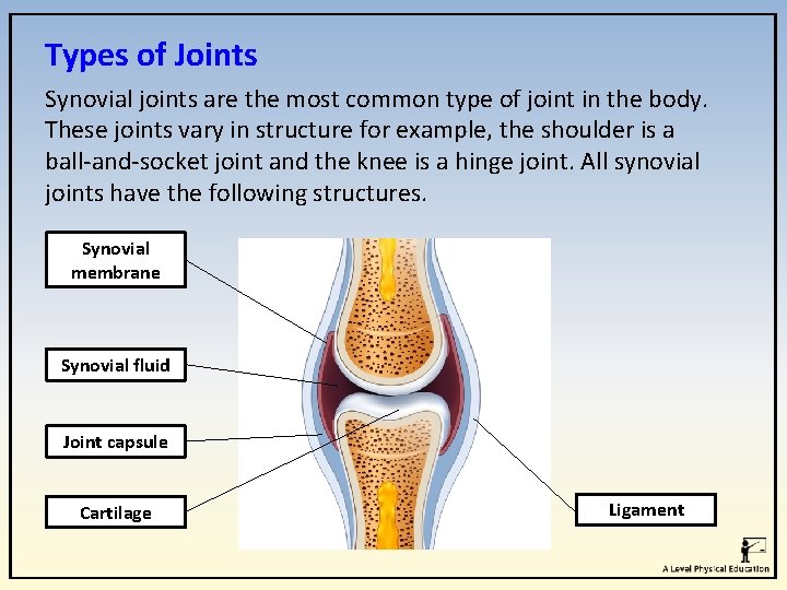
Types of Joints Synovial joints are the most common type of joint in the body. These joints vary in structure for example, the shoulder is a ball-and-socket joint and the knee is a hinge joint. All synovial joints have the following structures. Synovial membrane Synovial fluid Joint capsule Cartilage Ligament
Types of Joints The following structures help prevent injury. Cartilage prevents the ends of bones rubbing together at joints. Synovial fluid – lubricates the joint allowing the parts to move freely. Ligaments – these are tough, elastic fibres that link bones to bones. Tendons – These connect muscles to bones.
Types of Joints 1. Ball and socket joints allow movement in all directions and is the most mobile joints in the body. Examples: Shoulders and hips. Think. Pair. Share – Using examples, how are these joints used in sport? i. e. tennis serve
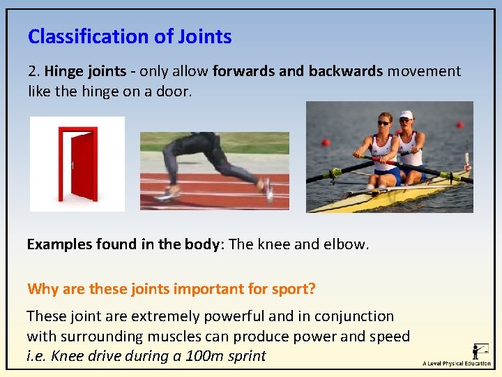
Classification of Joints 2. Hinge joints - only allow forwards and backwards movement like the hinge on a door. Examples found in the body: The knee and elbow. Why are these joints important for sport? These joint are extremely powerful and in conjunction with surrounding muscles can produce power and speed i. e. Knee drive during a 100 m sprint
Classification of Joints 3. Pivot joints have a ring of bone that fits over a bone sticking out. Pivot joints allow rotation only. Examples found in the body: The joint between the atlas and axis in the neck which allows turning and nodding of head Why are these joints important for sport? This joint allows for small movements that assist a larger sporting action i. e. breathing during a swimming stroke
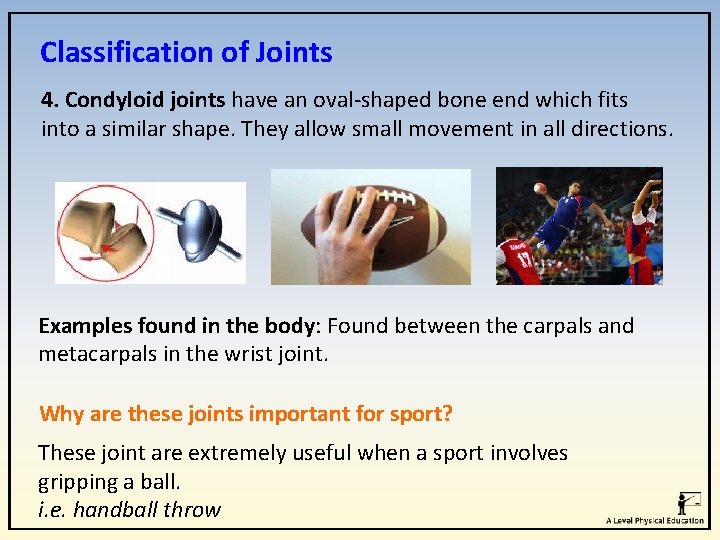
Classification of Joints 4. Condyloid joints have an oval-shaped bone end which fits into a similar shape. They allow small movement in all directions. Examples found in the body: Found between the carpals and metacarpals in the wrist joint. Why are these joints important for sport? These joint are extremely useful when a sport involves gripping a ball. i. e. handball throw
Classification of Joints 5. Gliding joints occur between the surfaces of two flat bones that are held together by ligaments. Examples found in the body: The bones in your wrists and ankles as well as the spine. Why are these joints important for sport? These joints are used to allow flexibility and movement in the hands, feet and back regions. i. e. a kicking or catching action or a boxing slip
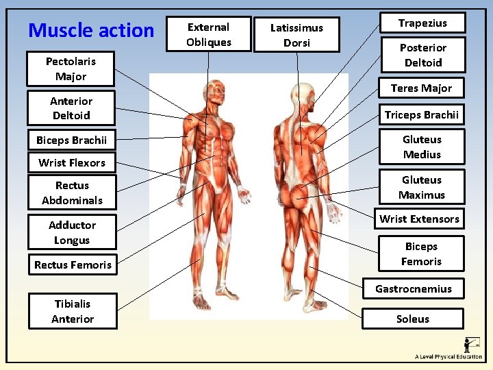
Muscle action Pectolaris Major Anterior Deltoid Biceps Brachii Wrist Flexors External Obliques Latissimus Dorsi Trapezius Posterior Deltoid Teres Major Triceps Brachii Gluteus Medius Rectus Abdominals Gluteus Maximus Adductor Longus Wrist Extensors Rectus Femoris Biceps Femoris Gastrocnemius Tibialis Anterior Soleus
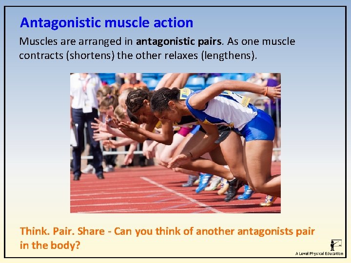
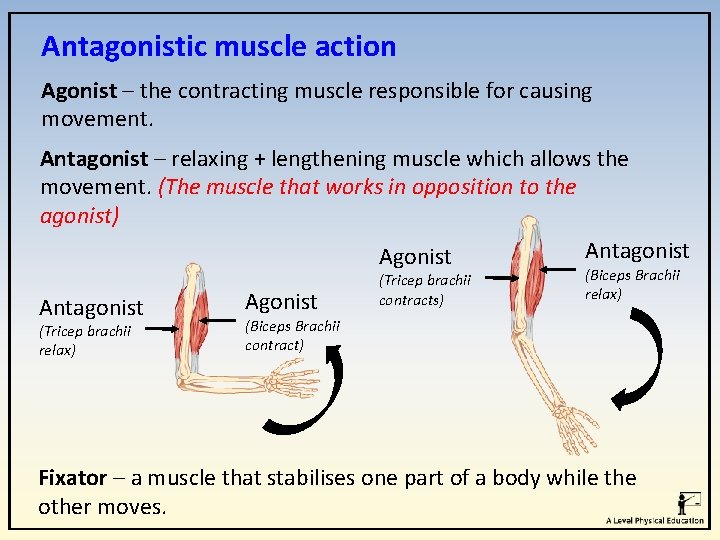
Antagonistic muscle action Muscles are arranged in antagonistic pairs. As one muscle contracts (shortens) the other relaxes (lengthens). Think. Pair. Share - Can you think of another antagonists pair in the body?
Antagonistic muscle action Agonist – the contracting muscle responsible for causing movement. Antagonist – relaxing + lengthening muscle which allows the movement. (The muscle that works in opposition to the agonist) Antagonist (Tricep brachii relax) Agonist (Tricep brachii contracts) (Biceps Brachii relax) (Biceps Brachii contract) Fixator – a muscle that stabilises one part of a body while the other moves.
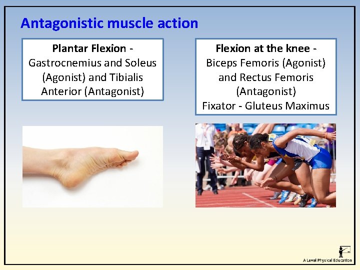
Antagonistic muscle action Plantar Flexion - Gastrocnemius and Soleus (Agonist) and Tibialis Anterior (Antagonist) Flexion at the knee - Biceps Femoris (Agonist) and Rectus Femoris (Antagonist) Fixator - Gluteus Maximus
Types of muscular contractions 1. Isometric 2. Isotonic a. Concentric b. Eccentric
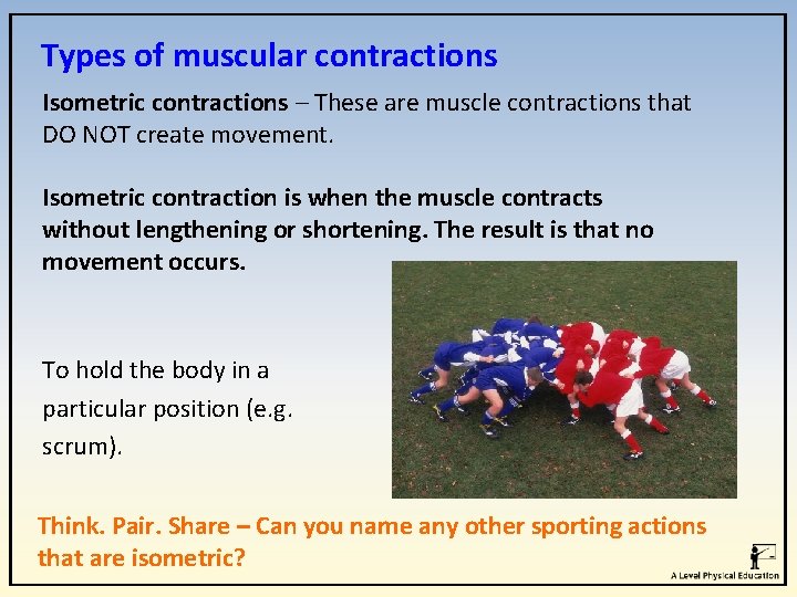
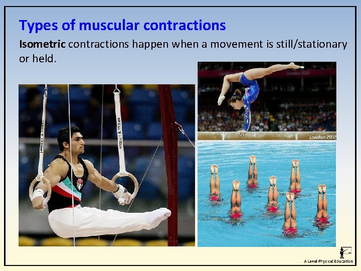
Types of muscular contractions Isometric contractions – These are muscle contractions that DO NOT create movement. Isometric contraction is when the muscle contracts without lengthening or shortening. The result is that no movement occurs. To hold the body in a particular position (e. g. scrum). Think. Pair. Share – Can you name any other sporting actions that are isometric?
Types of muscular contractions Isometric contractions happen when a movement is still/stationary or held.
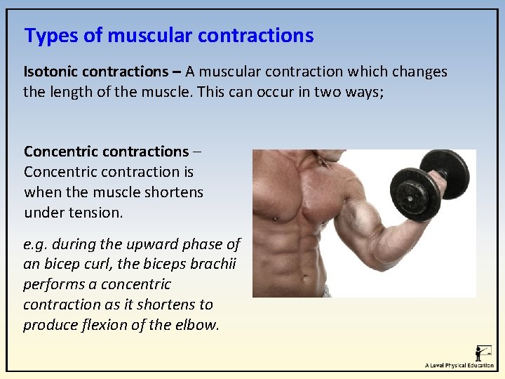
Types of muscular contractions Isotonic contractions – A muscular contraction which changes the length of the muscle. This can occur in two ways; Concentric contractions – Concentric contraction is when the muscle shortens under tension. e. g. during the upward phase of an bicep curl, the biceps brachii performs a concentric contraction as it shortens to produce flexion of the elbow.
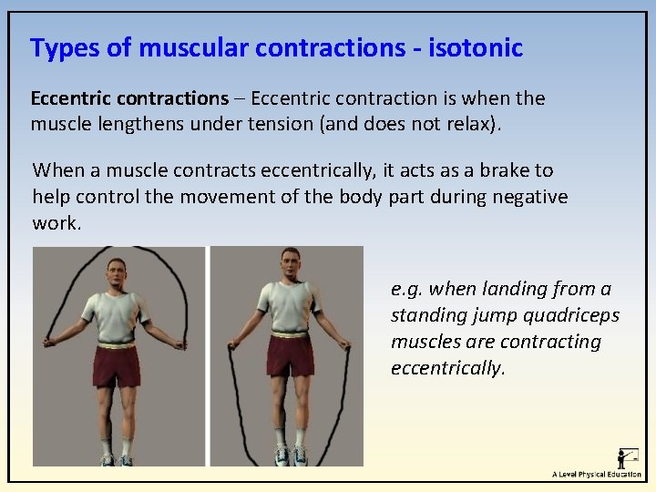
Types of muscular contractions - isotonic Eccentric contractions – Eccentric contraction is when the muscle lengthens under tension (and does not relax). When a muscle contracts eccentrically, it acts as a brake to help control the movement of the body part during negative work. e. g. when landing from a standing jump quadriceps muscles are contracting eccentrically.
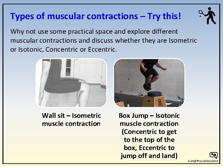
Types of muscular contractions – Try this! Why not use some practical space and explore different muscular contractions and discuss whether they are Isometric or Isotonic, Concentric or Eccentric. Wall sit – Isometric muscle contraction Box Jump – Isotonic muscle contraction (Concentric to get to the top of the box, Eccentric to jump off and land)
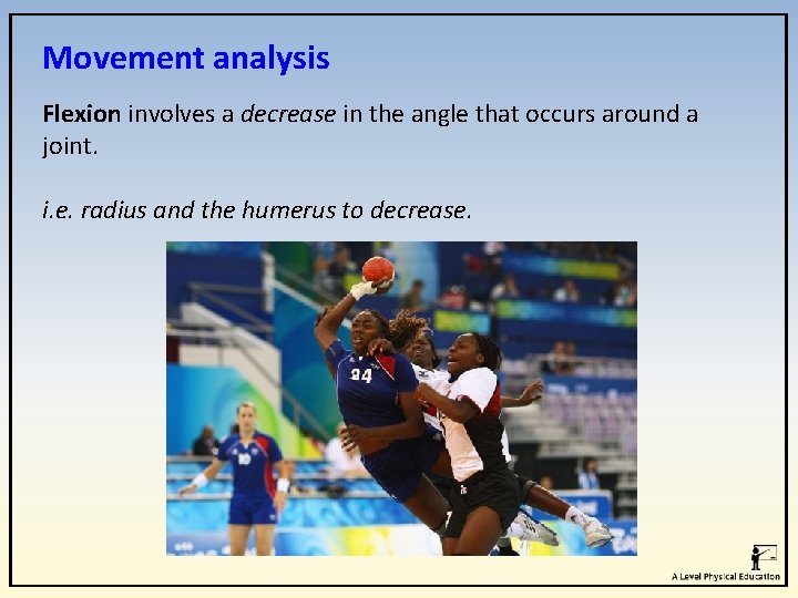
Movement analysis Flexion involves a decrease in the angle that occurs around a joint. i. e. radius and the humerus to decrease.
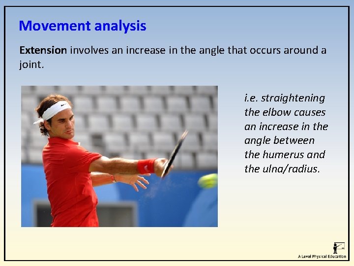
Movement analysis Extension involves an increase in the angle that occurs around a joint. i. e. straightening the elbow causes an increase in the angle between the humerus and the ulna/radius.
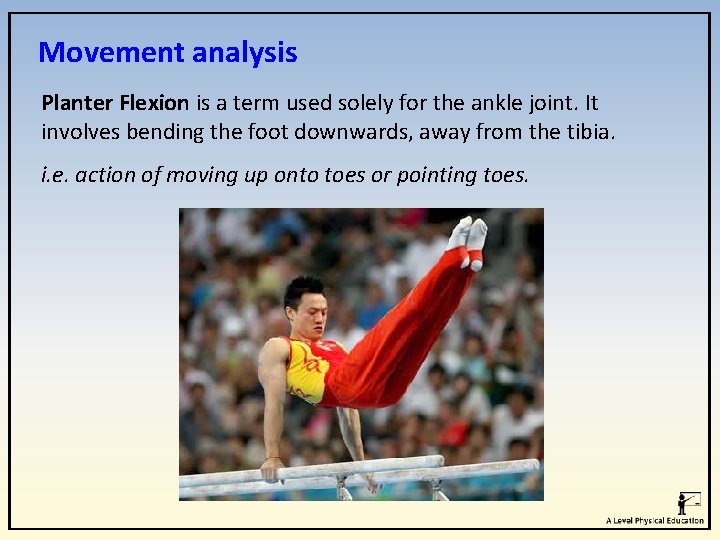
Movement analysis Planter Flexion is a term used solely for the ankle joint. It involves bending the foot downwards, away from the tibia. i. e. action of moving up onto toes or pointing toes.
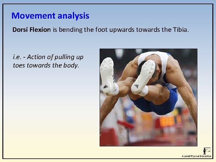
Movement analysis Dorsi Flexion is bending the foot upwards towards the Tibia. i. e. - Action of pulling up toes towards the body.
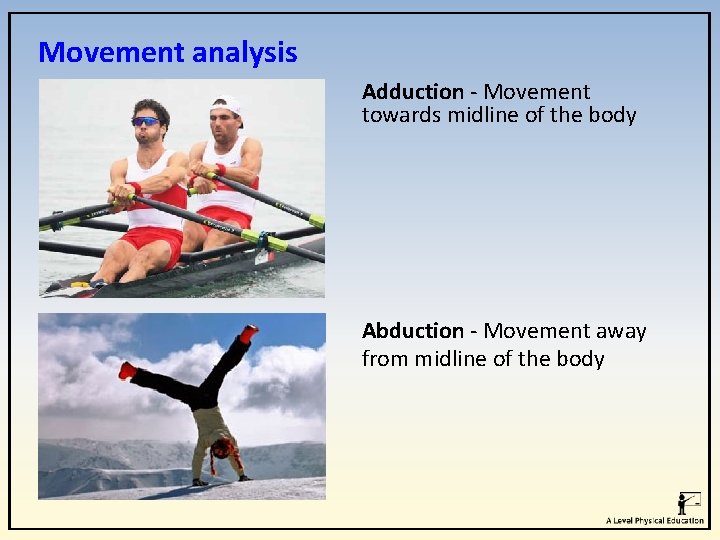
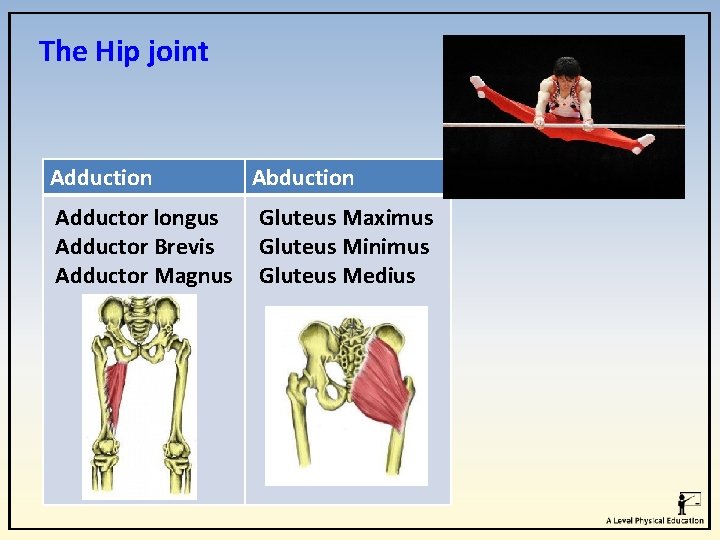
Movement analysis Adduction - Movement towards midline of the body Abduction - Movement away from midline of the body
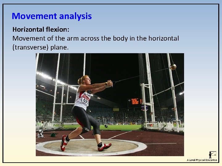
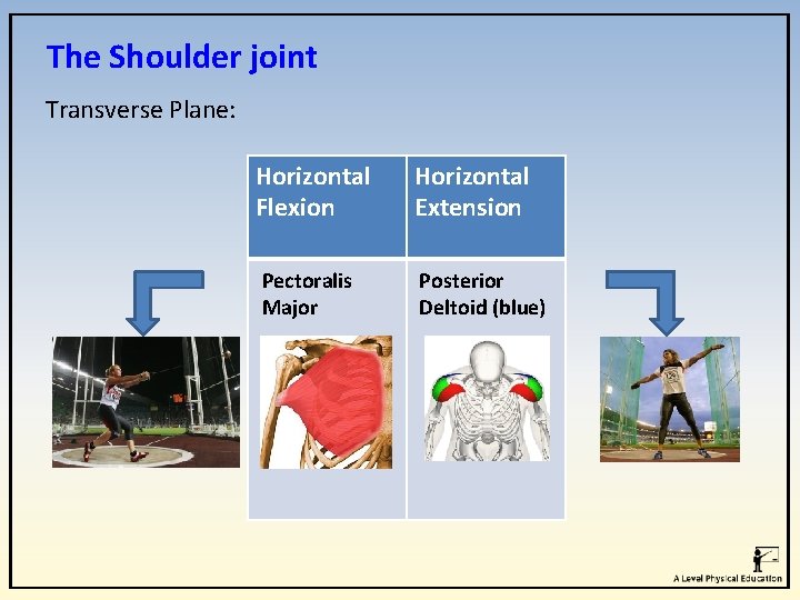
Movement analysis Horizontal flexion: Movement of the arm across the body in the horizontal (transverse) plane.
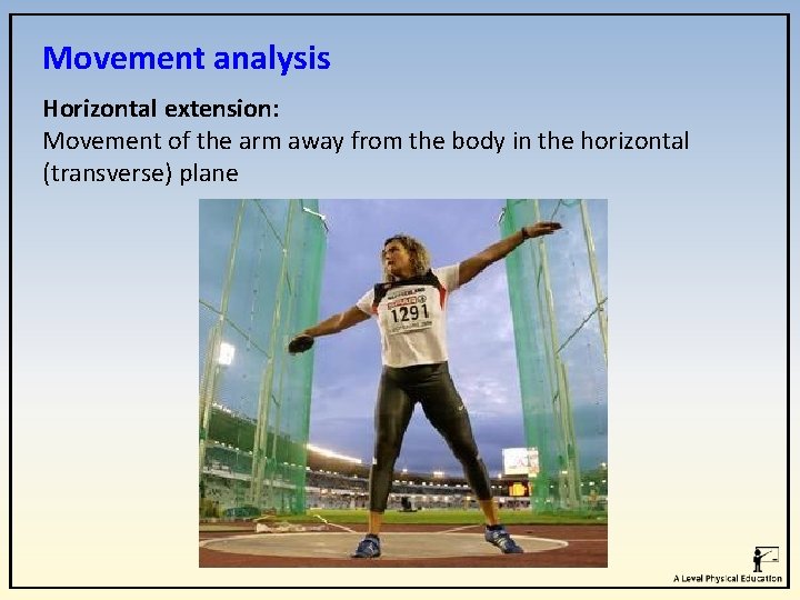
Movement analysis Horizontal extension: Movement of the arm away from the body in the horizontal (transverse) plane
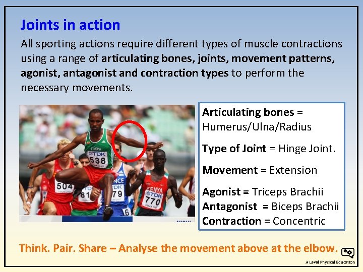
Joints in action All sporting actions require different types of muscle contractions using a range of articulating bones, joints, movement patterns, agonist, antagonist and contraction types to perform the necessary movements. Articulating bones = Humerus/Ulna/Radius Type of Joint = Hinge Joint. Movement = Extension Agonist = Triceps Brachii Antagonist = Biceps Brachii Contraction = Concentric Think. Pair. Share – Analyse the movement above at the elbow.
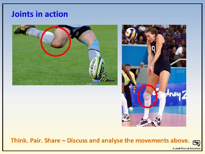
Joints in action Think. Pair. Share – Discuss and analyse the movements above.
The Shoulder joint The shoulder is a ball and socket joint where the head of the humerus fits into a cavity on the scapula. This type of joint allows the most movement. Its structure also makes it one of the least stable joints, so it is heavily reliant on ligaments and muscles to increase its stability.
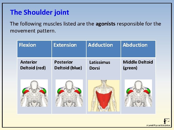
The Shoulder joint The following muscles listed are the agonists responsible for the movement pattern. Flexion Extension Adduction Abduction Anterior Deltoid (red) Posterior Deltoid (blue) Latissimus Dorsi Middle Deltoid (green)
The Shoulder joint Transverse Plane: Horizontal Flexion Horizontal Extension Pectoralis Major Posterior Deltoid (blue)
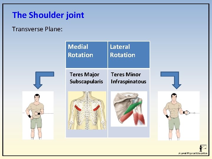
The Shoulder joint Transverse Plane: Medial Rotation Teres Major Subscapularis Lateral Rotation Teres Minor Infraspinatous
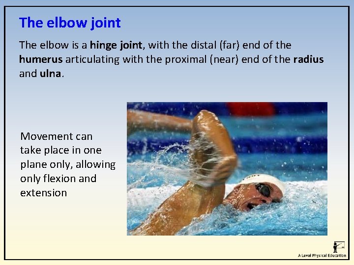
The elbow joint The elbow is a hinge joint, with the distal (far) end of the humerus articulating with the proximal (near) end of the radius and ulna. Movement can take place in one plane only, allowing only flexion and extension
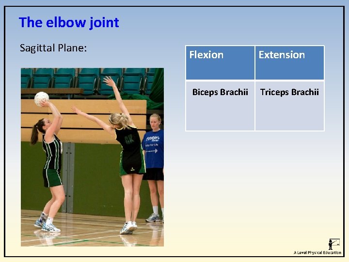
The elbow joint Sagittal Plane: Flexion Biceps Brachii Extension Triceps Brachii
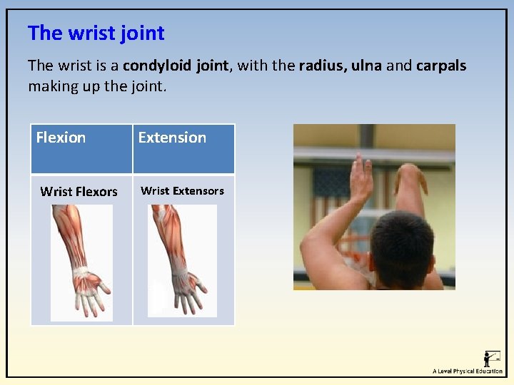
The wrist joint The wrist is a condyloid joint, with the radius, ulna and carpals making up the joint. Flexion Wrist Flexors Extension Wrist Extensors
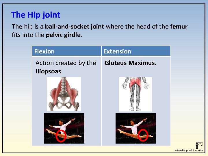
The Hip joint The hip is a ball-and-socket joint where the head of the femur fits into the pelvic girdle. Flexion Extension Action created by the Gluteus Maximus. Iliopsoas.
The Hip joint Adduction Adductor longus Adductor Brevis Adductor Magnus Abduction Gluteus Maximus Gluteus Minimus Gluteus Medius
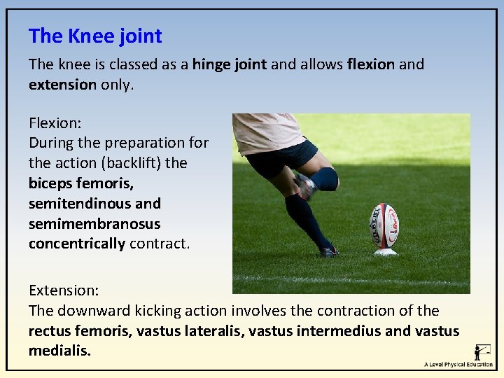
The Knee joint The knee is classed as a hinge joint and allows flexion and extension only. Flexion: During the preparation for the action (backlift) the biceps femoris, semitendinous and semimembranosus concentrically contract. Extension: The downward kicking action involves the contraction of the rectus femoris, vastus lateralis, vastus intermedius and vastus medialis.
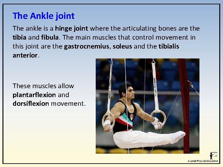
The Ankle joint The ankle is a hinge joint where the articulating bones are the tibia and fibula. The main muscles that control movement in this joint are the gastrocnemius, soleus and the tibialis anterior. These muscles allow plantarflexion and dorsiflexion movement.
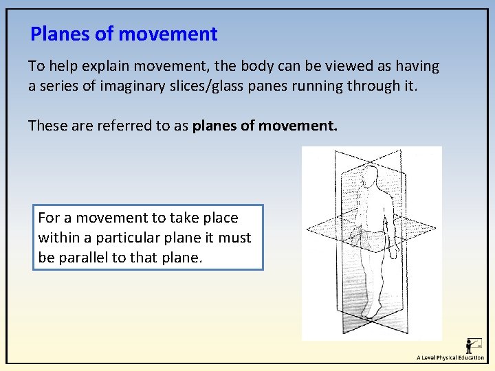
Planes of movement To help explain movement, the body can be viewed as having a series of imaginary slices/glass panes running through it. These are referred to as planes of movement. For a movement to take place within a particular plane it must be parallel to that plane.
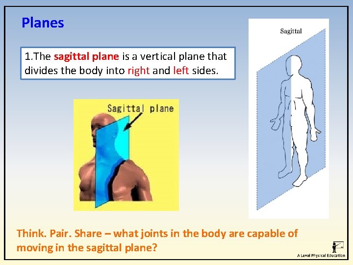
Planes 1. The sagittal plane is a vertical plane that divides the body into right and left sides. Think. Pair. Share – what joints in the body are capable of moving in the sagittal plane?
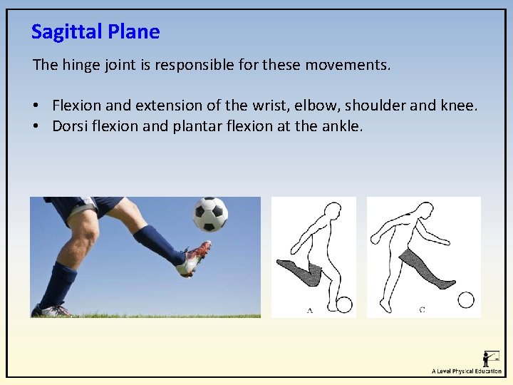
Sagittal Plane The hinge joint is responsible for these movements. • Flexion and extension of the wrist, elbow, shoulder and knee. • Dorsi flexion and plantar flexion at the ankle.
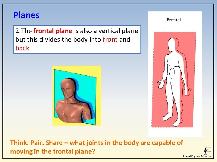
Planes 2. The frontal plane is also a vertical plane but this divides the body into front and back. Think. Pair. Share – what joints in the body are capable of moving in the frontal plane?
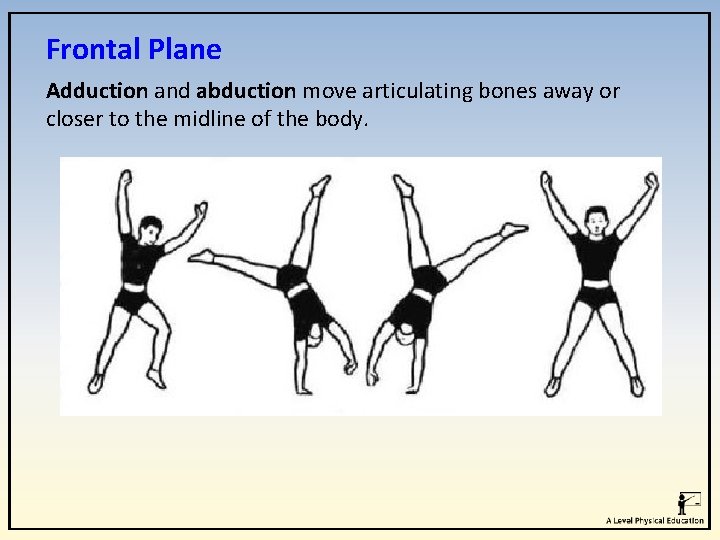
Frontal Plane Adduction and abduction move articulating bones away or closer to the midline of the body.
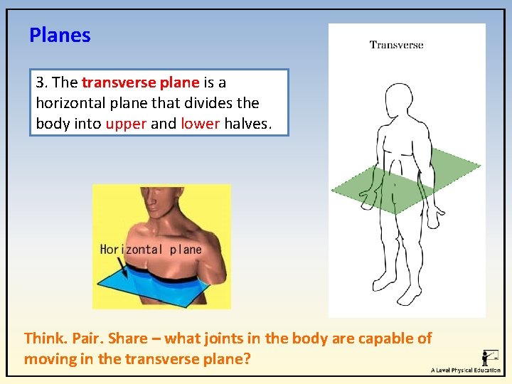
Planes 3. The transverse plane is a horizontal plane that divides the body into upper and lower halves. Think. Pair. Share – what joints in the body are capable of moving in the transverse plane?
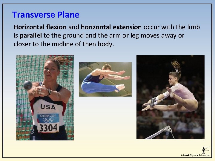
Transverse Plane Horizontal flexion and horizontal extension occur with the limb is parallel to the ground and the arm or leg moves away or closer to the midline of then body.
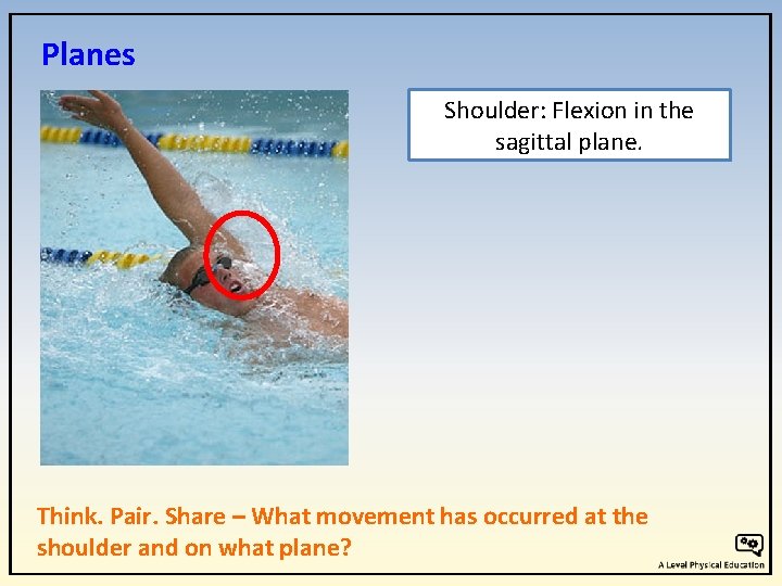
Planes Shoulder: Flexion in the sagittal plane. Think. Pair. Share – What movement has occurred at the shoulder and on what plane?
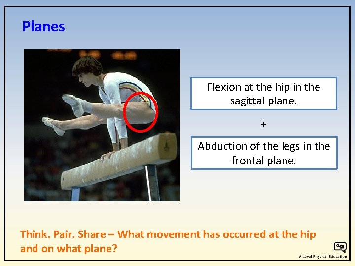
Planes Flexion at the hip in the sagittal plane. + Abduction of the legs in the frontal plane. Think. Pair. Share – What movement has occurred at the hip and on what plane?
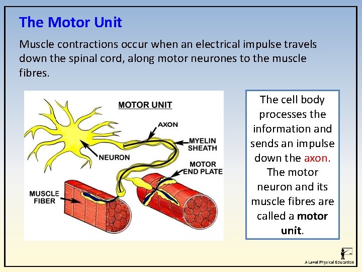
The Motor Unit Muscle contractions occur when an electrical impulse travels down the spinal cord, along motor neurones to the muscle fibres. The cell body processes the information and sends an impulse down the axon. The motor neuron and its muscle fibres are called a motor unit.
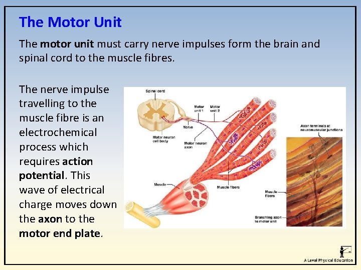
The Motor Unit The motor unit must carry nerve impulses form the brain and spinal cord to the muscle fibres. The nerve impulse travelling to the muscle fibre is an electrochemical process which requires action potential. This wave of electrical charge moves down the axon to the motor end plate.
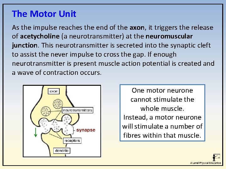
The Motor Unit As the impulse reaches the end of the axon, it triggers the release of acetycholine (a neurotransmitter) at the neuromuscular junction. This neurotransmitter is secreted into the synaptic cleft to assist the never impulse to cross the gap. If enough neurotransmitter is present muscle action potential is created and a wave of contraction occurs. One motor neurone cannot stimulate the whole muscle. Instead, a motor neurone will stimulate a number of fibres within that muscle.
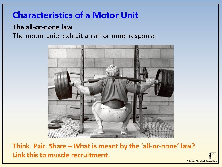
Characteristics of a Motor Unit The all-or-none law The motor units exhibit an all-or-none response. Think. Pair. Share – What is meant by the ‘all-or-none’ law? Link this to muscle recruitment.
Characteristics of a Motor Unit A minimum amount of stimulation is required to start a muscle contraction. If an impulse is strong enough then all the muscle fibres in a motor unit will contract. However, if the impulse is less than the threshold required then no muscle action will occur.
Muscle fibre types Watch me What is the difference between fast twitch and slow twitch muscle fibres?
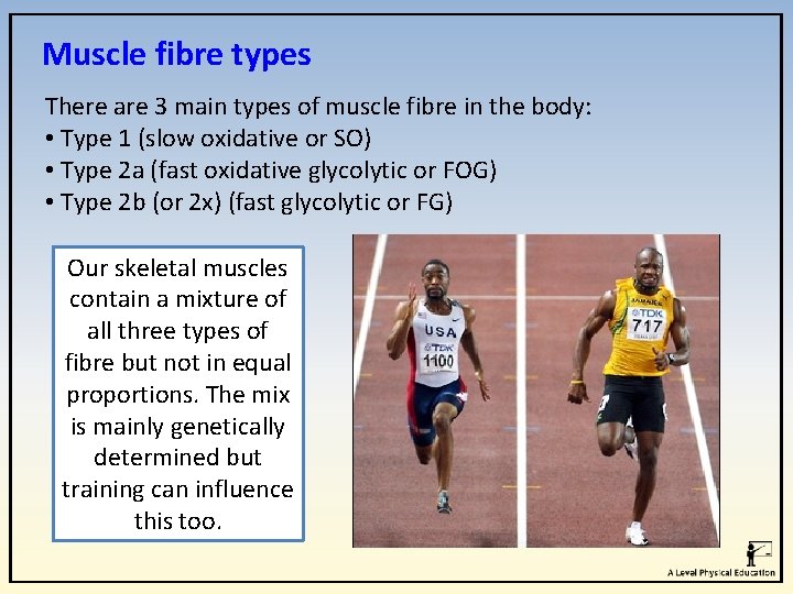
Muscle fibre types There are 3 main types of muscle fibre in the body: • Type 1 (slow oxidative or SO) • Type 2 a (fast oxidative glycolytic or FOG) • Type 2 b (or 2 x) (fast glycolytic or FG) Our skeletal muscles contain a mixture of all three types of fibre but not in equal proportions. The mix is mainly genetically determined but training can influence this too.
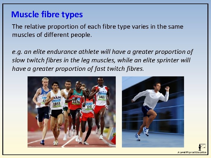
Muscle fibre types The relative proportion of each fibre type varies in the same muscles of different people. e. g. an elite endurance athlete will have a greater proportion of slow twitch fibres in the leg muscles, while an elite sprinter will have a greater proportion of fast twitch fibres.
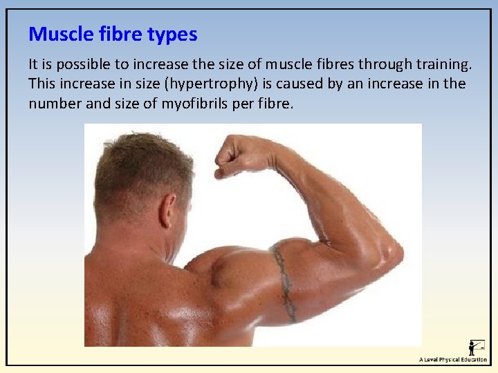
Muscle fibre types It is possible to increase the size of muscle fibres through training. This increase in size (hypertrophy) is caused by an increase in the number and size of myofibrils per fibre.
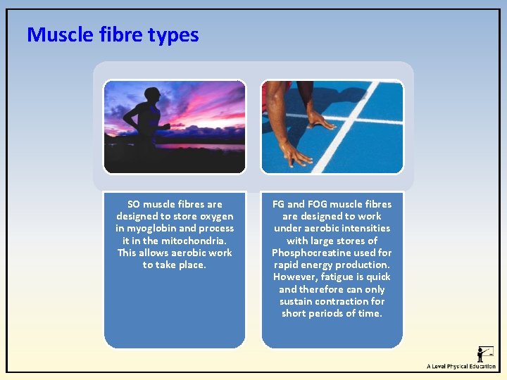
Muscle fibre types SO muscle fibres are designed to store oxygen in myoglobin and process it in the mitochondria. This allows aerobic work to take place. FG and FOG muscle fibres are designed to work under aerobic intensities with large stores of Phosphocreatine used for rapid energy production. However, fatigue is quick and therefore can only sustain contraction for short periods of time.
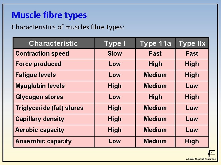
Muscle fibre types Characteristics of muscles fibre types: Characteristic Type I Type 11 a Type IIx Contraction speed Slow Fast Force produced Low High Fatigue levels Low Medium High Myoglobin levels High Medium Low Glycogen stores Low High Triglyceride (fat) stores High Medium Low Capillary density High Medium Low Aerobic capacity High Medium Low Anaerobic capacity Low Medium High
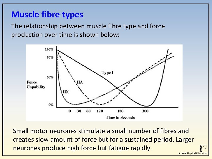
Muscle fibre types The relationship between muscle fibre type and force production over time is shown below: Small motor neurones stimulate a small number of fibres and creates slow amount of force but for a sustained period. Larger neurones produce high force but fatigue rapidly.
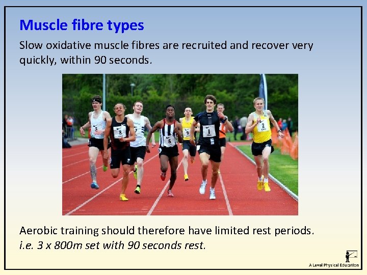
Muscle fibre types Slow oxidative muscle fibres are recruited and recover very quickly, within 90 seconds. Aerobic training should therefore have limited rest periods. i. e. 3 x 800 m set with 90 seconds rest.
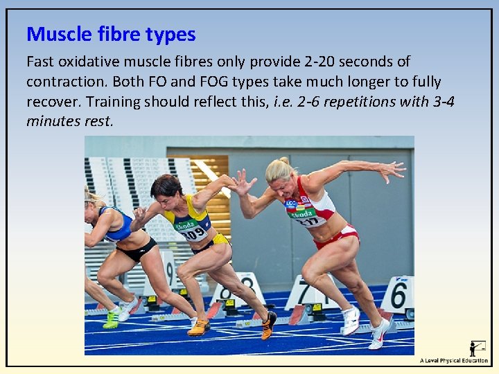
Muscle fibre types Fast oxidative muscle fibres only provide 2 -20 seconds of contraction. Both FO and FOG types take much longer to fully recover. Training should reflect this, i. e. 2 -6 repetitions with 3 -4 minutes rest.
Apply it! What has stuck with you? Describe the 3 planes of movement. Explain the movement possible at the shoulder and articulating bones, muscle actions and main agonist? Skeletal and muscular system Highlight the difference between Isometric and Isotonic muscle contractions Describe the role of motor units in the muscle contractions process.
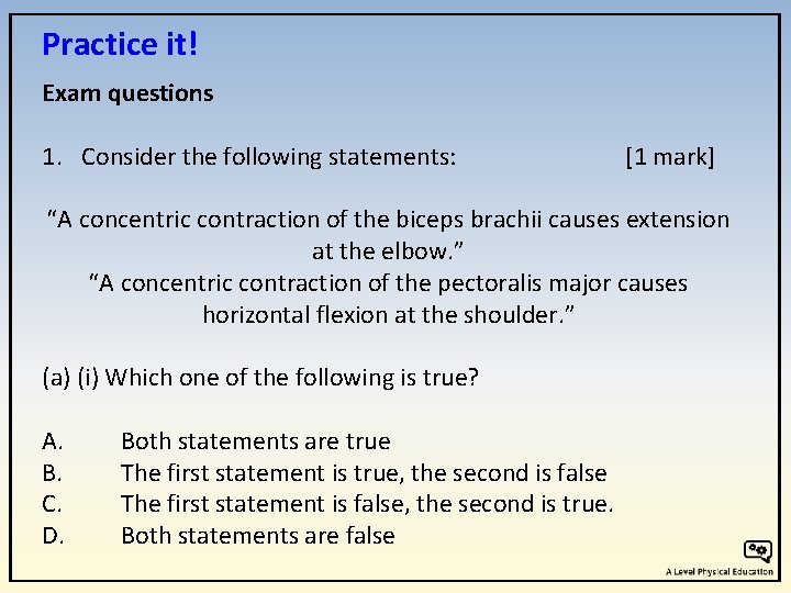
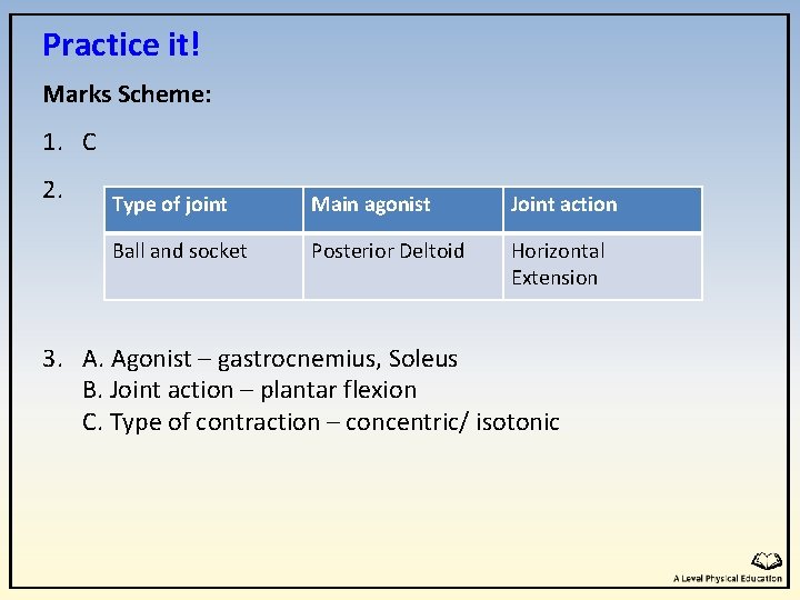
Practice it! Exam questions 1. Consider the following statements: [1 mark] “A concentric contraction of the biceps brachii causes extension at the elbow. ” “A concentric contraction of the pectoralis major causes horizontal flexion at the shoulder. ” (a) (i) Which one of the following is true? A. B. C. D. Both statements are true The first statement is true, the second is false The first statement is false, the second is true. Both statements are false
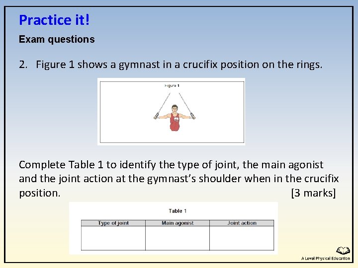
Practice it! Exam questions 2. Figure 1 shows a gymnast in a crucifix position on the rings. Complete Table 1 to identify the type of joint, the main agonist and the joint action at the gymnast’s shoulder when in the crucifix position. [3 marks]
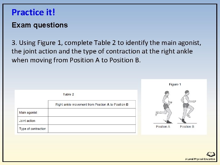
Practice it! Exam questions 3. Using Figure 1, complete Table 2 to identify the main agonist, the joint action and the type of contraction at the right ankle when moving from Position A to Position B.
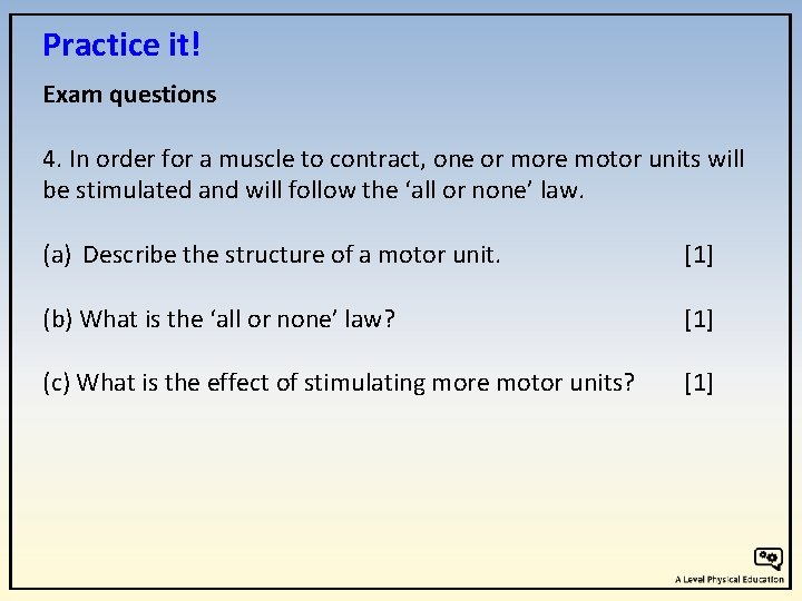
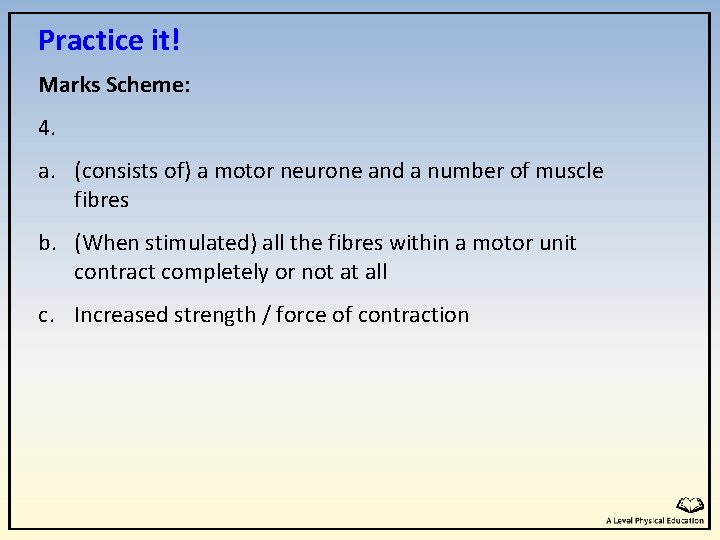
Practice it! Exam questions 4. In order for a muscle to contract, one or more motor units will be stimulated and will follow the ‘all or none’ law. (a) Describe the structure of a motor unit. [1] (b) What is the ‘all or none’ law? [1] (c) What is the effect of stimulating more motor units? [1]
Practice it! Marks Scheme: 1. C 2. Type of joint Ball and socket Main agonist Joint action Posterior Deltoid Horizontal Extension 3. A. Agonist – gastrocnemius, Soleus B. Joint action – plantar flexion C. Type of contraction – concentric/ isotonic
Practice it! Marks Scheme: 4. a. (consists of) a motor neurone and a number of muscle fibres b. (When stimulated) all the fibres within a motor unit contract completely or not at all c. Increased strength / force of contraction