
Chapter 10 Part A The Muscular System Annie






- Slides: 59
Chapter 10 Part A The Muscular System © Annie Leibovitz/Contact Press Images © 2016 Pearson Education, Inc. Power. Point® Lecture Slides prepared by Karen Dunbar Kareiva Ivy Tech Community College
I. Muscle Actions and Interactions A. This chapter focuses on skeletal muscle and looks at: 1. How muscles interact to bring about movement 2. Criteria for naming muscles 3. Identifying whole muscles, their origins and insertions, and their actions B. FYI 1. Muscles can only pull; never push 2. What one muscle group “does, ” another “undoes” © 2016 Pearson Education, Inc.
C. Functional groups 1. Prime mover (agonist) a. Major responsibility for producing specific movement 2. Antagonist a. Opposes or reverses particular movement b. Prime mover and antagonist are located on opposite sides of joint across which they act © 2016 Pearson Education, Inc.
3. Synergist helps prime movers a. Adds extra force to same movement b. Reduces undesirable or unnecessary movement 4. Fixator a. Synergist that immobilizes bone or muscle’s origin b. Gives prime mover stable base on which to act © 2016 Pearson Education, Inc.
D. Same muscle may be: 1. Prime mover of one movement 2. Antagonist for different movement 3. Synergist for third movement © 2016 Pearson Education, Inc.
II. Naming Skeletal Muscles A. Criteria Example Muscle Location – bone or body region Temporalis (over temporal bone) Muscle Shape Deltoid (Deltoid=Triangle) Muscle Size Maximus (Large) Minimus (Small) Longus (long) Direction of fibers or fascicles Rectus (Parallel) Transversus (Perpendicular) Oblique (at an angle) © 2016 Pearson Education, Inc.
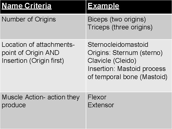
Name Criteria Example Number of Origins Biceps (two origins) Triceps (three origins) Location of attachmentspoint of Origin AND Insertion (Origin first) Sternocleidomastoid Origins: Sternum (sterno) Clavicle (Cleido) Insertion: Mastoid process of temporal bone (Mastoid) Muscle Action- action they produce Flexor Extensor © 2016 Pearson Education, Inc.
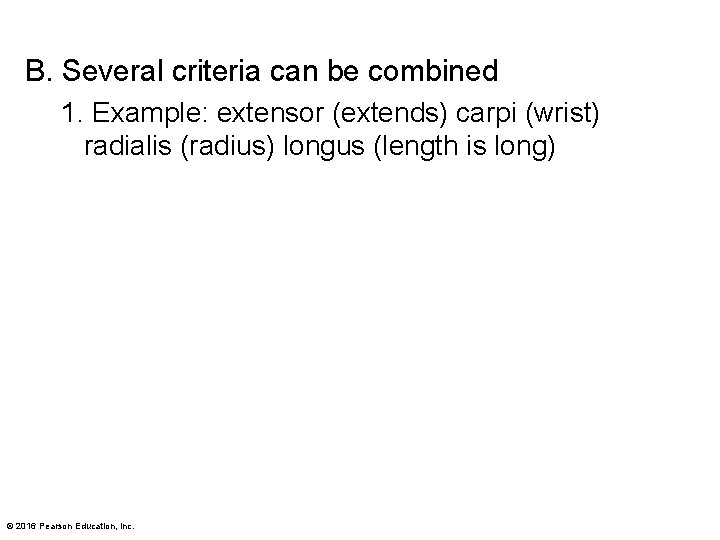
B. Several criteria can be combined 1. Example: extensor (extends) carpi (wrist) radialis (radius) longus (length is long) © 2016 Pearson Education, Inc.
Bell Ringer 10. 01 Which functional group immobilizes a bone? 10. 02 By which criteria is the Biceps Brachii named? 10. 03 Complete the following sentence using the proper Movement Terms (flashcards) Proper jumping jacks require you to begin with your arms straight down my your side. Next you will ___ your arms until they are straight out and then ___ your arms until they are raised above your head. © 2016 Pearson Education, Inc.
III. Fascicle Arrangements A. Fascicle arrangements vary, resulting in muscles with different shapes and functional capabilities 1. Fascicles determine muscle’s range of motion a. Amount of movement when muscle shortens 2. Fascicles determine muscle’s power a. Power depends on number of muscle fibers © 2016 Pearson Education, Inc.
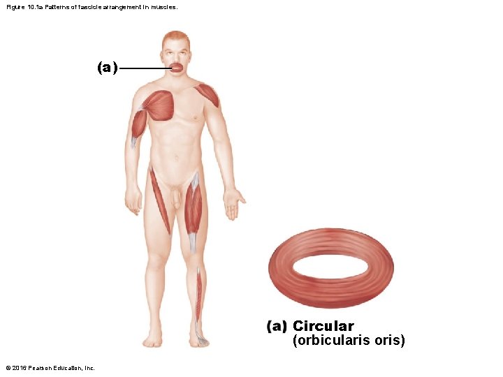
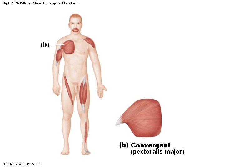
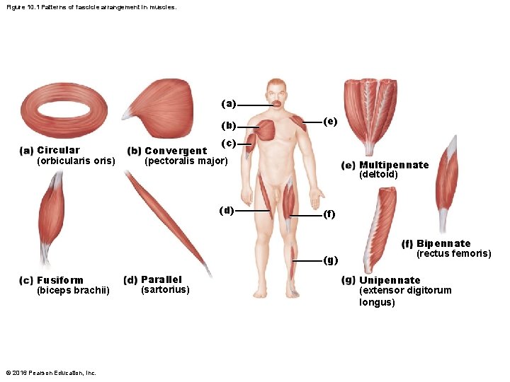
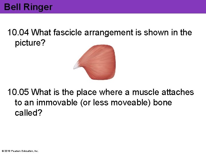
B. The most common patterns of arrangement 1. Circular: fascicles arranged in concentric rings (example: orbicularis oris) 2. Convergent: broad origin; fascicles converge toward single tendon insertion (example: pectoralis major) © 2016 Pearson Education, Inc.
Figure 10. 1 a Patterns of fascicle arrangement in muscles. (a) Circular (orbicularis oris) © 2016 Pearson Education, Inc.
Figure 10. 1 b Patterns of fascicle arrangement in muscles. (b) Convergent (pectoralis major) © 2016 Pearson Education, Inc.
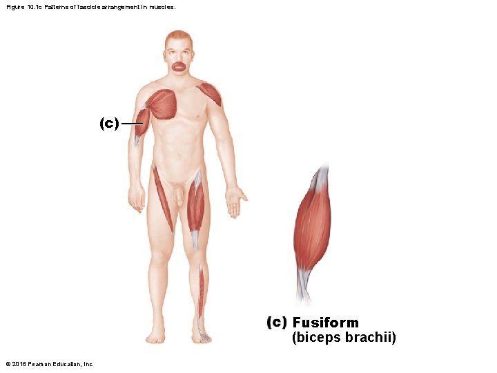
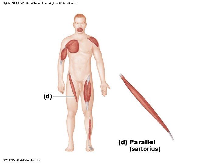
3. Fusiform: spindle-shaped muscles with parallel fibers (example: biceps brachii) 4. Parallel: fascicles parallel to long axis of straplike muscle (example: sartorius) © 2016 Pearson Education, Inc.
Figure 10. 1 c Patterns of fascicle arrangement in muscles. (c) Fusiform (biceps brachii) © 2016 Pearson Education, Inc.
Figure 10. 1 d Patterns of fascicle arrangement in muscles. (d) Parallel (sartorius) © 2016 Pearson Education, Inc.
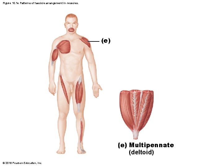
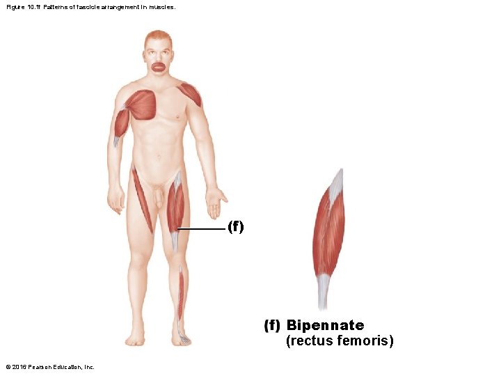
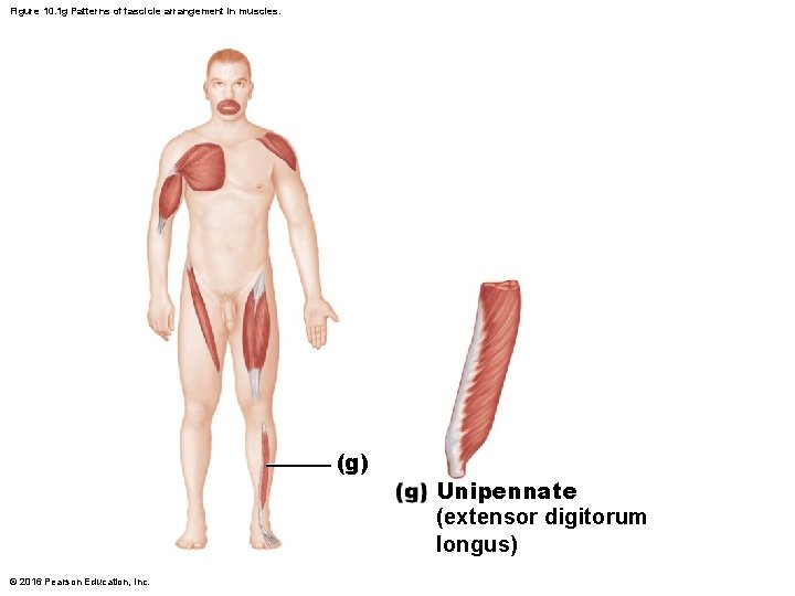
5. Pennate: short fascicles attach obliquely to central tendon running length of muscle (Pennate means “feather”) a. Three forms i. Unipennate: fascicles attach only to one side of tendon (example: extensor digitorum longus) ii. Bipennate: fascicles insert from opposite sides of tendon (example: rectus femoris) iii. Multipennate: appears as feathers inserting into one tendon (example: deltoid) © 2016 Pearson Education, Inc.
Figure 10. 1 e Patterns of fascicle arrangement in muscles. (e) Multipennate (deltoid) © 2016 Pearson Education, Inc.
Figure 10. 1 f Patterns of fascicle arrangement in muscles. (f) Bipennate (rectus femoris) © 2016 Pearson Education, Inc.
Figure 10. 1 g Patterns of fascicle arrangement in muscles. (g) © 2016 Pearson Education, Inc. Unipennate (extensor digitorum longus)
Figure 10. 1 Patterns of fascicle arrangement in muscles. (a) (b) Circular (orbicularis oris) (e) (c) Convergent (pectoralis major) (d) Multipennate (deltoid) (f) (g) Fusiform (biceps brachii) © 2016 Pearson Education, Inc. Parallel (sartorius) Bipennate (rectus femoris) Unipennate (extensor digitorum longus)
Bell Ringer 10. 04 What fascicle arrangement is shown in the picture? 10. 05 What is the place where a muscle attaches to an immovable (or less moveable) bone called? © 2016 Pearson Education, Inc.
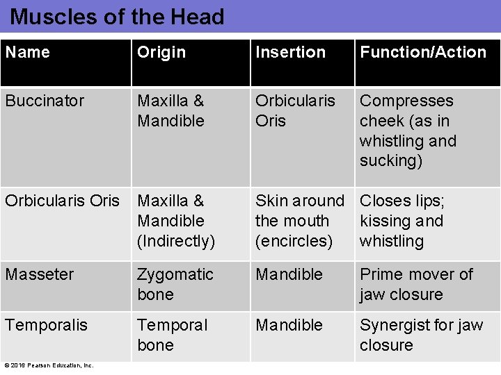
Muscles of the Head Name Origin Insertion Function/Action Buccinator Maxilla & Mandible Orbicularis Oris Compresses cheek (as in whistling and sucking) Orbicularis Oris Maxilla & Mandible (Indirectly) Skin around Closes lips; the mouth kissing and (encircles) whistling Masseter Zygomatic bone Mandible Prime mover of jaw closure Temporalis Temporal bone Mandible Synergist for jaw closure © 2016 Pearson Education, Inc.
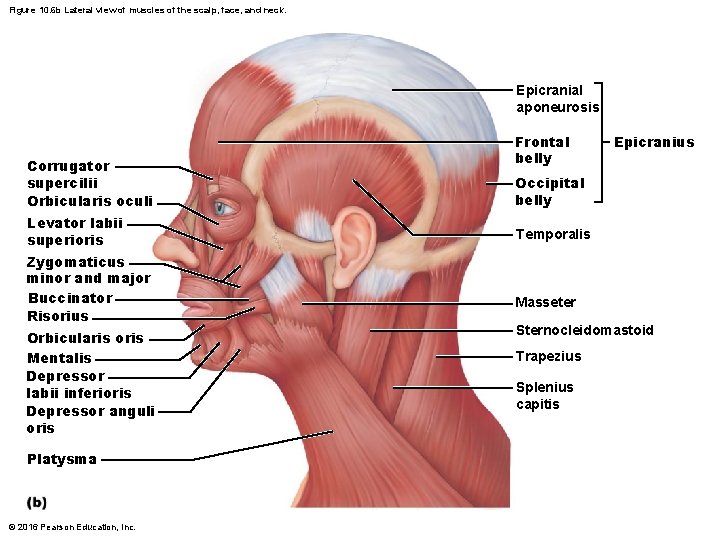
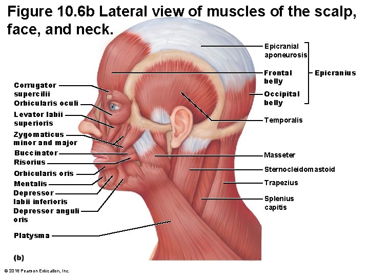
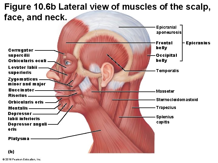
Figure 10. 6 b Lateral view of muscles of the scalp, face, and neck. Epicranial aponeurosis Corrugator supercilii Orbicularis oculi Levator labii superioris Zygomaticus minor and major Buccinator Risorius Orbicularis oris Mentalis Depressor labii inferioris Depressor anguli oris Platysma © 2016 Pearson Education, Inc. Frontal belly Epicranius Occipital belly Temporalis Masseter Sternocleidomastoid Trapezius Splenius capitis
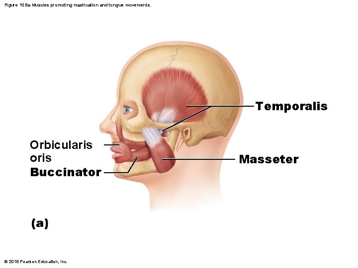
Figure 10. 8 a Muscles promoting mastication and tongue movements. Temporalis Orbicularis oris Buccinator © 2016 Pearson Education, Inc. Masseter
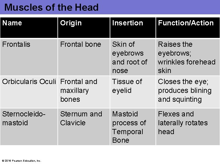
Muscles of the Head Name Origin Insertion Function/Action Frontalis Frontal bone Skin of eyebrows and root of nose Raises the eyebrows; wrinkles forehead skin Orbicularis Oculi Frontal and maxillary bones Tissue of eyelid Closes the eye; produces blining and squinting Sternocleidomastoid Mastoid process of Temporal Bone Flexes and laterally rotates head © 2016 Pearson Education, Inc. Sternum and Clavicle
Figure 10. 6 b Lateral view of muscles of the scalp, face, and neck. Epicranial aponeurosis Corrugator supercilii Orbicularis oculi Levator labii superioris Zygomaticus minor and major Buccinator Risorius Orbicularis oris Mentalis Depressor labii inferioris Depressor anguli oris Platysma © 2016 Pearson Education, Inc. Frontal belly Epicranius Occipital belly Temporalis Masseter Sternocleidomastoid Trapezius Splenius capitis
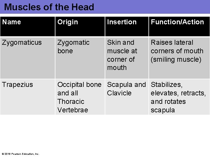
Muscles of the Head Name Origin Insertion Function/Action Zygomaticus Zygomatic bone Skin and muscle at corner of mouth Raises lateral corners of mouth (smiling muscle) Trapezius Occipital bone Scapula and all Clavicle Thoracic Vertebrae © 2016 Pearson Education, Inc. Stabilizes, elevates, retracts, and rotates scapula
Figure 10. 6 b Lateral view of muscles of the scalp, face, and neck. Epicranial aponeurosis Corrugator supercilii Orbicularis oculi Levator labii superioris Zygomaticus minor and major Buccinator Risorius Orbicularis oris Mentalis Depressor labii inferioris Depressor anguli oris Platysma © 2016 Pearson Education, Inc. Frontal belly Epicranius Occipital belly Temporalis Masseter Sternocleidomastoid Trapezius Splenius capitis
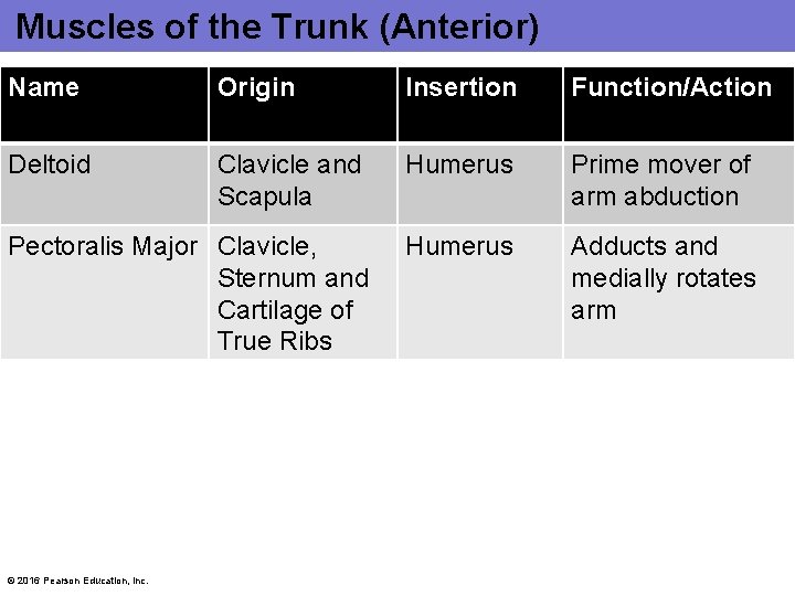
Muscles of the Trunk (Anterior) Name Origin Insertion Function/Action Deltoid Clavicle and Scapula Humerus Prime mover of arm abduction Pectoralis Major Clavicle, Sternum and Cartilage of True Ribs Humerus Adducts and medially rotates arm © 2016 Pearson Education, Inc.
Figure 10. 14 a Superficial muscles of the thorax and shoulder acting on the scapula and arm. Sternocleidomastoid Subclavius Clavicle Deltoid Subscapularis Pectoralis minor Pectoralis major Sternum Biceps brachii © 2016 Pearson Education, Inc. Coracobrachialis Serratus anterior Humerus
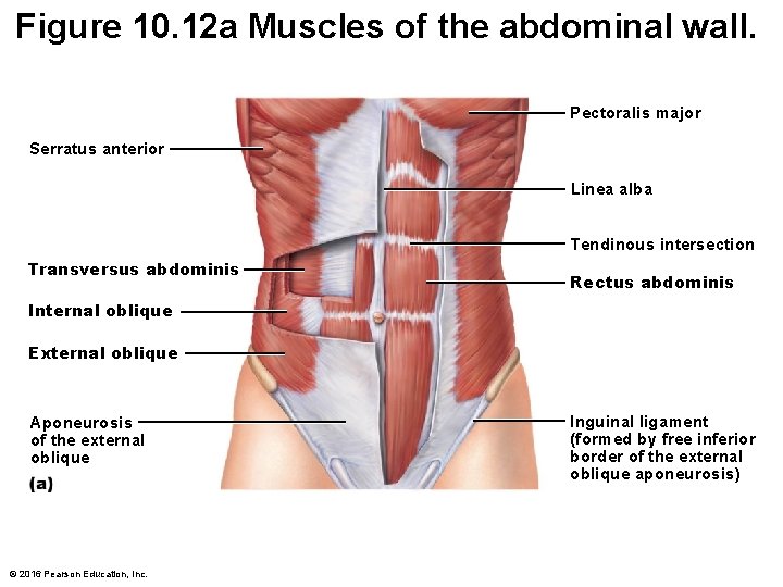
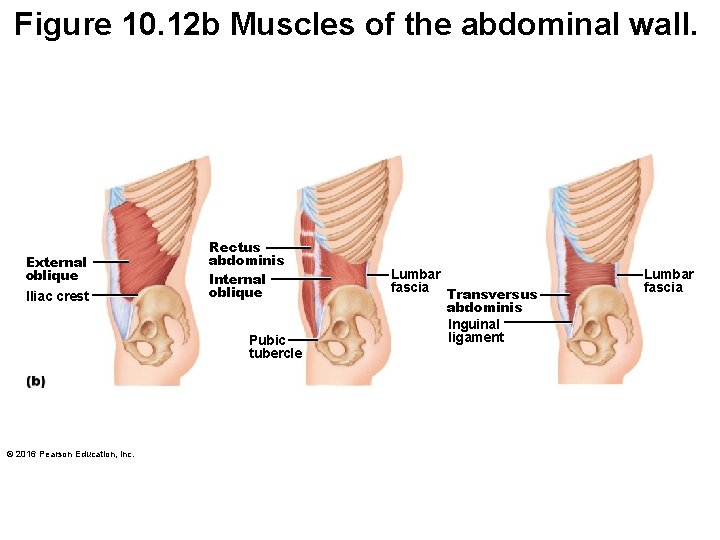
Muscles of the Trunk (Anterior) Name Origin Insertion Function/Action External Oblique Outer surfaces Pubic and of lower 8 ribs iliac crest Flex vertebral column and compress abdominal wall; rotates trunk and flexes laterally Rectus Abdominis Pubic Crest Flex and rotate lumbar region of vertebral column Transversus Abdominis Cartilages of last 6 ribs and iliac crest © 2016 Pearson Education, Inc. Xiphoid Process and cartilages of ribs 5 -7 Pubic crest Combresses abdonimal contents
Figure 10. 12 a Muscles of the abdominal wall. Pectoralis major Serratus anterior Linea alba Tendinous intersection Transversus abdominis Rectus abdominis Internal oblique External oblique Aponeurosis of the external oblique © 2016 Pearson Education, Inc. Inguinal ligament (formed by free inferior border of the external oblique aponeurosis)
Figure 10. 12 b Muscles of the abdominal wall. External oblique IIiac crest Rectus abdominis Internal oblique Pubic tubercle © 2016 Pearson Education, Inc. Lumbar fascia Transversus abdominis Inguinal ligament Lumbar fascia
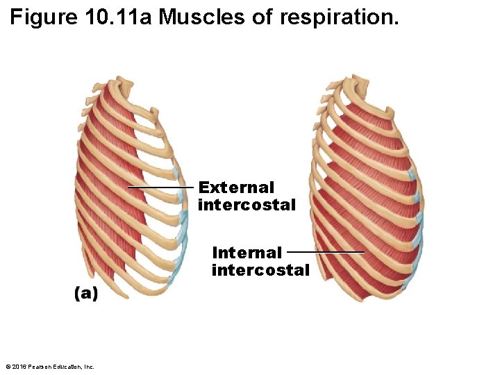
Muscles of the Trunk (Anterior) Name Origin Insertion Function/Action Diaphragm Sternum and rib cage Central tendon Prime mover of inspiration; flattens on contraction Internal Intercostal Superior border of rib below Inferior border of rib above Superior border of rib below Draw ribs together and depress rib cage Pull ribs toward one another to elevate rib cage External Intercostal Internal Oblique Iliac Crest © 2016 Pearson Education, Inc. Pubic Crest Same as external and Last 3 -4 oblique ribs
Figure 10. 11 a Muscles of respiration. External intercostal Internal intercostal © 2016 Pearson Education, Inc.
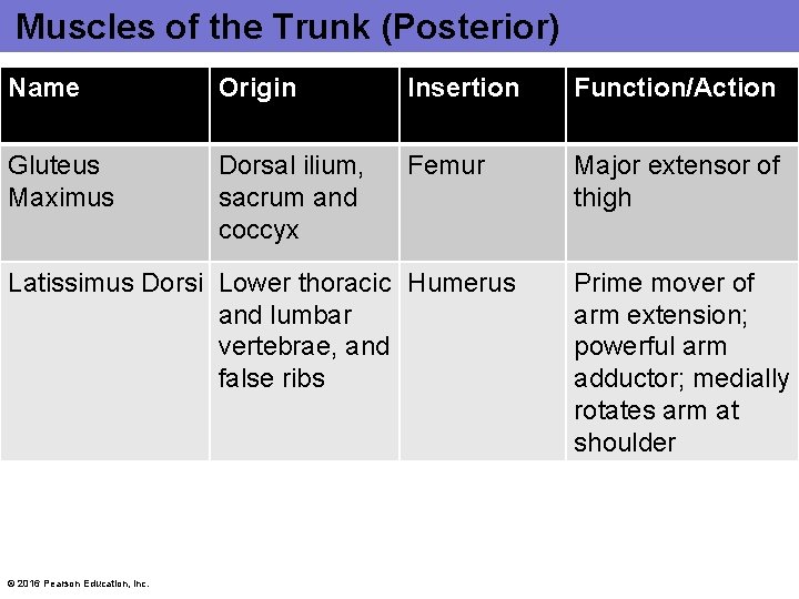
Muscles of the Trunk (Posterior) Name Origin Insertion Function/Action Gluteus Maximus Dorsal ilium, sacrum and coccyx Femur Major extensor of thigh Latissimus Dorsi Lower thoracic Humerus and lumbar vertebrae, and false ribs © 2016 Pearson Education, Inc. Prime mover of arm extension; powerful arm adductor; medially rotates arm at shoulder
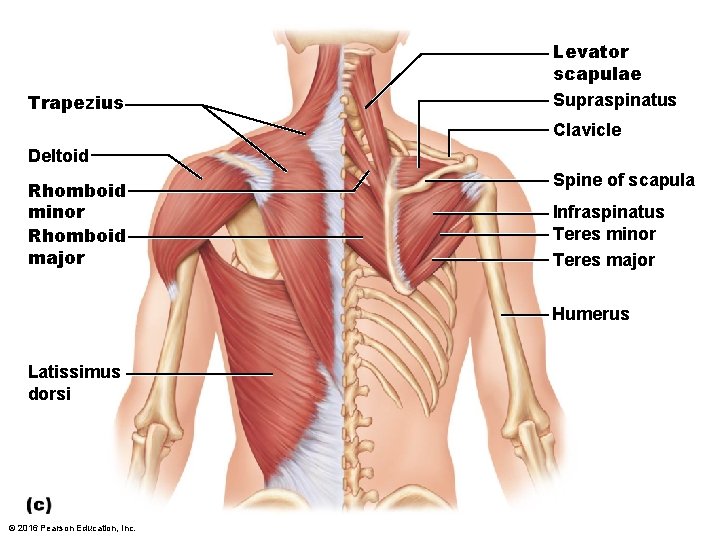
Trapezius Levator scapulae Supraspinatus Clavicle Deltoid Rhomboid minor Rhomboid major Spine of scapula Infraspinatus Teres minor Teres major Humerus Latissimus dorsi © 2016 Pearson Education, Inc.
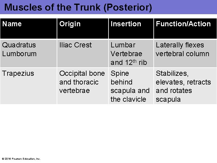
Muscles of the Trunk (Posterior) Name Origin Insertion Function/Action Quadratus Lumborum Iliac Crest Lumbar Vertebrae and 12 th rib Laterally flexes vertebral column Trapezius Occipital bone Spine and thoracic behind vertebrae scapula and the clavicle © 2016 Pearson Education, Inc. Stabilizes, elevates, retracts and rotates scapula
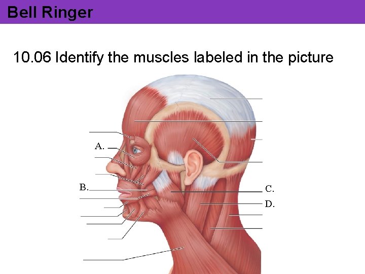
Bell Ringer 10. 06 Identify the muscles labeled in the picture A. B. C. D.
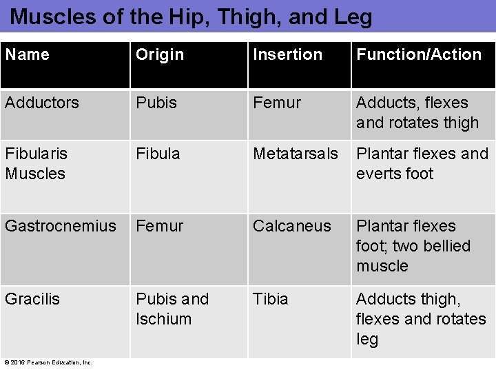
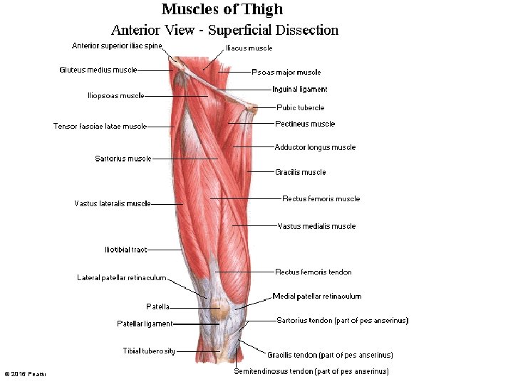
Muscles of the Hip, Thigh, and Leg Name Origin Insertion Function/Action Adductors Pubis Femur Adducts, flexes and rotates thigh Fibularis Muscles Fibula Metatarsals Plantar flexes and everts foot Gastrocnemius Femur Calcaneus Plantar flexes foot; two bellied muscle Gracilis Pubis and Ischium Tibia Adducts thigh, flexes and rotates leg © 2016 Pearson Education, Inc.
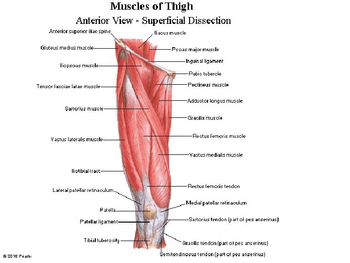
© 2016 Pearson Education, Inc.
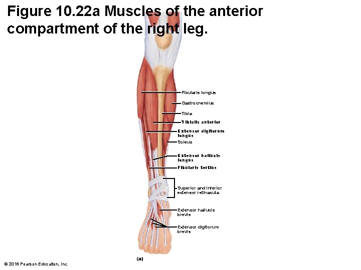
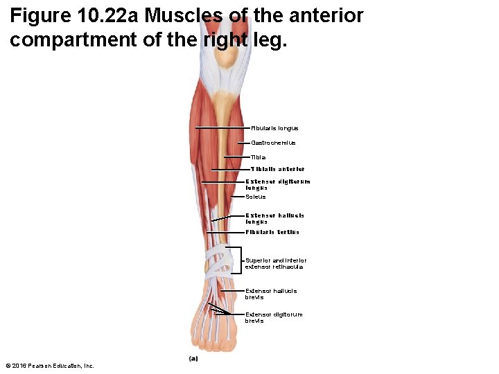
Figure 10. 22 a Muscles of the anterior compartment of the right leg. Fibularis longus Gastrocnemius Tibialis anterior Extensor digitorum longus Soleus Extensor hallucis longus Fibularis tertius Superior and inferior extensor retinacula Extensor hallucis brevis Extensor digitorum brevis © 2016 Pearson Education, Inc.
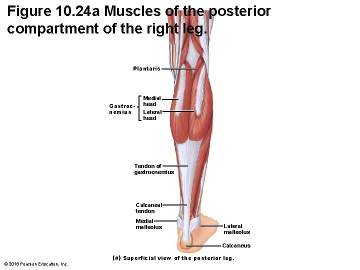
Figure 10. 24 a Muscles of the posterior compartment of the right leg. Plantaris Gastrocnemius Medial head Lateral head Tendon of gastrocnemius Calcaneal tendon Medial malleolus Lateral malleolus Calcaneus Superficial view of the posterior leg. © 2016 Pearson Education, Inc.
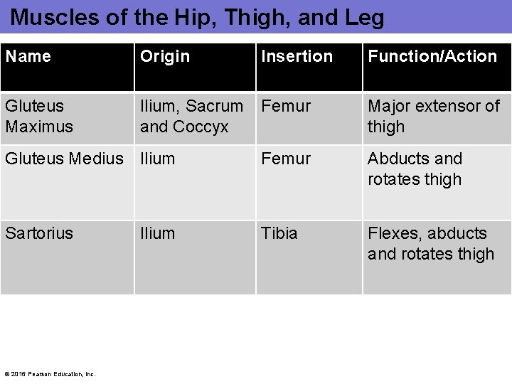
Muscles of the Hip, Thigh, and Leg Name Origin Insertion Gluteus Maximus Ilium, Sacrum Femur and Coccyx Function/Action Major extensor of thigh Gluteus Medius Ilium Femur Abducts and rotates thigh Sartorius Tibia Flexes, abducts and rotates thigh © 2016 Pearson Education, Inc. Ilium
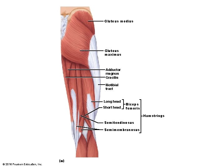
Gluteus medius Gluteus maximus Adductor magnus Gracilis Iliotibial tract Long head Short head Biceps femoris Hamstrings Semitendinosus Semimembranosus © 2016 Pearson Education, Inc.
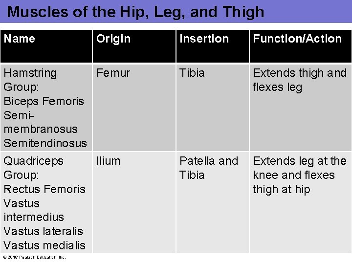
Muscles of the Hip, Leg, and Thigh Name Origin Hamstring Femur Group: Biceps Femoris Semimembranosus Semitendinosus Quadriceps Ilium Group: Rectus Femoris Vastus intermedius Vastus lateralis Vastus medialis © 2016 Pearson Education, Inc. Insertion Function/Action Tibia Extends thigh and flexes leg Patella and Tibia Extends leg at the knee and flexes thigh at hip
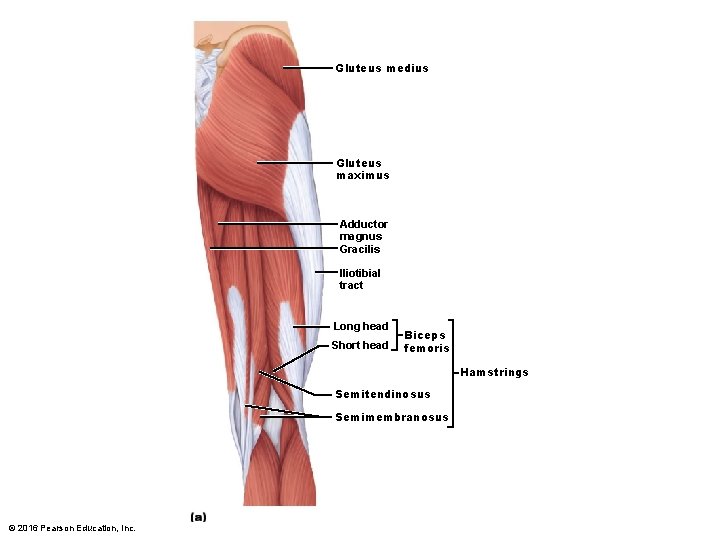
Gluteus medius Gluteus maximus Adductor magnus Gracilis Iliotibial tract Long head Short head Biceps femoris Hamstrings Semitendinosus Semimembranosus © 2016 Pearson Education, Inc.
© 2016 Pearson Education, Inc.
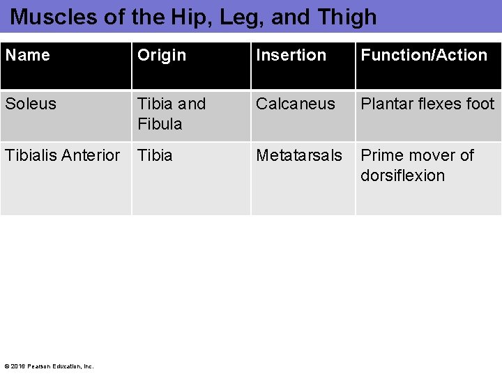
Muscles of the Hip, Leg, and Thigh Name Origin Insertion Function/Action Soleus Tibia and Fibula Calcaneus Plantar flexes foot Tibialis Anterior Tibia Metatarsals Prime mover of dorsiflexion © 2016 Pearson Education, Inc.
Figure 10. 22 a Muscles of the anterior compartment of the right leg. Fibularis longus Gastrocnemius Tibialis anterior Extensor digitorum longus Soleus Extensor hallucis longus Fibularis tertius Superior and inferior extensor retinacula Extensor hallucis brevis Extensor digitorum brevis © 2016 Pearson Education, Inc.
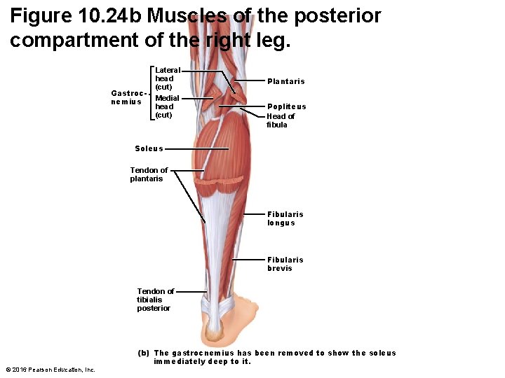
Figure 10. 24 b Muscles of the posterior compartment of the right leg. Gastrocnemius Lateral head (cut) Medial head (cut) Plantaris Popliteus Head of fibula Soleus Tendon of plantaris Fibularis longus Fibularis brevis Tendon of tibialis posterior © 2016 Pearson Education, Inc. The gastrocnemius has been removed to show the soleus immediately deep to it.
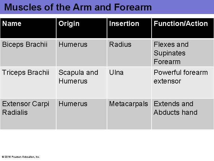
Muscles of the Arm and Forearm Name Origin Insertion Function/Action Biceps Brachii Humerus Radius Flexes and Supinates Forearm Triceps Brachii Scapula and Humerus Ulna Powerful forearm extensor Extensor Carpi Radialis Humerus Metacarpals Extends and Abducts hand © 2016 Pearson Education, Inc.
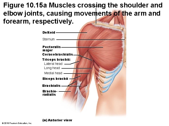
Clavicle Figure 10. 15 a Muscles crossing the shoulder and elbow joints, causing movements of the arm and forearm, respectively. Deltoid Sternum Pectoralis major Coracobrachialis Triceps brachii: Lateral head Long head Medial head Biceps brachii Brachialis Brachioradialis © 2016 Pearson Education, Inc. Anterior view
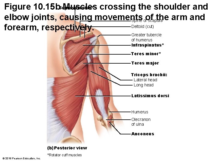
Figure 10. 15 b. Supraspinatus* Muscles crossing the shoulder and elbow joints, causing movements the arm and Spine ofof scapula Deltoid (cut) forearm, respectively. Greater tubercle of humerus Infraspinatus* Teres minor* Teres major Triceps brachii: Lateral head Long head Latissimus dorsi Humerus Olecranon of ulna Anconeus Posterior view © 2016 Pearson Education, Inc. *Rotator cuff muscles
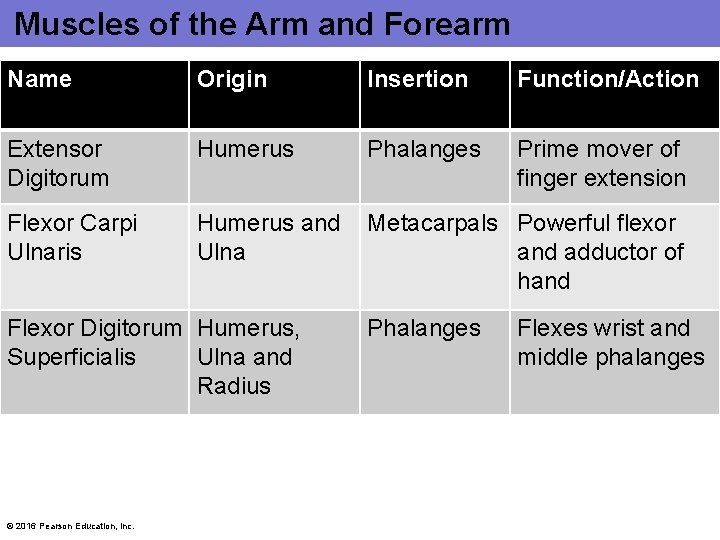
Muscles of the Arm and Forearm Name Origin Insertion Function/Action Extensor Digitorum Humerus Phalanges Prime mover of finger extension Flexor Carpi Ulnaris Humerus and Ulna Metacarpals Powerful flexor and adductor of hand Flexor Digitorum Humerus, Superficialis Ulna and Radius © 2016 Pearson Education, Inc. Phalanges Flexes wrist and middle phalanges
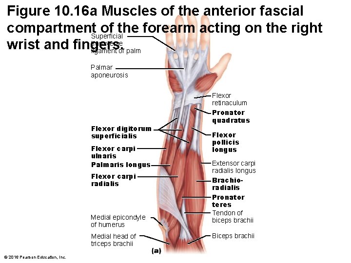
Figure 10. 16 a Muscles of the anterior fascial compartment. Superficial of the forearm acting on the right transverse wrist and fingers. ligament of palm Palmar aponeurosis Flexor digitorum superficialis Flexor carpi ulnaris Palmaris longus Flexor carpi radialis Medial epicondyle of humerus Medial head of triceps brachii © 2016 Pearson Education, Inc. Flexor retinaculum Pronator quadratus Flexor pollicis longus Extensor carpi radialis longus Brachioradialis Pronator teres Tendon of biceps brachii Biceps brachii
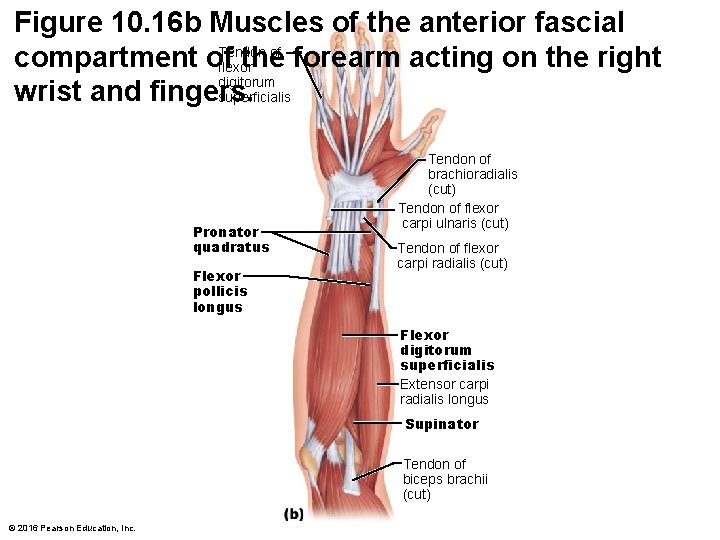
Figure 10. 16 b Muscles of the anterior fascial Tendon of compartment of the forearm acting on the right flexor digitorum wrist and fingers. superficialis Pronator quadratus Flexor pollicis longus Tendon of brachioradialis (cut) Tendon of flexor carpi ulnaris (cut) Tendon of flexor carpi radialis (cut) Flexor digitorum superficialis Extensor carpi radialis longus Supinator Tendon of biceps brachii (cut) © 2016 Pearson Education, Inc.
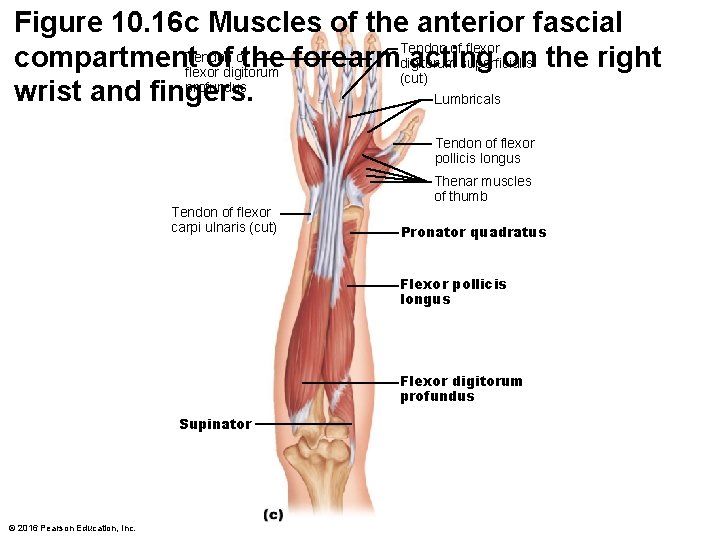
Figure 10. 16 c Muscles of the anterior fascial Tendon of flexor ofthe forearmdigitorum compartment. Tendon of acting on the right superficialis flexor digitorum (cut) profundus wrist and fingers. Lumbricals Tendon of flexor pollicis longus Thenar muscles of thumb Tendon of flexor carpi ulnaris (cut) Pronator quadratus Flexor pollicis longus Flexor digitorum profundus Supinator © 2016 Pearson Education, Inc.