VEINS ASSOCIATED WITH HEART Dr Mujahid Khan Development


- Slides: 28
VEINS ASSOCIATED WITH HEART Dr. Mujahid Khan
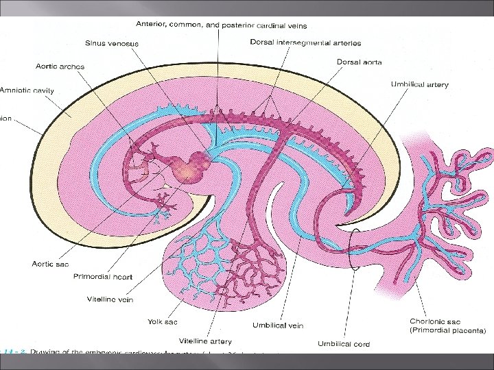
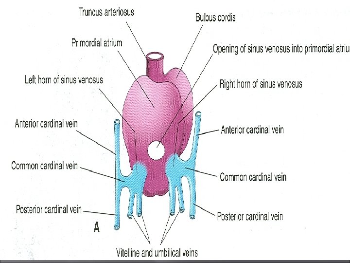
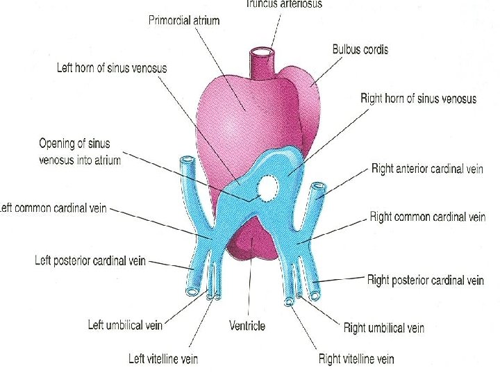
Development of Veins Ø Three paired veins drain into the tubular heart of a four week old embryo Ø Vitelline veins return poorly oxygenated blood from the yolk sac Ø Umbilical veins carry well-oxygenated blood from the primordial placenta Ø Common cardinal veins return poorly oxygenated blood from the body of the embryo
Vitelline Veins Ø The vitelline veins follow the yolk stalk into the embryo Ø Yolk stalk is a narrow tube connecting the yolk sac with the midgut Ø After passing through the septum transversum the vitelline veins enter the venous end of the heart, the sinus venosus
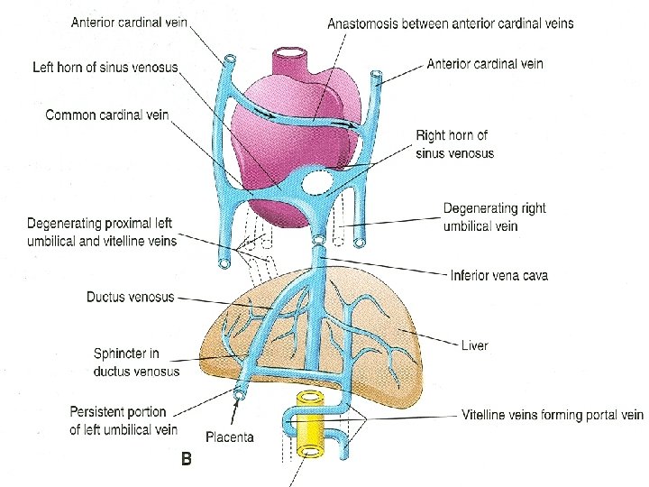
Vitelline Veins Ø As the liver primordium grows into the septum transversum, the hepatic cords anastomose around preexisting endothelium-lined spaces Ø These spaces, the primordia of the hepatic sinusoids later become linked to the vitelline veins Ø The hepatic veins form from the remains of the right vitelline vein in the region of the developing liver Ø The portal vein develops from an anastomotic network formed by the vitelline veins around the duodenum
Umbilical Veins Ø The umbilical veins run on each side of the liver and carry well-oxygenated blood from the placenta to the sinus venosus Ø As the liver develops the umbilical veins lose their connection with the heart and empty into the liver Ø The right umbilical vein disappears during the 7 th week Ø Now the left umbilical vein is the only vessel carrying well-oxygenated blood from the placenta to the embryo
Umbilical Veins Ø The right umbilical vein and the caudal part of the left umbilical vein between the liver and the sinus venosus degenerate Ø The persistent caudal part of the left umbilical vein becomes the umbilical vein Ø It carries all the blood from the placenta to the embryo
Umbilical Veins Ø A large venous shunt, the ductus venosus develops within the liver and connects the umbilical vein with the inferior vena cava Ø The ductus venosus forms a bypass through the liver Ø This enables most of the blood from the placenta to pass directly to the heart without passing through the capillary networks in the liver
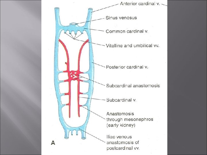
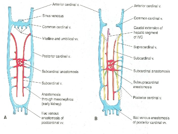
Cardinal Veins Ø The cardinal veins constitute the main venous drainage system of the embryo Ø The anterior and posterior cardinal veins drain cranial and caudal parts of the embryo Ø The anterior and posterior cardinal veins join the common cardinal veins which enter the sinus venosus
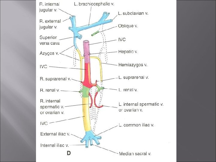
Cardinal Veins Ø During the eighth week the anterior cardinal veins become connected by an anastomosis Ø This anastomosis shunts blood from the left to the right anterior cardinal vein Ø This shunt becomes the left brachiocephalic vein when the caudal part of the left anterior cardinal vein degenerates Ø Superior vena cava forms from the right anterior cardinal vein and the right common cardinal vein
Posterior Cardinal Veins Ø The posterior cardinal veins develop primarily as the vessels of the mesonephroi and largely disappear with these transitory kidneys Ø The only adult derivatives of the posterior cardinal veins are the root of the azygos vein and the common iliac veins
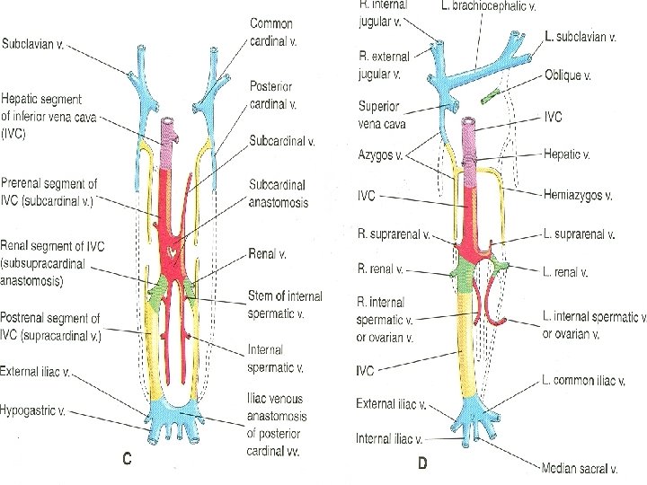
Subcardinal & Supracardinal Veins Ø The subcardinal and supracardinal veins gradually replace and supplement the posterior cardinal veins Ø The subcardinal veins appear first Ø They are connected with each other through the subcardinal anastomosis and with the posterior cardinal veins through the mesonephric sinusoids Ø The subcardinal veins form the stem of the left renal veins, suprarenal veins, gonadal veins and a segment of IVC
Subcardinal & Supracardinal Veins Ø The supracardinal veins are the last pair of vessels to develop Ø They become disrupted in the region of the kidneys Ø Cranial to this they become united by an anastomosis that is represented in the adult by azygos and hemiazygos veins Ø Caudal to the kidneys the left supracardinal vein degenerates Ø The right supracardinal vein becomes the inferior part of the IVC
Inferior Vena Cava Ø The inferior vena cava forms during a series of changes in the primordial veins of the trunk Ø This occurs as blood returning from the caudal part of the embryo is shifted from the left to the right side of the body Ø The IVC is composed of four main segments
Inferior Vena Cava Ø A hepatic segment derived from the hepatic vein (proximal part of right vitelline vein) and hepatic sinusoids Ø A prerenal segment derived from the right subcardinal vein Ø A renal segment derived from the subcardinal supracardinal anastomosis Ø A postrenal segment derived from the right supracardinal vein