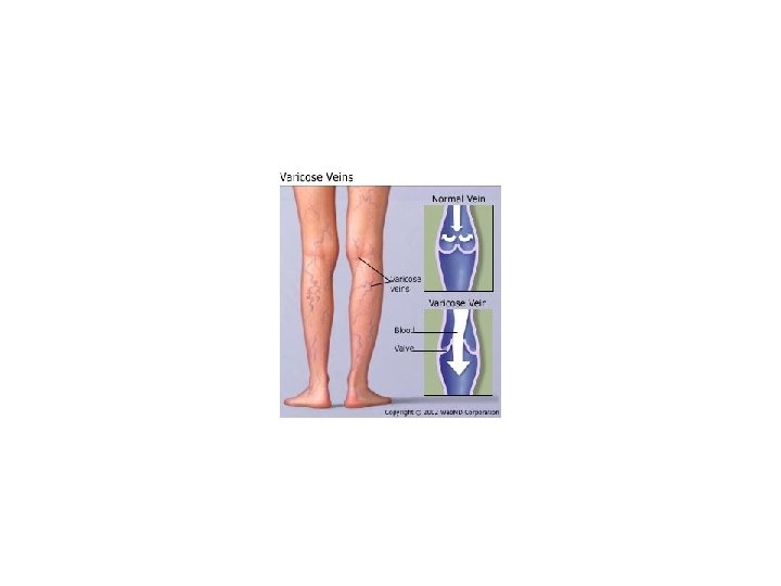
Varicose Veins Vascular Tumors Varicose veins Dilated tortuous


 ---")


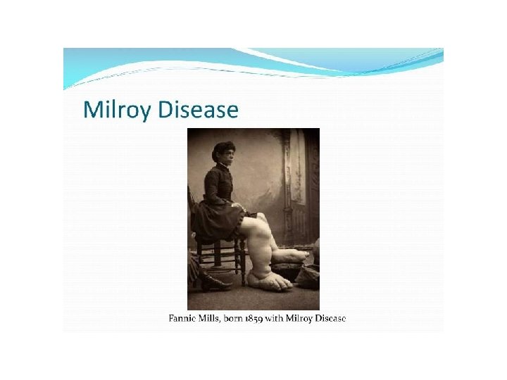
 - familial Milroy disease")


 lymphangiomas - Cavernous lymphangiomas")
. - modified smooth muscle cells of the glomus bodies,")
 - Classic KS: older")


- Slides: 28
Varicose Veins Vascular Tumors
Varicose veins: Dilated tortuous increased intraluminal pressure incompetence of the venous valves.
• superficial veins of the upper and lower leg (m. c, high venous pressures, prolonged dependent posture) • Risk factors: obesity pregnancy familial
Clinical Features: Stasis --- congestion --- edema --- pain --thrombosis. stasis dermatitis (ischemia) --- ulcerations --superimposed infections. embolism from these superficial veins is very rare
Liver cirrhosis /portal vein obstruction/ hepatic vein thrombosis --portal vein hypertension --- portosystemic shunts the gastroesophageal junction (forming esophageal varices), the rectum (forming hemorrhoids), periumbilical veins of the abdominal wall (forming a caput medusa). Esophageal varices are the most important since their rupture can lead to massive (even fatal) upper gastrointestinal hemorrhage.
Thrombophlebitis and Phlebothrombosis Venous thrombosis and inflammation deep leg veins accounts for more than 90% of cases. periprostatic venous plexus /pelvic venous plexus /large veins in the skull and the dural sinuses / Portal vein thrombosis (peritonitis, appendicitis, salpingitis, and pelvic abscesses, platelet hyperactivity e. g. , polycythemia vera) Risk: - Prolonged immobilization --- venous stasis --- DVT --- PE, pain, edema, erythema, cyanosis, heat - Systemic hypercoagulability (genetic, cancer/adenocarcinomas/migratory thrombophlebitis (Trousseau sign).
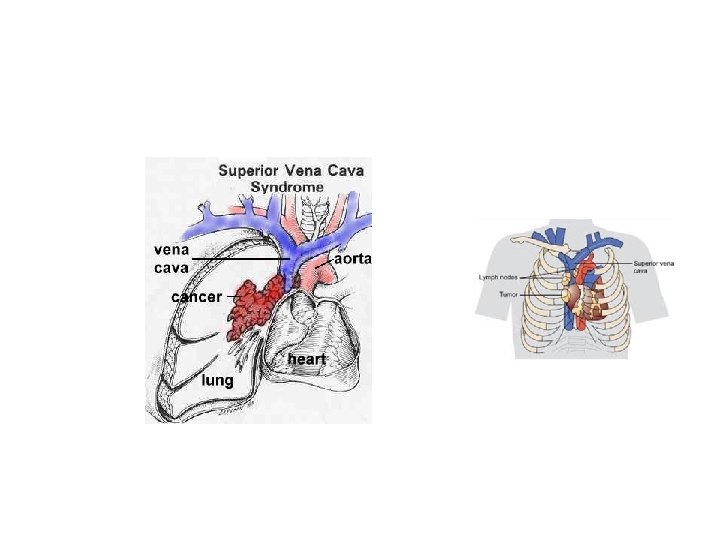
Superior and Inferior Vena Cava Syndromes • The superior vena cava syndrome Neoplasms/mass/aneurysm --- superior vena cava --- dilation of the veins of the head, neck, and arms with cyanosis • The inferior vena cava syndrome Neoplasms/thrombosis (RCC, HCC)--- inferior vena cava -----lower extremity edema
Lymphangitis • Lymphangitis: group A β-hemolytic streptococci are the most common agent red, painful subcutaneous streaks painful enlargement of the draining lymph nodes (lymphadenitis). If bacteria are not successfully contained within the lymph nodes --- venous circulation --- bacteremia or sepsis
Lymphedema Primary lymphedema: - isolated congenital defect (simple congenital lymphedema) - familial Milroy disease (heredofamilial congenital lymphedema) lymphatic agenesis or hypoplasia. Secondary or obstructive lymphedema: - Malignant tumors - Surgical procedures that remove regional groups of lymph nodes (e. g. , axillary lymph nodes in radical mastectomy) - Postirradiation fibrosis - Filariasis - Postinflammatory thrombosis and scarring blockage of a previously normal lymphatic +hydrostatic pressure in the lymphatics --- edema --- peau d’orange (orange peel). chylous ascites (abdomen), chylothorax, and chylopericardium
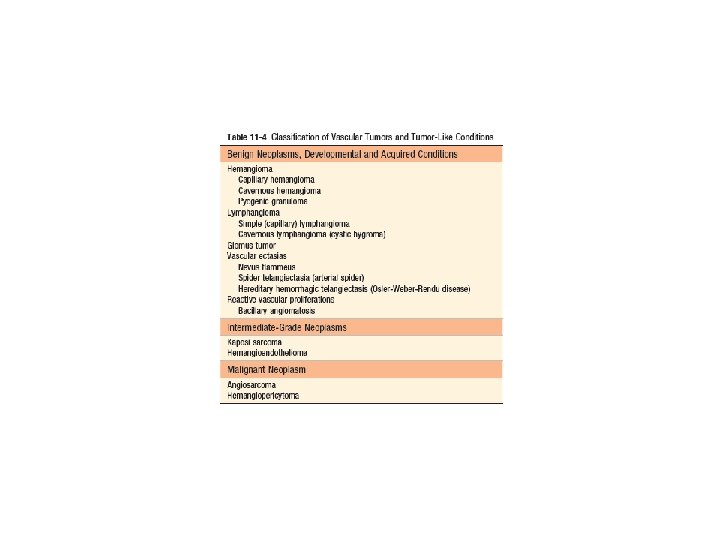
Vascular Tumors
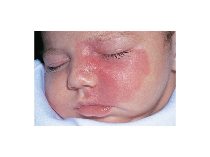
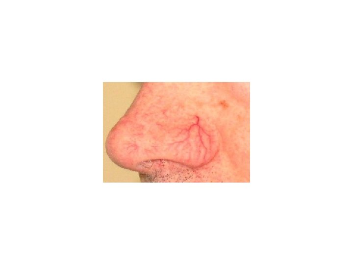
Benign Tumors and Tumor-Like Conditions • • Vascular Ectasias ---------- dilation Telangiectasia ----- dilation small vessels (capillaries, venules, and arterioles) --skin /mm. not true neoplasms ------ malformations/hamartomas - Nevus flammeus (a “birthmark”): m. c, head or neck , regress spontaneously. - port wine stain: do not fade with time. Sturge-Weber syndrome ( facial port wine nevi, ipsilateral venous angiomas in the cortical leptomeninges, mental retardation, seizures, hemiplegia, and skull radio-opacities) - Spider telangiectasias : face, neck, or upper chest, hyperestrogenic states (pregnancy / liver cirrhosis). - Hereditary hemorrhagic telangiectasia (Osler-Weber-Rendu disease): AD, skin /mm/respiratory, gastrointestinal, and urinary tracts. spontaneously rupture
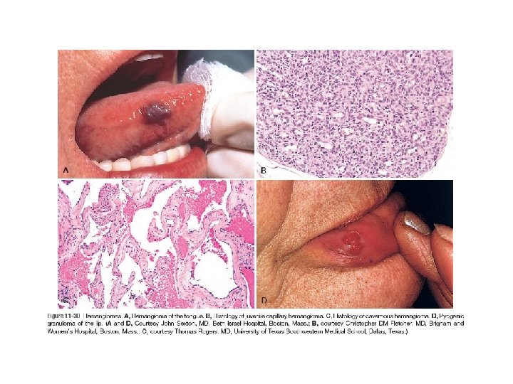
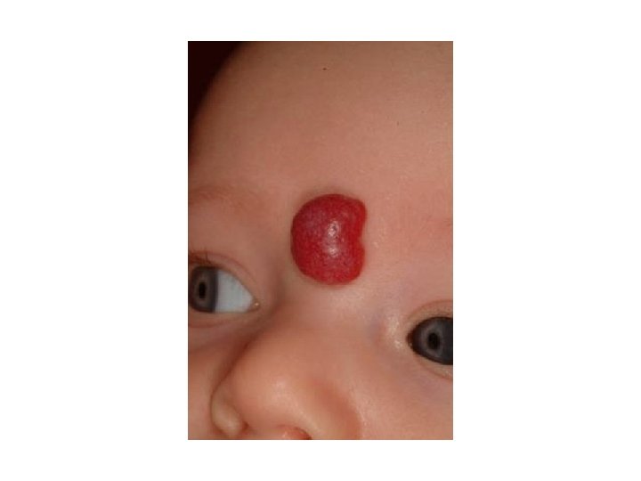
• Hemangioma: common infancy and childhood initially increase in size, but many eventually regress spontaneously head and neck, liver. - Capillary hemangiomas - Juvenile hemangiomas “strawberry type” hemangiomas): newborn , fade by 1 to 3 years of age and completely regress by age 7 - Cavernous hemangiomas : deep structures, do not spontaneously regress. On histologic examination - Pyogenic granulomas: capillary hemangiomas , rapidly growing , skin/gingival/oral mucosa. Pregnancy tumor (granuloma gravidarum) is a pyogenic granuloma that occurs infrequently (1% of patients) in the gingiva of pregnant women.
• Lymphangiomas. lymphatic counterparts of hemangiomas. - Simple (capillary) lymphangiomas - Cavernous lymphangiomas (cystic hygromas) Turner syndrome.
• Glomus Tumor (Glomangioma). - modified smooth muscle cells of the glomus bodies, arteriovenous structures involved in thermoregulation. - distal portion of the digits, especially under the fingernails. Bacillary Angiomatosis. - immunocompromised hosts (e. g. , patients with AIDS) opportunistic gram-negative bacilli of the Bartonella family. - induction of host hypoxia-inducible factor-1 (HIF-1) by the bacteriafactor (VEGF) production. - cleared by macrolide antibiotics (including erythromycin).
• Kaposi Sarcoma. - human herpesvirus 8 (HHV 8) - Classic KS: older men , Mediterranean, malignancy or altered immunity, nodules, distal lower extremities - Endemic African KS: younger than age 40 , lymph nodes much more frequently than the classic variant. - Transplant-associated KS: solid organ transplant recipients /immunosuppression. lymph nodes, mucosa, and viscera - AIDS-associated (epidemic) KS : lymph nodes , viscera
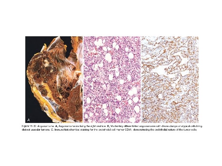
Malignant Tumors • Angiosarcoma. older adults skin, soft tissue, breast, and liver. Hepatic angiosarcoma : arsenic (e. g. , in pesticides), Thorotrast (a radioactive contrast agent formerly used for radiologic imaging), and polyvinyl chloride (a widely used plastic). Angiosarcoma can also arise in the setting of lymphedema, - ipsilateral upper extremity /radical mastectomy /breast lymphangiosarcom).