TUMORS OF EXTERNAL AND MIDDLE EAR Tumors of


 � Females – 5")


- Slides: 34
TUMORS OF EXTERNAL AND MIDDLE EAR
Tumors of auricle � Pre auricular sinus or cyst � Due to faulty union of hillocks of 1 st and 2 nd branchial arches � Tract lined by squamous epithelium
Tumors of auricle � Sebaceous cyst – post auricular sulcus � Dermoid cyst – over upper part of mastoid � Keloid follows trauma Surgical excision with injection of triamcinolone
Tumors of auricle � Hemangioma � 1. capillary – port wine stain � 2. cavernous – also called strawberry tumor
Tumors of auricle � PAPILLOMA
Malignant tumors � SQUAMOUS CELL CA � Helix � Painless nodule or ulcer � Males , fifties � Small lesions – excised locally � Large lesions with nodal mets – total amputation of pinna with en bloc removal of parotid and cervical nodes
Malignant tumors � BASAL CELL CA � Men beyond 50 y � Helix and tragus � Nodule with central crust/ ulcer � No lymphatic spread � Superficial lesions – RT � Deep - Surgery
Malignant tumors � MELANOMA � Light complexion � Exposure to sunlight � Superficial <1 cm – wedge resection � Rest – Resection of pinna, parotidectomy and radical neck dissection
Benign Tumors of EAC � OSTEOMA
Benign Tumors of EAC � EXOSTOSES � Multiple, bilateral � Smooth sessile bony swelling in deep meatus � Divers, swimmers � Large – remove with a high speed drill
Benign Tumors of EAC � CERUMINOMA � Smooth skin covered swelling in outer part of meatus � Recurrence � Wide surgical excision
Malignant tumors of EAC � Squamous cell CA � Long standing ear discharge � Blood stained ear discharge , ear ache � Ulcer, polyp / granulation � Facial palsy � Lymphatic spread � Surgery with post op RT
Malignant tumors of EAC � Basal cell CA � Malignant ceruminoma � Malignant melanoma
Tumors of middle ear 1. Primary Benign – glomus tumor Malignant – Carcinoma, sarcoma 2. Secondary From adjacent areas – eg: nasopharynx, external meatus, parotid Metastatic- From CA bronchus, breast, thyroid, prostrate, GIT
Glomus tumor �Most common benign neoplasm of middle ear �Origin – glomus bodies �Paraganglionic cells from neural crest
Aetiology and pathology � Middle age (40 – 50 y) � Females – 5 times more common � Benign non encapsulated extremely vascular � Slow growth
Pathology
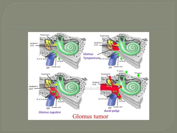
Types �Glomus jugulare �Glomus tympanicum
Spread of glomus tumor
Clinical features �Hearing loss �Tinnitus �Profuse bleeding from the ear �Dizziness or vertigo
Clinical features � Rising sun sign � Brown’s sign � Red vascular polyp filling the meatus
�Cranial nerve palsies �Audible bruit �Rule of 10 – 10% familial, 10% multicentric, 10 % functional ie they secrete catecholamines
Investigations
Treatment �Surgical removal �Radiation �Combination of the above
INITIAL CAROTID ARTERIOGRAM
Surgical approaches � Transcanal � Hypotympanic � Extended facial recess � Mastoid neck � Infratemporal fossa ( Fisch) � Transcondylar
�Radiation �Reduces vascularity �Done for inoperable cases, residual and recurrent cases, elderly
Carcinoma of middle ear �Rare � 40 – 60 y �Females
Pathology � Squamous cell CA � Adenocarcinoma
Clinical features � Chronic foul smelling blood stained ear discharge � Pain � Facial palsy � Granulations or polyp � Hearing loss or vertigo
Diagnosis � Biopsy � CT scan
Treatment � Surgery Radical mastoidectomy, subtotal or total petrosectomy � Radiotherapy
Sarcomas �Rare �Children �Arises from pleuripotent stem cells �Ear discharge, facial palsy �Radiotherapy and chemotherapy