Total Serum Proteins Dr Aida Ahmed Lecturer of



- Slides: 53
Total Serum Proteins Dr/ Aida Ahmed Lecturer of Clinical Pathology 28/11/2015
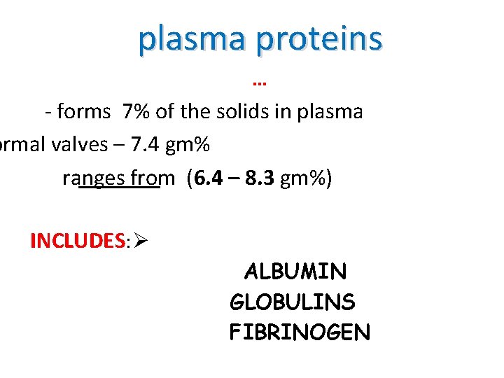
plasma proteins … ‐ forms 7% of the solids in plasma ormal valves – 7. 4 gm% ranges from (6. 4 – 8. 3 gm%) INCLUDES: Ø ALBUMIN GLOBULINS FIBRINOGEN
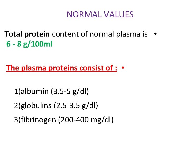
NORMAL VALUES Total protein content of normal plasma is • 6 - 8 g/100 ml The plasma proteins consist of : • 1)albumin (3. 5‐ 5 g/dl) 2)globulins (2. 5‐ 3. 5 g/dl) 3)fibrinogen (200‐ 400 mg/dl)
The albumin : globulin ratio is • usually between 1. 2 : ; 1 to 1. 5 : 1 Almost all plasma proteins , except • immunoglobulins are synthesized in liver
• Clinical Significance: The two general causes of alteration of serum total protein are : A‐ change in the volume of plasma water and B‐a change in the concentration of one ore more of specific proteins in the plasma.
1‐Impact of Haemoconcentration and Haemodilution a‐ Relative hyperproteinemia: Decrease in the volume of plasma water(haemoconcentration)is reflected as relative hyperproteinemia , concentrations of all the individual plasma proteins are increased to the same degree.
Hemoconcentration is noted in dehydration due to inadequate water intake or to excessive water loss as in severe vomiting, diarrhoea. addison, s disease, or diabetic ketoacidosis
b‐Relative hypoproteinemia: Hemodilution(Increase in plasma water volume) s reflected as relative hypoproteinemia. concentrations ll the individual plasma proteins are decreased to the same degree.
Haemodilution occurs with 1‐water intoxication • or 2‐ salt retention syndromes, 3 during massive intravenous infusions and physiologically when a 4 -recumbent position is assumed. A recumbent position decreases total protein concentration by 0. 3 -0. 5 g/dl and many individual proteins by up to 10%.
• 2‐ Impact of hypoalbuminaemia: • Of the individual proteins, albumin is present in such high concentrations that low levels of it alone may cause hypoproteinemia, such hypoproteinemia is quite common and has many causes.
Causes of Hypoalbumineamia: a‐ Impaired synthesis: Either primary or genetic(e. g. , in analbuminemia) or aquired(e. g. , due to diminished protein intake, ver disease, or inflammatory processes due to direct inhibition by cytokines as tumour necrosis factor‐œ(TNF-œ), nterleukin‐ 1(IL-1), and interleukin‐ 6(IL-6)
B‐ Increased catabolism: AS a result of tissue damage and inflammation. C‐ Reduced absorption of amino acids: Caused by malabsorption syndromes D‐ Protein loss: In urine, due to nephrotic syndrome, chronic glomerulonephritis, diabetes mellitus, systemic lupus erythematosus, in faeces, due to protein‐ losing enteropathy arising from inflammatory or neoplastic disease, or from the skin itself thruogh burns.
3‐ Imact of Increased Levels of Specific Serum Proteins: Mild hyperproteinaemia may be caused by an increase in the concentration of specific proteins normally present in relatively low concentration such as : a‐ œ 1 –Acid glycoprotein (Orosomucoid): An œ 1‐globulin, increased by 3‐ 4 folds in conditions associated with inflammation or tissue necrosis( acute phase response), and by glucocorticoid effect(cushing syndrome or steroid therapy.
b ‐œ 1 - Antitrypsin(œ 1 – proteinase inhibitor): Increased as an acute phase response, and by estrogen therapy and late pregnancy). C‐Ceruloplasmin : An œ 2‐ globulin, increased in the acute phase response, and its synthesis is also stimulated by oestrogen
d- C-Reactive protin: Has long been recognized as one of the most , or even the most sensitive acute phase reactant. Serum levels rise dramatically after myocardial infarction, srtress, trauma , infection, inflammation, surgery, or neoplastic prolifration.
œ 1 –Fetoprotin(AFP) • Its elevation in maternal serum idicates the possoibility of neural tube defect in the fetus AFPis also an important marker whose rise in indicative of hepatocellular and germ cell carcinomas.
F- Haptoglobin: An œ 2 - glycoprotein, whose synthesis is increased as an acute phase response in the presence of acute inflammatory processes, tissue necrosis or malignancy. It is also increased in protein –losing syndromes as nephrotic syndrome, and protein – losing enteropathies (inceased synthesis as a compansatory mechanism), and by the effect of androgen and glucocorticoids.
G -œ 2 -Macroglobulin: Its levels are increased in nephrotic syndrome, in part because of volume loss secondary to decreased plasma oncotic (osmotic) pressure , and in part because of increased synthesis of all proteins to compansate for loss by the kidnys. The elevated levels compansate renal loss of lower molecular physiologically for –weight proteins inhibitors. Levels are also increased by estrogens.
h- β 2 -Microglobulin: High plasma levels of which occur in renal failure, inflammation, neoplasms, especially those associated with B- lymphocytes. Its prencipal clinical value is to test renal tubular function paricularely in kidney transplant recipients. In whom rejection of the allograft manifests as diminished tubular function.
i- Complement protein C 3: Increased plasma levels of which are reported as an acute phase response after trauma , surgery, and during inflammatory processes. In biliary obstruction , levels are elevated in direct proportional to the levels of hyperbilirubinemia. About 30% of pateints with idiopathic focal glomerulosclerosis have elevated C 3 levels.
4‐ Impact of Increased Levels of Immunoglobulins: Mild, moderate or severe hyperproteinemia may be caused by high levels of the immunoglobulins. This is either of polyclonal or monoclonal nature. Polyclonal hyperimmunoglobulins: (Polyclonal Gammopathy): Polyclonal increases In serum immunoglobulins are the normal response to infections.
Ig. G tends to predominate in autoimmune • responses, Ig. A in skin, gut, respiratory and renal infections, and Ig. M in primary viral infections and blood stream infections such as malaria. Chronic bacterial infections cause an increase • serum levels of all immunoglobulins. ,
In primary biliary cirrhosis, the Ig. M level is • markedly elevated, in active hepatitis, Ig. G, and sometimes Ig. M are increased , and In portal cirrhosis, Ig. A and sometimes Ig. M are increased. In intrauterine infections, production of Ig. M by the • fetus increases and at birth the Ig. M Level in the cord blood is increased. Estimation of Ig. E are used in management of the asthema and other allergic conditions, especially in children.
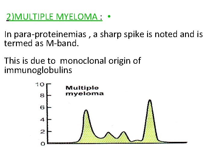
b- Monoclonal immunoglobulins(Monoclonal Gammopathy): A single clone of plasma cells produces immunoglobulin molecules with identical structure. If the clone is permitted to multiply, the concentration of its particular protein in the pateint, s serum become so great that on electrophoresis, .
It often produces anarrow Sharply spike. These also referred to monoclonal immunoglobulins are as paraproteins. About 60% of paraproteins are due to multiple myloma or a solitary plasmacytoma , about 15% are due to overproduction of B –lymphocytes, mainly in lymph nodes , lymphomas, chronic lymphatic leukemia, Waldenstrom, s macroglobulinemia, or rarely in Heavy chain disease. Probably up to 25% of paraproteins are benign.
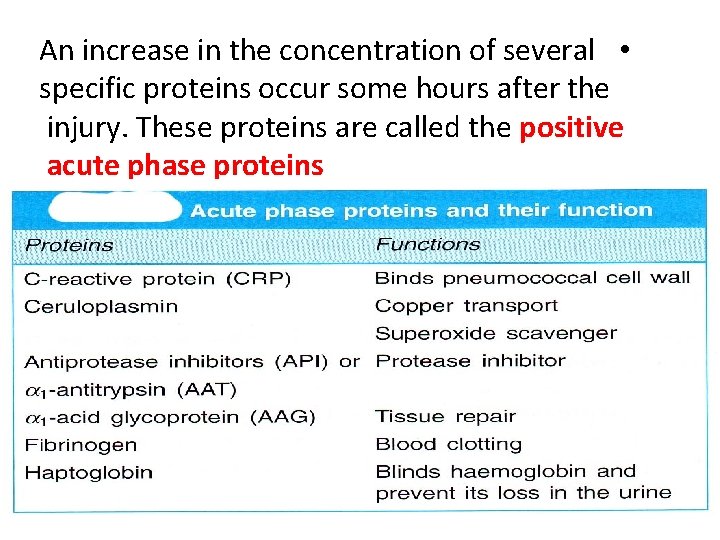
An increase in the concentration of several • specific proteins occur some hours after the injury. These proteins are called the positive acute phase proteins
ESTIMATION OF PLASMA PROTEINS In clinical laboratory , total proteins of patients are • estimated by Biuret method. Albumin is quantitated by Bromo cresol green • (BCG) method , in which the dye is preferentially bound with albumin , and the colour intensity is measured colourimetrically.
Specimen: Serum, urine, cerebrospinal fluid(CSF) , amniotic fluid, saliva, faeces
Biuret method Lowry’s method Terbidimetry and Neplelometry Kjeldahl’s method Dye‐binding method UV‐absorption method(Direct photometric ) Refractometry method Electrophoresis
1 -Biuret Method principle: This method depends on the presence of peptide bonds in all proteins. These peptide bonds react with Cu 2+ ions in alkaline solution to form a colored product whose absorbance is measured spectrophotometrically at 540 nm.
The biuret reagent contains sodium potassium tartarate to complex cupric ions and maintain their solubility in alkaline solution, iodide is included as an antioxidant. In the reaction , a colord chelate is formed between the Cu+2 ion and the carbonyl oxygen (C=Oand amide nitrogen atoms(NH) of the peptide bond.
The sensitivity of the reaction can be increased by the addition of phosphotungstic phosphomolybdic acid (Folin‐ Ciocalteu reagent, phenol reagent) (Lowry method), which, together with other modifications, results in increased absorption for a given amount of protein.
2‐Folin‐ Ciocalto(Lowry) method Advantage of this method than biuret method is lowest detection limit 100 times lower than biurete so can measure the very low concentration of proteins. Disadvantage not wholly satisfactory for urine , CSFproteins because the reagent reacts with nonspecifically with nonprotein compounds resulting in positive errors
the reactivity of the various plasma proteins to the biuret reagent is not significantly different, since it is dependent on the number of peptide linkages, not on amino acid composition
Interference Causes include: . bilirubin: [bilirubin] 500 μmol/L causes negative interference. haemoglobin: [Hb] 10 g/L causes up to 6% positive interference lipaemia causes negligible interference
3 -Dye –Binding methods: These methods depend on the ability of proteins to bind dyes such as Amido Black 10 B And Coomassie brilliant blue This proparty is involved to stain protein bands after electrophoresis for assay of serum albumin, also the dye can also be used in spectrophotometric methods for total protein
4 -Kjeldal Method: Based on acid digetion used to convert nitrogen in the protein to ammonium ion The concentration of ammonia nitrogen was then evaluated by titeration or nesslerization Advantage well defined and reproducible Disadvantage Time consuming
Kjeldal Method still remains a means of • defining referance material for the biuret method in use today. 5‐ Refractometry: ‐Refractometry is a quick alternative to chemical analysis for serum total protein when a rabid estimate is required.
Principle • The refractive index of water at 20 o. C is 1. 33. • If solute is added to the water the refractive index • of a dilute solution is increased linearly and proprtionally to the solute concentration Disadvantage • Large positive error with azotemia, and • hyperglycemia, altration of A/G ratio, hyperbilirubinmia an specially lipemia •
4‐ Direct photometric methods: principle: Absorption of ultraviolet (UV) light at 200‐ 225 nm and 270‐ 290 nm can be used to measure the protein content of biological samples This depends mainly on the aromatic rings of tyrosine and tryptophan at PH 8 ,
Disadvantage: accuracy and specificity cannot be guaranteed because of the uneven distribution of these amino acids among the individual proteins, also there are interference with( UA), Bbilirubin
5 -Turbidimetric and Nephelometric Methods Principle Precipetation of total protein assay depend on formation of a fine precipitate of uniform , insoluble protein particles, which in suspension scatter incident light).
Precipitation of protein for turbidimetric or nephelometric assay can achieved with sulfosalicilic acid alone or with SSA in combination with sodium sulfate or trichloroacetic acid(TCA) or TCA alone. This method is suited for tatal protein measurement in urine , CSF.
ELECTROPHORESIS The most common method of analyzing plasma • proteins is by electrophoresis. The term electrophoresis refers to the movement • of charged particles through an electrolyte when subjected to an electric field.
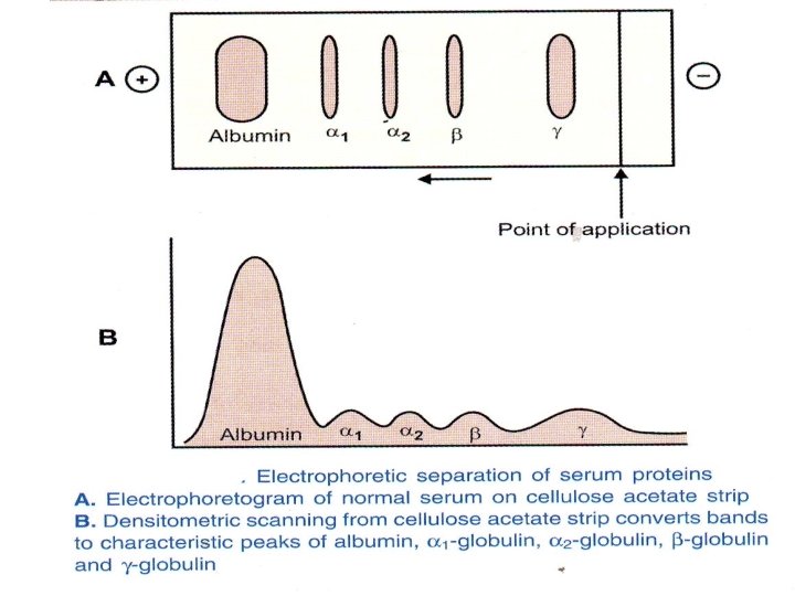
In clinical laboratory , cellulose acetate is widely • used as a supporting medium. Its use permits resolution , after staining , of plasma • proteins into five bands , designated albumin , α 1 , α 2 , β and γ fractions, respectively
The stained strip of cellulose acetate is called • electrophoretogram. The amounts of these five bands can be • conveniently quantified by use of Densitometric scanning machines. Characteristic changes in the amounts of one or • more of these five bands are found in many diseases.
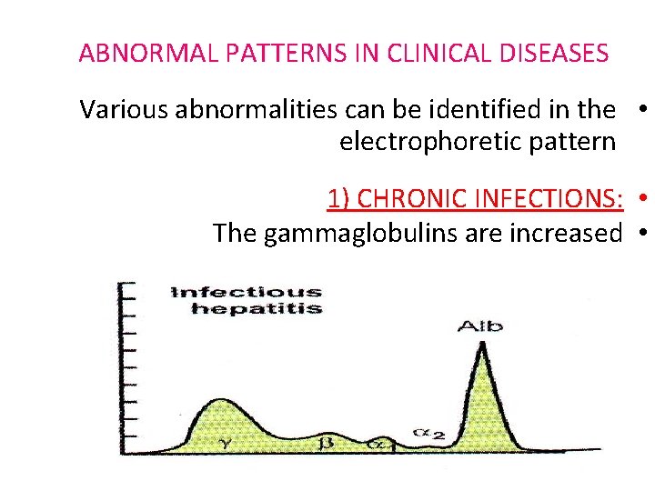
ABNORMAL PATTERNS IN CLINICAL DISEASES Various abnormalities can be identified in the • electrophoretic pattern 1) CHRONIC INFECTIONS: • The gammaglobulins are increased •
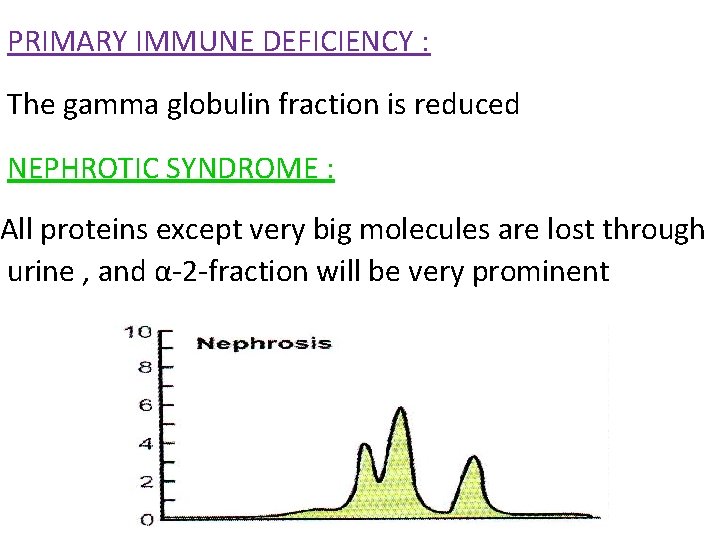
PRIMARY IMMUNE DEFICIENCY : The gamma globulin fraction is reduced NEPHROTIC SYNDROME : All proteins except very big molecules are lost through urine , and α‐ 2‐fraction will be very prominent
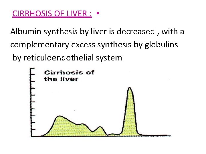
CIRRHOSIS OF LIVER : • Albumin synthesis by liver is decreased , with a complementary excess synthesis by globulins by reticuloendothelial system
2)MULTIPLE MYELOMA : • In para‐proteinemias , a sharp spike is noted and is termed as M‐band. This is due to monoclonal origin of immunoglobulins
Thank you